Eur. J. Biochem. 269, 1162–1170 (2002) (cid:211) FEBS 2002
Repression of FasL expression by retinoic acid involves a novel mechanism of inhibition of transactivation function of the nuclear factors of activated T-cells
Mi-Ock Lee1,*, Hyo-Jin Kang1,*, Young Mi Kim1, Ji-Hyun Oum2 and Jungchan Park2 1Department of Bioscience and Biotechnology, Institute of Bioscience, Sejong University, Seoul, Korea; 2Department of Bioscience and Biotechnology, Hankuk University of Foreign Studies, Kyounggi-do, Korea
tion of the distal NFAT binding motif present in the inter- leukin (IL)-2 promoter, suggesting that the inhibition of NFAT function by all-trans-RA was not specific to the FasL promoter. Gel shift assays corroborated the results of the gene reporter studies by showing that all-trans-RA decreased the NFAT binding to DNA. All-trans-RA blocked trans- location of NFATp from the cytosol into the nucleus, which was induced by PMA/ionomycin treatment in HeLa cells transfected with a Flag-tagged NFATp. Taken together, our results indicate that FasL inhibition by all-trans-RA involves a novel mechanism whereby the transcriptional function of NFAT is blocked.
Keywords: retinoic acid; NFAT; FasL.
Retinoids are potent immune modulators that inhibit Fas ligand (FasL) expression and thereby repress the activation- induced apoptosis of immature thymocytes and T-cell hybridomas. In this study, we demonstrate that all-trans- retinoic acid (all-trans-RA) directly represses the transcrip- tional activity of the nuclear factors of activated T-cells (NFAT), which is an important transactivator of the FasL promoter. The analysis of reporter constructs containing the FasL promoter and wild-type or mutant NFAT binding- sites indicated that all-trans-RA repression was mediated via an NFAT binding element located in the promoter. A reporter construct comprising the NFAT binding sequence linked to a heterologous SV-40 promoter showed that NFAT transcriptional activity was significantly inhibited by all-trans-RA. Furthermore, all-trans-RA inhibited activa-
factors of
receptors (RXRs), both of which are ligand-dependent transcriptional the steroid/thyroid hormone receptor superfamily [9–11]. However, the molecular details of RA-mediated repression of FasL gene expression have not been elucidated.
The CD95 (Fas) ligand (FasL) is a type-II transmembrane protein expressed on highly activated T-lymphocytes [1,2]. Activated T-lymphocytes undergo apoptosis following homotypic interaction of FasL and its receptor, Fas [3–5]. Thus, the elimination of highly activated T-cells by the Fas/ FasL system is critical for the downregulation of immune responses, the homeostasis of lymphocytes, and the main- tenance of peripheral tolerance. Retinoids, vitamin A and its natural and synthetic derivatives, regulate a wide array of biological processes, including cellular proliferation, differ- entiation, and immune modulation. All-trans-retinoic acid (RA) and 9-cis-RA inhibit FasL expression, and thereby suppress the activation-induced apoptosis of immature thymocytes and T-cell hybridomas [6–9]. The inhibitory effects of RA are mediated through two classes of nuclear receptors, retinoic acid receptors (RARs) and retinoid X
Nuclear factors of activated T-cells (NFAT) is a family of related transcription factors that play a central role in regulating the immune response by modulating the expres- sion of important cytokines such as interleukin (IL)-2 in the activated T-cells [12]. Five members of the NFAT family are currently known, NFATp (NFAT1, NFATc2), NFATc (NFATc4), NFAT4 (NFAT2, NFATc1), NFAT3 (NFATc3, NFATx), and NFAT5, which share homology within a region of the DNA binding domain that is distantly related to the Rel domain [13–17]. Moreover, various lines of biochemical evidence, including knock-out studies and tissue distribution patterns of the proteins, indicate that three of the NFAT family members, NFATp, NFATc, and NFAT4, play important roles in the modulation and development of the immune system [12,18]. Although NFAT5 appears to be constitutively localized in the nucleus and under the regulation of osmotic shock, the other NFAT family members are primarily controlled by their subcellular localization depending on their phosphorylation status. In resting T-cells, NFAT proteins are present in the cytoplasm in a phosphorylated state. Activation via the T-cell receptor (TCR) or other stimulus results in an influx of calcium and induces the dephosphorylation of NFAT, and rapid trans- location of the protein into the nucleus [19,20]. Dephos- phorylated NFAT binds to specific response elements and including those thereby activates a number of genes,
Correspondence to M.-O. Lee, Department of Bioscience and Biotechnology, Sejong University, 98 Kunja-dong, Kwangjin-gu, Seoul 143-747, Korea. Fax: + 82 2 3408 3768, Tel.: + 82 2 3408 3768, E-mail: molee@sejong.ac.kr Abbreviations: FasL, Fas ligand; RA, retinoic acid; RARs, retinoic acid receptors; RXRs, retinoid X receptors; NFAT, nuclear factors of activated T-cells; TCR, T-cell receptor; CsA, cyclosporin A; PBMCs, peripheral blood mononuclear cells; PMA, 4b-phorbol 12-myristate 13-acetate; b-gal, b-galactosidase; IL, interleukin; VDR, vitamin D receptor. *Note: both authors contributed equally to this work. (Received 30 July 2001, revised 18 December 2001, accepted 19 December 2001)
Repression of NFAT by retinoic acid (Eur. J. Biochem. 269) 1163
(cid:211) FEBS 2002
encoding cytokines, cell surface receptors, signaling mole- cules, and other, as yet unidentified, targets. As NFAT dephosphorylation is mediated by the Ca2+/calmodulin- dependent phosphatase, calcineurin, NFAT-regulated genes are sensitive to inhibition by immunosuppressive agents that inhibit calcineurin, such as cyclosporin A (CsA) and FK506 [21].
Recently, several studies have demonstrated the involve- ment of NFAT in the transcriptional activation of FasL [22–25]. Therefore, we speculated that NFAT inhibition might be an important mechanism through which RA inhibited the expression of FasL. In this study, we show that all-trans-RA inhibits FasL expression by blocking tran- scriptional activation by NFAT. Our results suggest the therapeutic potential of targeting NFAT function with RA to achieve immunosuppression.
E X P E R I M E N T A L P R O C E D U R E S
Cells and reagents
(DNFAT) or SP1 (DSP-1) sites, were previously described [22]. The luciferase reporter constructs containing deleted promoter fragments (nucleotides )1783 to )2) and (nucleo- tides )1703 to )2), were constructed by restricting the 2.3-kb full promoter using XhoI and NcoI/XhoI, respectively. The NFAT-Luc reporter was constructed by inserting an oligonucleotide encoding the NFAT binding site of the FasL promoter (5¢-ATTGTGGGCGGAAACTTCCAG-3¢) with additional GATC motifs at the 5¢ end into the BglII site of the pGL2-promoter (Promega, Madison, WI) that carries an SV40 promoter. The eukaryotic expression vectors carrying Flag-NFATp, RARa, RARb, RARc, and RXRa have been reported previously [27,28]. Jurkat cells (1–2 · 107 cells) were transfected with reporter plas- mids (7.5 lg) or with a b-galactosidase (b-gal) expression vector (2.5 lg) by electroporation. CV-1 cells were seeded in a 24-well culture plate at 5 · 104 cells per well, and transfected with DNA mixtures (1 lg per well) containing reporter plasmids (0.1 lg), the eukaryotic expression vector encoding Flag-NFATp (25 ng), the retinoid receptor expression plasmid (25 ng), or the b-gal expression vector (0.15 lg) with carrier DNA (pBluescript). The cell cultures were incubated for 6 h with PMA (10 ngÆmL)1) and ionomycin (0.5 lM), in the presence or absence of all- trans-RA. At the end of the incubation period, luciferase activity was determined using a luminometer according to the manufacturer’s instructions. The luciferase activity was normalized for transfection efficiency using the correspond- ing b-gal activity.
The Jurkat human T-cell leukemia (ATCC, CRL1990), and HeLa human cervical carcinoma (ATCC, CCL-2) cell lines were obtained from the American Type Culture Collection. Cells were maintained in RPMI 1640 medium containing 10% fetal bovine serum. Human peripheral blood mono- nuclear cells (PBMCs) were isolated from healthy donors by density gradient centrifugation of heparinized blood on a layer of Ficoll/Hypaque (Sigma, St. Louis, MO, USA). All- trans-RA, 9-cis-RA, 4b-phorbol 12-myristate 13-acetate (PMA) and CsA were purchased from Sigma. Ionomycin was obtained from Calbiochem (La Jolla, CA, USA). All other chemicals used were of the purest grade available from Sigma.
To examine the effects of all-trans-RA on IL-2 NFAT site-dependent transcription, we employed a Jurkat cell line that was stably transfected with the NFATZH reporter construct (Oum, J.-H. & Park, J., unpublished results). The reporter construct contained three copies of the distal NFAT binding site in the human IL-2 promoter and a minimal IL-2 promoter, upstream of the b-gal gene [29]. The Jurkat-NFAT cells (1 · 105 cells per well) were cultured in a 24-well plate and stimulated for 6 h with PMA (10 ngÆmL)1) and ionomycin (0.5 lM), in the presence or absence of all-trans-RA (2.0 lM). The b-gal activity was determined using the fluorogenic substrate 4-methyl-lum- bellifery-b-galactoside, and was normalized for protein content [30]. A one-way analysis of variance was performed using GraphPad INSTAT(cid:210) (GraphPad Software, San Diego, CA, USA). A value of P < 0.05 was considered statistically significant.
RT-PCR for FasL Jurkat cells (2 · 106 cells) were treated with a mixture of PMA (10 ngÆmL)1) and ionomycin (0.5 lM) for 6 h with or without a 24-h pretreatment with various concentrations of all-trans-RA. Total RNA was prepared using Qiagen RNeasy kit (Qiagen Inc., Chatsworth, CA, USA) following the manufacturer’s instructions. RT-PCR was performed essentially as described previously [26]. cDNA was synthe- sized from 4 lg total RNA using 100 ng random hexamer (Pharmacia, Uppsala, Sweden). The PCR primer sequences used were as follows. FasL (forward: 5¢-ATGTTTCAGC TCTTCCACCTACAGAAGGA-3¢, reverse: 5¢-CAGAGA GAGCTCAGATACGTTGAC-3¢); and b-actin (forward: 5¢-CGTGGGCCGCCCTAGGCACCA-3¢,reverse: 5¢-TTG GCCTTAGGGTTCAGGGGGG-3¢. PCR cycling condi- tions were: de-naturation at 94 (cid:176)C for 30 s, annealing at 52 (cid:176)C for 30 s and extension at 72 (cid:176)C for 30 s. Twenty-eight cycles were carried out for amplification of FasL and 22 cycles for b-actin.
Plasmids and reporter gene assay
The luciferase reporter constructs containing a 2.3-kb fragment (from nucleotides )2365 to )2) and a 320-bp fragment (nucleotides )318 to )2) of genome region located 5¢ upstream of the FasL translation initiation site, and the luciferase reporters containing mutations in the NFAT
Electrophoretic mobility shift assay (EMSA) PBMCs (7 · 106 cells) obtained from healthy donors were stimulated in a 100-cm2 plates precoated with anti-CD3 Ig (100 lgÆmL)1) for 4 h with or without various concentra- tions of all-trans-RA pretreatment. A mouse antibody against human CD3 was prepared from the supernatants of OKT3 hybridoma cell cultures [28]. Nuclear extracts were prepared from the PBMCs and gel-shift assays were carried out using previously described methods [28]. Nuclear extracts (5 lg) were incubated for 20 min at 25 (cid:176)C with 32P-labeled oligonucleotides encoding either the NFAT or SP-1 binding sequences in a 20-lL reaction mixture containing 10 mM Tris buffer (pH 7.5), 100 mM KCl, 1 mM dithiothreitol, 1 mM EDTA, 0.2 mM phenyl- methanesulfonyl fluoride, 1 mgÆmL)1 BSA, and 5%
1164 M.-O. Lee et al. (Eur. J. Biochem. 269)
(cid:211) FEBS 2002
glycerol. The sequences of oligonucleotides used as probe in the experiments were: NFAT, 5¢-GATCATTGTGGGCG GAAACTTCC AG-3¢; and SP-1, 5¢-GATCGATCGGGG CGGGGCGAG-3¢.
Immunofluorescence studies For the subcellular localization studies, HeLa cells (1 · 106 per well) were transiently transfected with 4 lg Flag- NFATp using LipofectaminePlusTM (Gibco BRL, Grand Island, NY, USA) according to the manufacturer’s instruc- tions. The transfected HeLa cells were cultured for 24 h on poly L-lysine-coated 11-mm coverslips. The cells were stimulated with PMA (10 ngÆmL)1) and ionomycin in the presence or absence of all-trans-RA (0.5 lM), (1.0 lM). Following treatment, the cells were fixed overnight at )20 (cid:176)C in a methanol/acetone (1 : 1) solution. The cells were then stained with an anti-(Flag M2) Ig (Upstate Biotech., Lake Placid, NY, USA) at a concentration of 1 lgÆmL)1 in NaCl/Pi and 1% bovine serum albumin, followed by a biotin-labeled, anti-(mouse Ig) Ig (1 : 1000, Vector Laboratories, Inc., Burlingame, CA, USA), and streptavidin–fluorescein isothiocyanate (1 : 200, Vector Laboratories). Fluorescent cells were washed with NaCl/Pi and visualized by confocal microscopy (Nikon, Japan).
R E S U L T S
All-trans-RA represses FasL expression
four reporter genes that were induced by PMA and ionomycin treatment. These results suggested that the putative RA-responsive elements were located within the nucleotides )318 to )2 region of the FasL promoter.
As RA has been shown to inhibit the expression of FasL in the immature thymocytes and T-cell hybridomas [6–8], we confirmed these data using a human leukemia cell line, Jurkat. The addition of PMA and ionomycin into culture media remarkably induced the expression of FasL in Jurkat cells and the induction was repressed by all-trans-RA treatment in a dose-dependent manner (Fig. 1A). FasL transcription was decreased at all-trans-RA concentrations as low as 0.01 lM, and was almost completely abolished at 1.0 lM. To further establish the inhibitory effect of all-trans- RA on FasL gene expression, we employed a luciferase reporter system containing the 2.3-kb genomic DNA fragment that is sufficient for transcriptional activation of the FasL gene [22]. Transient transfection of the reporter into Jurkat cells produced a 3.25-fold increase in reporter gene activity in response to PMA and ionomycin treatment, a finding that was consistent with previously reported results [22]. Approximately 80% of the reporter gene activity was repressed in the presence of all-trans-RA (Fig. 1B). In summary, the results from RT-PCR and reporter gene analyses clearly showed that RA decreased the transcrip- tional expression of FasL in Jurkat cells.
The NFAT binding motif in the FasL promoter confers responsiveness to all-trans-RA
We studied the RA-responsive, cis-regulatory elements in the FasL promoter, in order to elucidate the molecular mechanism through which RA represses FasL expression. First, we tested the responsiveness to all-trans-RA of four reporter constructs containing serially deleted FasL pro- moters (Fig. 2A). As shown in Fig. 2B, all-trans-RA significantly repressed the transcriptional induction of the
The FasL promoter (nucleotides )318 to )2) contains several potential cis-acting regulatory elements, including binding sites for NFAT and SP-1 [22–25]. However, there are no consensus retinoid-responsive elements present in this region, suggesting that retinoid receptors may not bind directly to this portion of the FasL promoter. Therefore, it is possible that the activities of RA are mediated through transcriptional modulation by other nuclear transcriptional factors, such as NFAT and SP-1. To test this hypothesis, we encoding mutated DNA-binding employed reporters sequences for NFAT or SP-1 (Fig. 3A). When the wild- type or SP-1-mutated reporter was transfected into Jurkat cells, PMA and ionomycin treatment induced an approxi- mately 3.5-fold increase in reporter gene activation (Fig. 1B). Co-treatment with all-trans-RA of cells carrying either of these reporter constructs repressed the PMA and ionomycin-induced reporter gene activity by approximately 80% (Fig. 3B). In contrast, neither PMA and ionomycin nor all-trans-RA treatment meaningfully modulated the transcriptional activity of a reporter gene containing the
Fig. 1. All-trans-RA represses the induction of FasL expression. (A) The effects of all-trans-RA on FasL transcription were examined using RT-PCR. Jurkat cells were incubated with the indicated concentra- tions of all-trans-RA for 24 h and then treated with PMA (10 ngÆmL)1) and ionomycin (0.5 lM) for 6 h. The expression of b-actin was monitored as a control. (B) The FasL (nucleotides )2306 to )2)-Luc reporter, together with the b-gal expression vector, was transiently transfected into Jurkat cells as described in the Experi- mental procedures. Transfected cells were treated with PMA (10 ngÆmL)1) and ionomycin (0.5 lM) in the presence or absence of 1.0 lM RA for 6 h. The luciferase activity was measured and nor- malized by b-gal activity. Data are shown as the mean (cid:139) SE of three independent measurements.
Repression of NFAT by retinoic acid (Eur. J. Biochem. 269) 1165
(cid:211) FEBS 2002
Fig. 2. Delineation of all-trans-RA-responsive cis-acting elements in the FasL promoter. (A) Schematic representation of the deletions in the 5¢ terminus of the FasL promoter that were cloned upstream of a luciferase reporter gene. The 3¢ end of the FasL promoter contains nucleotide )2, counted from the translation initiation site, and tran- scription starts from nucleotide )181 [22]. (B) Each reporter construct was transiently transfected into Jurkat cells. Transfected cells were stimulated with PMA (25 ngÆmL)1) and ionomycin (0.5 lM) in the absence (empty bar) or presence (filled bar) of all-trans-RA (2.0 lM) for 6 h. Luciferase activity was measured and normalized by b-gal activity. To establish the reporter construct basal expression, pTK-luc, which contains a minimal promoter of thymidine kinase, was also used in the transfection assay.
supporting our contention that RA modulates the transac- tivation function of NFAT.
mutated NFAT sequence. These results indicated that all- trans-RA repressed the FasL promoter, mainly through the inhibition of NFAT activity. To further confirm the involvement of NFAT, we generated a reporter construct, NFAT(FasL)-Luc, in which an NFAT binding site from the FasL promoter was subcloned upstream of a heterologous SV40 promoter and luciferase. When this construct was transfected into Jurkat cells, the reporter gene activity was increased about threefold by PMA and ionomycin treat- ment; approximately 60% and 70% of the PMA and ionomycin-induced reporter gene activity was repressed by the addition of all-trans-RA and 9-cis-RA, respectively (Fig. 3C). We then tested whether all-trans-RA inhibited the transcriptional activation driven by NFAT binding sequences present in other NFAT target genes. For this purpose, we employed a Jurkat cell line in which b-galacto- sidase expression was under the control of three copies of the distal IL-2 NFAT site upstream of the minimal IL-2 promoter. As shown in Fig. 3D, approximately 65% and 85% of the reporter gene transcriptional activity induced by PMA and ionomycin was repressed by treatment with all- trans-RA and 9-cis-RA, respectively (Fig. 3D), indicating that all-trans-RA-induced repression of NFAT binding motifs was not specific for the FasL promoter, and further
We also cotransfected the NFAT(FasL)-Luc reporter, along with the retinoid receptor expression plasmid, into CV-1 cells, in order to investigate whether the modulatory activities of all-trans-RA were mediated by retinoid recep- tors. As shown in Fig. 4, NFAT-Luc was strongly induced by PMA and ionomycin in the presence of NFATp. Although all-trans-RA did not induce a significant repres- sion of the reporter gene activity in the absence of cotransfection with the retinoid receptor plasmid, repression was greater when plasmids containing RARa, RARb,
Fig. 3. The effect of all-trans-RA is mediated by an NFAT binding motif present in the FasL promoter region. (A) Schematic representation of the FasL promoter (nucleotides )318 to )2) reporter construct, along with NFAT and SP-1 binding sites. The nucleotide sequences of the NFAT- and SP-1- binding sites and of mutations in these sites are shown. (B) The indicated reporter constructs together with a b-gal expression vector were transiently transfected into Jurkat cells as described in the Experimental procedures. Transfected cells were treated with PMA (10 ngÆmL)1) and ionomycin (0.5 lM) in the pres- ence or absence of RA (1.0 lM) for 6 h. Luciferase activity was mea- sured and normalized by b-gal activity. (C) The NFAT(FasL)-Luc construct was transfected into Jurkat cells and incubated for 6 h with PMA (10 ngÆmL)1) and ionomycin (0.5 lM) in the absence or presence of all-trans-RA (1.0 lM). Luciferase activity was measured and nor- malized by b-gal activity. (D) Jurkat-NFAT cells were treated with PMA (25Æng mL)1)/ionomycin (0.5 lM), CsA (1 lgÆmL)1), and RA (2.0 lM) for 6 h, as indicated. b-Gal activity was measured and nor- malized with the protein concentrations of cell extracts. All data from the reporter gene assays are shown as the mean (cid:139) SE of more than three independent measurements.
1166 M.-O. Lee et al. (Eur. J. Biochem. 269)
(cid:211) FEBS 2002
and/or RXRa were cotransfected (Fig. 4). Interstingly, ligand-dependent repression was observed when RXRa or RARa/RXRa was cotransfected, implicating that RXR plays an important role in repressing NFAT activity. However, RARc expression did not induce a significant change in the reporter gene activity. These results indicated that the inhibition of FasL expression by RA involves a novel mechanism of NFAT blockage that is mediated by a subset of the retinoid receptors.
All-trans-RA inhibits the DNA binding activity of NFAT
Fig. 4. Retinoid receptors repress the transcriptional activity of the NFAT response element. The NFAT(FasL)-Luc was cotransfected, along with the indicated retinoid receptor expression vector (25 ng) and NFATp (25 ng), into CV-1 cells, as described in the Experimental procedures. Transfected cells were incubated for 6 h with PMA (10 ngÆmL)1) and ionomycin (0.5 lM) in the absence or presence of all- trans-RA (1.0 lM) or 9-cis-RA (1.0 lM). Luciferase activity was measured and normalized by b-gal activity. Data are shown as the mean (cid:139) SE of three independent measurements.
To understand the molecular mechanism of RA-induced inhibition of NFAT activity, we investigated whether the DNA-binding activity of NFAT was changed by RA treatment. When PBMCs were stimulated with anti-CD3 Ig, binding to the NFAT binding sequence from the FasL promoter was significantly increased (Fig. 5A). However, the induced NFAT binding activity was significantly inhibited by all-trans-RA treatment, whereas binding to the consensus SP-1 binding sequence was unchanged. A 100-fold excess of unlabeled probe or of an unlabeled oligonucleotide encompassing the NFAT binding sequence from the IL-2 promoter, completely abolished the protein– DNA complexes, whereas a 100-fold excess of a nonspecific oligonucleotide had no effect, indicating that the complex was specific. As shown in Fig. 5B, the repression of NFAT– DNA binding by all-trans-RA was dose-dependent; the repression was observed with all-trans-RA concentrations as low as 0.1 lM, and NFAT binding was abolished in the presence of 1.0 lM all-trans-RA. In contrast, SP-1 binding was similar at all concentrations of all-trans-RA tested.
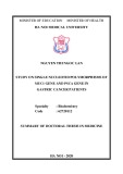
All-trans-RA blocks NFAT translocation to the nucleus
into the nucleus. To test this hypothesis, we analyzed the effects of all-trans-RA on the nuclear shuttling of NFATp. We performed immunocytochemistry on HeLa cells that had been transiently transfected with Flag- tagged recombinant NFATp. The Flag-tagged NFATp was found in the cytoplasm of unstimulated cells, and all-trans-RA treatment did not induce significant changes in the recombinant protein localization (Fig. 6). Following stimulation with PMA and ionomycin, NFATp was translocated to the nucleus in the majority of the cells. PMA and ionomycin-induced translocation was reduced by approximately 70% when the cells received cotreat- ment with all-trans-RA, and was almost completely inhibited by the addition of CsA.
Activation via the T-cell receptor (TCR) or stimuli such as ionomycin results in the rapid dephosphorylation of NFAT and its translocation into the nucleus [19,20]. Therefore, we speculated that the observed decrease in NFAT–DNA binding might be due to a decrease in the amount of NFAT proteins translocated from the cytosol
Fig. 5. All-trans-RA represses the DNA-binding activity of NFAT. A, PBMCs (7 · 106 cells) obtained from a healthy donor were stimulated in a 100-cm2 plate that was precoated with anti-CD3 Ig for 4 h with or without 1.0 lM all-trans-RA. B, Jurkat cells (3 · 106 cells) were treated with PMA (10 ngÆmL)1) and ionomycin (0.5 lM) for 4 h, in the presence or absence of a 24-h pretreatment with all-trans-RA, as indicated. The reaction mixture containing 5 lg nuclear extract was incubated with 32P-labeled oligonucleotide and analyzed by gel shift assay, as described in the Experimental procedures. The designations for cNFAT(FasL), cNFAT(IL-2), and cSP-1 indicate a 100-fold excess of the competing unlabeled oligonucleotides.
Repression of NFAT by retinoic acid (Eur. J. Biochem. 269) 1167
(cid:211) FEBS 2002
Fig. 6. All-trans-RA blocks nuclear translocation of NFAT. A, HeLa cells, transfected with an expression vector encoding the Flag epitope-tagged NFATp, were cultured on poly L-lysine-coated coverslips for 24 h. Cells were treated with PMA and ionomycin or vehicle for 30 min in the presence or absence of all-trans-RA or CsA. The cells were fixed and stained with anti-Flag Ig, followed by mouse-biotin and streptavidin–FITC, as described in the Experimental procedures. (A) no treatment; (B) all-trans-RA (1.0 lM); (C) PMA (10 ngÆmL)1) and ionomycin (0.5 lM); (D) all- trans-RA (1.0 lM) with PMA (10 ngÆmL)1) and ionomycin (0.5 lM); E, CsA (1 lgÆmL)1) with PMA (10 ngÆmL)1) and ionomycin (0.5 lM).
D I S C U S S I O N
Although it has been convincingly documented that RA induces the repression of T-cell apoptosis and FasL gene expression, the underlying molecular mechanism has not been clarified. In this study, we demonstrated that the repression of FasL transcription by RA was mediated through the inhibition of NFAT function. Both reporter gene analyses and DNA binding assays indicated that all- trans-RA mediated this repression through the NFAT binding sequence in the FasL promoter. In addition, we showed that all-trans-RA inhibited NFAT–DNA binding, as well as NFAT entry into the nucleus from the cytosol. Therefore, our results indicate that FasL expression inhibi- tion by RA involves a novel mechanism of NFAT transcription inhibition.
transactivation domain of NFATc was coactivated by CBP/ p300, well-characterized coactivators of RAR/RXR [36]. Therefore, competition for CBP/p300 between these tran- scriptional factors might result in the inhibition of the NFAT activity. Similarly, the cross-talk between retinoid receptors and NFAT might take place at the protein level, as NFAT inhibition by all-trans-RA was greater in the presence of retinoid receptors (Fig. 4). Therefore, further investigations into each of these potential mechanisms are in order to further understand the retinoid warranted, receptor-induced inhibition of NFAT. Interestingly, Szondy and others have shown that RARa stimulation inhibited, whereas RARc enhanced, activation-induced apoptosis [37,38]. Similarly, we showed that RARa repressed NFAT function, while RARc did not (Fig. 4). Thus, balanced RARa/RARc stimulation may decide whether all-trans-RA enhances or inhibits the transcriptional activity of NFAT and thereby FasL expression, which controls activation- induced apoptosis.
The biological functions of RA are mainly mediated by the ligand-dependent transcriptional factors RAR and RXR, which belong to the steroid/thyroid receptor super- family [7–9]. Several studies indicate that protein–protein interactions between nuclear retinoid receptors mediate cellular cross-talk, thus generating diverse gene-regulatory pathways. For instance, it was found that RXR could physically interact with either NF-jB or IjBb, resulting in the repression of IL-12 production in macrophages or altered LPS responses, respectively [31,32]. Furthermore, it has been shown that PPARc, another member of the steroid/thyroid receptor superfamily, interacts with NFAT at the protein level in T-lymphocytes, resulting in decreased IL-2 production [33]. Another potential mechanism involves competition for DNA binding at the NFAT site in the FasL promoter. In this regard, RXR was reported to play a crucial role in immunosuppression induced by 1a, 25 (OH)2D3, the active metabolite of vitamin D, by forming heterodimers with the vitamin D receptor (VDR), which can compete with NFAT-AP-1 binding on the IL-2 promoter NFAT site [34,35]. In addition, it has recently been reported that the activity of the inducible N-terminal
Activation via the TCR or some other stimulus induces calcium influx and leads to the dephosphorylation and rapid translocation into the nucleus of NFAT, where it activates a number of target genes. The dephosphorylated NFAT may be rephosphorylated at serine residues by either removing the stimulus or treating cells with a calcineurin inhibitor such as CsA, whereby it is translocated back to the cytoplasm [19,39]. While NFAT dephosphorylation is mediated by calcineurin, rephosphorylation is catalyzed by a variety of serine kinases, such as glycogen synthase kinase- 3, ERK, p38, casein kinase-2, and c-Jun N-terminal kinase [40–43]. These enzymatic activities may be targeted by RA in order to block NFAT dephosphorylation by repressing calcineurin and/or activating the specific serine kinases. For example, dithiocarbamate, a powerful inhibitor of NF-jB, inhibited NFAT dephosphorylation by inducing a pro- longed activation of the c-Jun N-terminal kinase [44]. Our preliminary results indicated that all-trans-RA inhibited dephosphorylation of NFAT, which could be an important
1168 M.-O. Lee et al. (Eur. J. Biochem. 269)
(cid:211) FEBS 2002
mechanism for RA-induced repression of nuclear translo- cation of NFAT (Kang, H.-J. & Lee, M.-O., unpublished results). Therefore, further studies are required to establish whether RA modulates the activities of the enzymes that affect nuclear translocation and transcriptional activity of NFAT.
& Green, D.R. (1995) Cell-autonomous Fas (CD95) /Fas–ligand interaction mediates activation-induced apoptosis in T-cell hybri- domas. Nature 373, 441–444.
5. Ju, S.-T., Panka, D.J., Cui, H., Ettinger, R., El-Khatib, M., Sherr, D.H., Stanger, B.Z. & Marshak-Rothstein, A. (1995) Fas (CD95) /FasL interactions required for programmed cell death after T-cell activation. Nature 373, 444–448.
6. Iwata, M., Mukai, M., Nakai, Y. & Iseki, R. (1992) Retinoic acids inhibit activation-induced apoptosis in T cell hybridomas and thymocytes. J. Immunol. 149, 3302–3308.
7. Szondy, Z., Reichert, U., Bernardon, J.-M., Michel, S., To´ th, R., Kara´ szi, E. & Fe´ su¨ s, L. (1998) Inhibition of activation-induced apoptosis of thymocytes by all-trans- and 9-cis-retinoic acid is mediated via retinoic acid receptor b. Biochem. J. 331, 767– 774.
inhibition of activation-induced fas
8. Yang, Y., Minucci, S., Ozato, K., Heyman, R.A. & Ashwell, J.D. ligand (1995) E(cid:129)cient up-regulation and T cell apoptosis by retinoids requires occupancy of both retinoid X receptors and retinoic acid receptors. J. Biol. Chem. 270, 18672–18677.
The NFAT proteins regulate the expression of FasL and a discrete set of cytokines involved in the regulation of immune responses, such as proliferation and differentiation, as well as in multiple effector functions of immune cells. The promoters of the IL-2, GM-CSF, IL-3, IL-4 and tumor necrosis factor alpha genes contain different types of NFAT binding elements that are independently active or combine with AP-1 binding sites [12]. The previous observations that all-trans-RA repressed IL-2 production and IL-2 gene transcription [45,46] correlate with our present findings (Fig. 3D). Currently, CsA and FK506 are the most powerful immunosuppressive drugs available that target calcineurin function. However, their clinical use is limited because of the toxic side-effects caused by inhibition of the many biological pathways controlled by calcineurin. There- fore, there is considerable therapeutic interest in drugs that directly target NFAT and allow reductions in CsA/FK506 dosage. In this regard, RA, or its more potent and receptor subtype-selective analogues, may sub serve the role of such agents.
Recently, the physiological
9. Bissonnette, R.P., Brunner, T., Lazarchik, S.B., Yoo, N.J., Boehm, M.F., Green, D.R. & Heyman, R.A. (1995) 9-cis Retinoic acid inhibition of activation-induced apoptosis is mediated via regulation of fas ligand and requires retinoic acid receptor and retinoid X receptor activation. Mol. Cell. Biol. 15, 5576– 5585.
10. Yang, Y., Merc´ ep, M., Ware, C.F. & Ashwell, J.D. (1995) Fas and activation-induced Fas ligand mediate apoptosis of T cell hybri- domas: Inhibition of Fas Ligand expression by retinoic acid and glucocorticoids. J. Exp. Med. 181, 1673–1682.
11. Mangelsdorf, D.J., Thummel, C., Beato, M., Herrlich, P., Schutz, G., Umesono, K., Blumberg, B., Kastner, P., Mark, M., Cham- bon, P. & Evans, R.M. (1995) The nuclear receptor superfamily: the second decade. Cell 83, 835–839.
retinoid receptor
12. Rao, A., Luo, C. & Hogan, P.G. (1997) Transcription factors of the NFAT family: regulation and function. Annu. Rev. Immunol. 15, 707–747.
13. McCaffrey, P.G., Luo, C., Kerppola, T.K., Jain, J., Badalian, T.M., Ho, A.M., Burgeon, E., Lane, W.S., Lambert, J.N., Curran, T., Verdine, G.L., Rao, A. & Hogan, P.G. (1993) Isolation of the cyclosporin-sensitive T cell transcription factor NFATp. Science 262, 750–754.
importance of NFAT in cells other than those of the immune system has been uncovered. The widespread distribution of NFAT mRNA and/or proteins in nonlymphoid tissues, including the heart, testis, brain, ovary, small intestine, prostate, colon, muscle, placenta, lung, and kidney, as well as in skin [47–50], suggests that NFAT family members might control cellular differentiation programs in these organ systems. Indeed, recent evidence suggests that NFAT may participate in adipogenesis and myogenesis [49,50]. Interestingly, expression has been implicated in cardiomyopathy and congestive heart link between suggesting a potential failure [51–53], RA-induced repression of NFAT and the pathophysiol- ogy of these diseases. Given the importance of NFAT in fundamental physiology, the inhibition of NFAT func- tion by retinoids may be a critical factor in NFAT- mediated biological signaling.
14. Northrop, J.P., Ho, S.N., Chen, L., Thomas, D.J., Timmerman, L.A., Nolan, G.P., Admon, A. & Crabtree, G.R. (1994) NF-AT component define a family of transcription factors targeted in T-cell activation. Nature 369, 497–502.
A C K N O W L E D G E M E N T S
15. Hoey, T., Sun, Y.-L., Williamson, K. & Xu, X. (1995) Isolation of two new members of the NF-AT gene family and functional characterization of the NF-AT proteins. Immunity 2, 461–472. 16. Masuda, E.S., Naito, Y., Tokumitsu, H., Campbell, D., Saito, F., Hannum, C., Arai, K. & Arai, N. (1995) NF-ATx, a novel member of the nuclear factor of activated T cells family that is expressed predominantly in the thymus. Mol. Cell. Biol. 15, 2697–2706.
We thank Dr Carlos V. Paya (The Mayo Clinic, Rochester, MN, USA) for the luciferase reporter constructs. We also thank Dr Crabtree (Stanford University, Stanford, CA, USA) for Flag-NFATp and NFATZH. This work was supported by a grant (KRF-99–015- DP0398) from the Korea Research Foundation to M.-O. L. and J. P.
17. Lopez-Rodriguez, C., Aramburu, C.J., Rakeman, A.S. & Rao, A. (1999) NFAT5, a constitutively nuclear NFAT protein that does not cooperate with Fos and June. Proc. Natl Acad. Sci. USA 96, 7214–7219.
R E F E R E N C E S
1. Pinkoski, M.J. & Green, D.R. (1999) Fas ligand, death gene. Cell Death Differ. 6, 1174–1181. 18. Kiani, A., Rao, A. & Aramburu, J. (2000) Manipulating immune responses with immunosuppressive agents that target NFAT. Immunity 12, 359–372. 2. Nagata, S. (1999) Fas ligand-induced apoptosis. Annu. Rev. Genet. 33, 29–55.
19. Beals, C.R., Clipstone, N.A., Ho, S.N. & Crabtree, G.R. (1997) Nuclear localization of NF-ATc by a calcineurin-dependent, cyclosporin–sensitive intramolecular interaction. Genes Dev. 11, 824–834. 3. Dhein, J., Walczak, H., Baumier, C., Debatin, K.-M. & Krammer, P.H. (1995) Autocrine T-cell suicide mediated by APO-1/ (Fas/ CD95). Nature 373, 438–441. 20. Shaw, K.T.-Y., Ho, A.M., Raghavan, A., Kim, J., Jain, J., Park, J., Sharma, S., Rao, A. & Hogan, A.G. (1995) Immunosuppres- 4. Brunner, T., Mogil, R.J., LaFace, D., Yoo, N.J., Mahboubl, A., Echeverri, F., Martin, S.J., Force, W.R., Lynch, D.H., Ware, C.F.
Repression of NFAT by retinoic acid (Eur. J. Biochem. 269) 1169
(cid:211) FEBS 2002
sive drugs prevent a rapid dephosphorylation of transcription factor NFAT1 in stimulated immune cells. Proc. Natl Acad. Sci. USA 92, 11205–11209.
36. Avots, A., Buttmann, M., Chuvpilo, S., Escher, C., Smola, U., Bannister, A.J., Rapp, U.R., Kouzarides, T. & Serfling, E. (1999) CBP/p300 integrates Raf/Rac-signaling pathways in the tran- scriptional induction of NFATc during T cell activation. Immunity 10, 515–524.
21. Ho, S., Clipstone, N., Timmermann, L., Northrop, J., Graef, I., Fiorentino, D., Nourse, J. & Crabtree, G.R. (1996) The mecha- nism of action of cyclosporin A and FK506. Clin. Immunol. Immunopathol. 80, S40–S45. 37. Szondy, Z., Reichert, U. & Fesus, L. (1998) Retinoic acids regulate apoptosis of T lymphocytes through an interplay between RAR and RXR receptors. Cell Death Differ. 5, 4–10.
22. Holtz-Heppelmann, C.J., Algeciras, A., Badley, A.D. & Paya, C.V. (1998) Transcriptional regulation of the human FasL promoter-enhancer region. J. Biol. Chem. 273, 4416–4423.
38. Szondy, Z., Reichert, U., Bernardon, J.M., Michel, S., Toth, R., Karaszi, E. & Fesus, L. (1998) Inhibition of activation-induced apoptosis of thymocytes by all-trans- and 9-cis-retinoic acid is mediated via retinoic acid receptor alpha. Biochem. J. 331, 767–774. 23. Rengarajan, J., Mittelstadt, P.R., Mages, H.W., Gerth, A.J., Kroczek, R.A., Ashwell, J.D. & Glimcher, L.H. (2000) Sequential involvement of NFAT and Egr transcription factors in FasL regulation. Immunity 12, 293–300.
39. Loh, C., Shaw, K.T.-Y., Carew, J., Viola, J.P.B., Luo, C., Perrino, B.A. & Rao, A. (1996) Calcineurin binds the transcription factor NFAT1 and reversibly regulates its activity. J. Biol. Chem. 271, 10884–10891. 24. Xiao, S., Matsui, K., Fine, A., Zhu, B., Marshak-Rothstein, A., Widom, R.L. & Ju, S.T. (1999) FasL promoter activation by IL-2 through SP1 and NFAT but not Egr-2 and Egr-3. Eur. J. Immunol. 29, 3456–3465.
40. Beals, C.R., Sheridan, C.M., Turck, C.W., Gardner, P. & Crab- tree, G.R. (1997) Nuclear export of NF-Atc enhanced by glycogen synthase kinase-3. Science 28, 1930–1934.
25. Latinis, K.M., Norian, L.A., Eliason, S.L. & Koretzky, G.A. (1997) Two NFAT transcription factor binding sites participate in the regulation of CD95 (Fas) ligand expression in activated human T cells. J. Biol. Chem. 272, 31423–31434.
41. Zhu, J., Shibasaki, F., Price, R., Guillemot, J.C., Yano, T., Dotsch, V., Wagner, G., Ferrar, P. & McKeon, F. (1998) Intracellular masking of nuclear import signal on NF-AT4 by casein kinase I and MEKK1. Cell 93, 851–861.
26. Shin, E.-C., Shin, J.-S., Park, J.-H., Kim, H. & Kim, S.-J. (1999) Expression of fas ligand in human hepatoma cell lines: role of hepatitis-B virus X (HBx) in induction of fas ligand. Int. J. Cancer 82, 587–591. 42. Chow, C.W., Rincon, M., Cavanagh, J., Dickens, M. & Davis, R.J. (1997) Nuclear accumulation of NFAT4 opposed by the JNK signal transduction pathway. Science 278, 1638–1641.
43. Porter, C.M., Havens, M.A. & Clipstone, N.A. (2000) Identifi- cation of amino acid residues and protein kinases involved in the regulation of NFATc subcellular localization. J. Biol. Chem. 275, 3543–3551.
27. Ho, S.N., Thomas, D.J., Timmerman, L.A., Li, X., Francke, U. & Crabtree, G.R. (1995) NFATc3, a lymphoid-specific NFATc family member that is calcium-regulated and exhibits distinct DNA binding specificity. J. Biol. Chem. 270, 19898–19907. 28. Kang, H.-J., Song, M.-R., Lee, S.-K., Shin, E.-C., Choi, Y.-H., Kim, S.J., Lee, J. & Lee, M.-O. (2000) Retinoic acid and its receptors repress the expression and transactivation function of Nur77: a possible mechanism for the inhibition of apoptosis by retinoic acid. Exp. Cell Res. 256, 545–554. 44. Martinez, S., Gomez, P., Armesilla, A.L., Aramburu, J., Luo, C., Rao, A. & Redondo, J.M. (1997) Blockade of T cell activation by dithiocarbamates involves novel mechanisms of inhibition of nuclear factor of activated T cells. Mol. Cell. Biol. 17, 6437–6447.
29. Mattila, P.S., Ullman, K.S., Fiering, S., Emmel, E.A., McCutch- eon, M., Crabtree, G.R. & Herzenberg, L.A. (1990) The actions of cyclosporin A and FK506 suggest a novel step in the activation of T lymphocytes. EMBO J. 9, 4425–4433.
45. Felli, M.P., Vacca, A., Meco, D., Screpanti, I., Farina, A.R., Maroder, M., Martinotti, S., Petrangeli, E., Frati, L. & Gulino, A. (1991) Retinoic acid-induced down-regulation of the interleukin-2 promoter via cis-regulatory sequences containing an octamer motif. Mol. Cell. Biol. 11, 4771–4778.
30. Oda, Y., Kinoshota, M. & Kakehi, K. (1997) Fluorometric assay of binding specificity of plant lectins to yeast cells by biotin-avidin system and its application to the classification of yeast cells. Anal. Biochem. 254, 41–48. (1994) Positive and negative regulation of
46. Grazia, U., Felli, M.P., Vacca, A., Farina, A.R., Maroder, M., Cappabianca, L., Meco, D., Farina, M., Screpanti, I., Frati, L. & Gulino, A. the composite octamer motif of the interleukin 2 enhancer by AP-1, Oct-2, and retinoic acid receptor. J. Exp. Med. 180, 1485– 1497. 31. Na, S.Y., Kang, B.Y., Chung, S.W., Han, S.J., Ma, X., Trinchieri, G., Im, S.Y., Lee, J.W. & Kim, T.S. (1999) Retinoids inhibit interleukin-12 production in macrophages through physical associations of retinoid X receptor and NFkappaB. J. Biol. Chem. 274, 7674–7680.
47. Armesilla, A.L., Lorenzo, E., Gomez, P., Martinez, S., Alfranca, A. & Redondo, J.M. (1999) Vascular endotherial growth factor activates nuclear factor of activated T cells in human endothelial cells: a role for tissue factor gene expression. Mol. Cell. Biol. 19, 2032–2043. 32. Na, S.Y., Kim, H.J., Lee, S.K., Choi, H.S., Na, D.S., Lee, M.O., Chung, M., Moore, D.D. & Lee, J.W. (1998) IjBb interacts with the retinoid X receptor and inhibits retinoid-dependent transacti- vation in lipopolysaccharide-treated cells. J. Biol. Chem. 273, 3212–3215.
48. Mosieniak, G., Pyrzynska, B. & Kaminska, B. (1998) Nuclear factor of activated T cells (NFAT) as a new component of the signal transduction pathway in glioma cells. J. Neurochem. 71, 134–141.
33. Yang, X.Y., Wang, L.H., Chen, T., Hodge, D.R., Resau, J.H., DaSilva, L. & Farrar, W.L. (2000) Activation of human T lymphocytes is inhibited by peroxisome proliferator-activated receptor gamma (PPARgamma) agonists. PPARc co-asso- ciation with transcription factor NFAT. J. Biol. Chem. 275, 4541– 4544.
34. Alroy, I., Towers, T.L. & Freedman, L.P. (1995) Transcriptional repression of the interleukin-2 gene by vitamine D3: direct inhib- tion of NFATp/AP-1 complex formation by nuclear hormone receptor. Mol. Cell. Biol. 15, 5789–5799. 49. Ho, I.C., Kim, J.H., Rooney, J.W., Spiegelman, B.M. & Glimcher, L.H. (1998) A potential role for the nuclear factor of activated T cells family of transcriptional regulatory proteins in adipogenesis. Proc. Natl Acad. Sci. USA 95, 15537–15541. 50. Ranger, A.M., Grusby, M.J., Hodge, M.R., Gravallese, E.M., Charles, F., Hoey, T., Mickanin, C., Baldwin, H.S. & Glimcher, L.H. (1998) The transcription factor NF-ATc is essential cardiac valve formation. Nature 392, 186–190.
35. Takeuchi, A., Reddy, G.S., Kobayashi, T., Okano, T., Park, J.C. & Sharma, S. (1998) Nuclear factor of activated T cells (NFAT) as a molecular target for 1a,25-dihydroxyvitamine D3-mediated effects. J. Immunol. 160, 209–218. 51. Colbert, M.C., Hall, D.G., Kimball, T.R., Witt, S.A., Lorenz, J.N., Kirby, M.L., Hewett, T.E., Kievitsky, R. & Robbins, J. (1997) Cardiac compartment-specific overexpression of a modified
1170 M.-O. Lee et al. (Eur. J. Biochem. 269)
(cid:211) FEBS 2002
retinoic acid receptor produces dilated cardiomyopathy and congestive heart failure in transgenic mice. J. Clin. Invest. 100, 1958–1968.
53. Gruber, P.J., Kubalak, S.W., Pexieder, T., Sucov, H.M., Evans, R.M. & Chien, K.R. (1996) RXR alpha deficiency confers genetic susceptibility for aortic sac, conotruncal, atrioventricular cushion, and ventricular muscle defects in mice. J. Clin. Invest. 98, 1332– 1343. 52. Zhou, M.D., Sucov, H.M., Evans, R.M. & Chien, K.R. (1995) Retinoid-dependent pathways suppress myocardial cell hypertro- phy. Proc. Natl Acad. Sci. USA 92, 7391–7395.