Cyclic GMP induced apoptosis via protein kinase G in oestrogen receptor-positive and -negative breast cancer cell lines Faranak Fallahian1, Fatemeh Karami-Tehrani1, Siamak Salami2 and Mahmoud Aghaei1
1 Department of Clinical Biochemistry, Cancer Research Laboratory, School of Medical Science, Tarbiat Modares University, Tehran, Iran 2 Department of Biochemistry, School of Medical Science, Urmia University of Medical Sciences, Iran
Keywords apoptosis; cyclic guanosine 3,5-monophosphate (cGMP); MCF-7; MDA-MB-468; protein kinase G
Correspondence F. Karami-Tehrani, Clinical Biochemistry Department, School of Medical Sciences, Tarbiat Modarres University, PO Box 14115-331, Tehran, Iran Fax: +98 (21) 82884555 Tel: +98 (21) 82883567 E-mail: karamitf@modares.ac.ir
(Received 7 March 2011, revised 20 June 2011, accepted 18 July 2011)
doi:10.1111/j.1742-4658.2011.08260.x
The activation of protein kinase G (PKG) by cyclic guanosine 3,5-mono- phosphate (cGMP) has become of considerable interest as a novel molec- ular approach for the induction of apoptosis in cancer cells. The present study was designed to examine the effects of cGMP and PKG on cell growth and apoptosis in the human breast cancer cell lines, MCF-7 and MDA-MB-468. To achieve this, 1-benzyl-3-(5P-hydroxymethyl-2P-furyl) indazole (YC-1), a soluble guanylyl cyclase activator, and 8-bromo-cGMP (8-br-cGMP), a membrane-permeant and phosphodiesterase-resistant ana- logue of cGMP, were employed in MCF-7 and MDA-MB-468 cells. Then, the role of PKG in the induction of apoptosis was evaluated using specific inhibitors of PKG. The KT5823 and Rp-8-pCPT-cGMP as expression of PKG isoforms in these cell lines was also investigated. KT5823 and Rp-8-pCPT-cGMP significantly attenuated the loss of cell viability caused by YC-1 and 8-br-cGMP in these cells. This study pro- the activation of PKG by cGMP induces vides direct evidence that growth inhibition and apoptosis in MCF-7 and MDA-MB-468 breast cancer cell lines.
Introduction
It
in mammalian cells.
smooth-muscle
in vascular
Cyclic guanosine 3¢,5¢-monophosphate (cGMP), an ubiquitous second messenger, mediates several signal- transduction pathways is involved in the regulation of various physiological functions, including neurotransmission and platelet aggregation. cGMP also modulates intracellular cal- cium levels cells and thereby modulates its tone [1–3]. There is increasing evidence that cGMP can play an important role in cel- lular proliferation, differentiation and apoptosis [4,5]. The intracellular levels of cGMP are regulated through a dynamic balance between the rate of synthesis by
guanylate cyclases and hydrolysis by specific phospho- diesterases (PDEs) especially PDE 2 and PDE 5 [6–8]. cGMP has several intracellular targets: it can bind to specific PDEs, cGMP-gated cation channels and pro- tein kinase G (PKG) [8–10]. PKG belongs to the fam- ily of serine ⁄ threonine kinases [11]. Two major forms of PKG have been identified in mammalian cells: PKG I and PKG II. In addition, there are two splice variants of PKG I, which are designated as Ia and Ib [12]. Both PKG Ia and PKG Ib are cytosolic enzymes that differ only in their amino-terminal sequences, while PKG II is a distinct membrane-bound enzyme.
Abbreviations 8-br-cGMP, 8-bromo-cGMP; cGMP, cyclic guanosine 3,5-monophosphate; ER, oestrogen receptor; FITC, fluorescein isothiocyanate; MTT, 3-(4,5-dimethyl-2-thiazolyl)-2,5-diphenyl-2H-tetrazolium bromide; PDE, phosphodiesterase; PI, propidium iodide; PKG, protein kinase G; YC-1, 1-benzyl-3-(5P-hydroxymethyl-2P-furyl) indazole.
FEBS Journal 278 (2011) 3360–3369 ª 2011 The Authors Journal compilation ª 2011 FEBS
3360
F. Fallahian et al.
Cyclic GMP induced apoptosis via protein kinase G
A
B
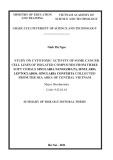
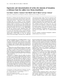
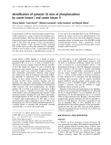
Fig. 1. RT-PCR analysis of the expression of PKG isoforms in the human breast cancer cell lines MCF-7 (A) and MDA-MB-468 (B). Total RNA was extracted from the cells and the mRNA of the PKG isoforms was amplified using RT-PCR. The PCR products were elec- trophoresed in a 2% agarose gel and stained with ethidium bromide. Lane 1, PKG II (205 bp); lane 2, PKG Ib (292 bp); lane 3, PKG Ia (301 bp); lane 4, 50-bp DNA ladder; Lane 5, GAPDH (496 bp). The mRNAs of PKG isoforms were detectable in both cell lines.
in
PKGIa, PKGIb and PKGII mRNA transcripts breast cancer cells (Fig. 1).
The effects of YC-1 and 8-br-cGMP on cell growth
PKG I is expressed in vascular smooth-muscle cells, platelets, lung, certain endothelial cells, fibroblasts, heart and the cerebellum. PKG II is expressed mainly in the brush border of intestinal mucosa and in specific regions of the brain, and mediates electrolyte balance in the intestine [13–15]. PKG is the central effector for cGMP that was initially recognized for its role in the regulation of platelet aggregation and vascular tone but studies over the past decade have shown that PKG can control several cellular processes [12,16]. Recently, activation of the cGMP ⁄ PKG pathway has been impli- cated as a novel molecular approach for the induction of apoptosis in cardiomyocytes, pancreatic B cells and cultured smooth muscle cells [4,5,17]. Evidence has also been obtained that the increase in cellular levels of cGMP leads to the activation of PKG and to the induction of apoptosis in human colon cancer cells [18–23]. It is of interest that colon cancer cells have rel- atively high levels of PDE 5 when compared with nor- mal colonic mucosa [24]. In addition, PKG Ib is expressed at reduced levels in colon adenoma and ade- nocarcinoma when compared with normal mucosa [21,24]. These findings suggest that cGMP-mediated pathways are suppressed in colon cancer cells, presum- ably to inhibit the process of apoptosis [24]. Several mechanisms have been proposed for the apoptotic effect of PKG, but the precise mechanism by which PKG activation leads to apoptosis is not known.
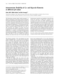
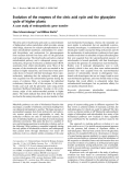
As shown in Fig. 2, YC-1 significantly inhibited the via- bility of both MCF-7 (Fig. 2A) and MDA-MB-468 (Fig. 2C) cells in a time- and dose-dependent manner. inhibitory effects of YC-1 in The most significant MCF-7 cells were 68% and 72% at 100 lm after treat- ment for 48 and 72 h, respectively. Similar effects were observed in MDA-MB-468 cells; treatment with 100 lm YC-1 caused a reduction of approximately 70% and 78% in cell viability after 48 and 72 h, respectively. Treatment of MCF-7 and MDA-MB468 cell
Breast cancer is the most common malignancy in women and therefore finding new molecular targets for the induction of apoptosis can be used as an important approach. Accordingly, the present study was designed to examine the effects of cGMP and PKG on cell growth inhibition and cell apoptosis in the human breast cancer cell lines, MCF-7 and MDA-MB-468. To achieve this, 1-benzyl-3-(5P-hydroxymethyl-2P-furyl) indazole (YC-1), a soluble guanylyl cyclase activator, and 8-bromo-cGMP (8-br-cGMP), a membrane-per- meant and phosphodiesterase-resistant analogue of cGMP, were employed in MCF-7 and MDA-MB-468 cells. Then, the role of PKG in the induction of apop- tosis was evaluated using KT5823 and Rp-8-pCPT- cGMP as selective inhibitors of PKG. In addition, the expression of PKG isoforms in these cell lines was also investigated.
lines with 8-br-cGMP, a PKG activator, resulted in a dose- dependent reduction in cell viability when compared with control cells (Fig. 2B,D). The cytotoxic effect was more evident at 48 and 72 h in both cell lines. YC-1 and 8-br-cGMP appeared to display similar inhibition activ- ities in both cell lines. The effective doses of YC-1 that inhibited 50% of growth (IC50) of MCF-7 and MDA- MB-468 cells after 48 h of treatment were 47.77 lm and 46.61 lm, respectively. The IC50 values were 56.57 lm and 55.97 lm, respectively, for MCF-7 and MDA-MB- 468 cells after 48 h of treatment with 8-br-cGMP.
Results
Expression of PKG isoforms
Involvement of cGMP and the PKG pathway in mediating the effects of YC-1 and 8-br-cGMP
To examine the expression of PKG isoforms in MCF-7 lines, RT-PCR analysis was and MDA-MB-468 cell show detectable levels of performed. The results
To examine the possible effects of YC-1 on the cGMP ⁄ PKG pathway in MCF-7 and MDA-MB-468
FEBS Journal 278 (2011) 3360–3369 ª 2011 The Authors Journal compilation ª 2011 FEBS
3361
F. Fallahian et al.
Cyclic GMP induced apoptosis via protein kinase G
C
A
24 h 48 h 72 h
24 h 48 h 72 h
) l o r t n o c f o %
) l o r t n o c f o %
( y t i l i
( y t i l i
b a i v l l e C
b a i v l l e C
YC-1 (µM)
YC-1 (µM)
B
D
24 h 48 h 72 h
24 h 48 h 72 h
) l o r t n o c
) l o r t n o c
f o %
f o %
(
y t i l i
( y t i l i
b a
i
i
v
b a v
l l
l l
e C
e C
8-br-cGMP (µM)
8-br-cGMP (µM)
, 48 h
Fig. 2. The effect of YC-1 and 8-br-cGMP on cell viability was measured using the MTT assay. MCF-7 cells were treated with different con- centrations of YC-1 (A) and 8-br-cGMP (B) for 24 h . MDA-MB-468 cells were also treated with YC-1 (C) and 8-br- and 72 h cGMP (D). The MTT assay was carried out as described in the Materials and methods. Each value is presented as mean ± SD of three experiments. Each experiment was conducted in triplicate. *P < 0.05; **P < 0.01 compared with the untreated control group.
MCF-7
MDA-MB-468
) 1 – L m
· l
o m P
(
P M G c
YC-1 (µM)
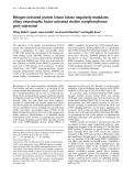
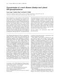
cells, the intracellular levels of cGMP were assessed. MCF-7 and MDA-MB-468 cells were treated for 2 h with increasing concentrations of YC-1 (2.5–100 lm). Extracts were then prepared and the intracellular levels of cGMP were determined using a colorimetric com- petitive ELISA. After treatment with YC-1, a detect- able rise in the cGMP level was observed in both cell types, which progressively increased as the concentra- tion was raised (Fig. 3). Up to 10 lm YC-1, no signifi- cant increase in the cGMP levels was observed in either MCF-7 cells or MDA-MB-468 cells (P > 0.05, compared with control group); however, when the con- increased to 100 lm, an centration of YC-1 was increase of approximately fourfold was observed in the cellular levels of cGMP (P < 0.01, compared with the control group).
and MDA- Fig. 3. The effect of YC-1 on the cGMP levels in MCF-7 MB-468 cells. Cells were treated with increasing concentrations of YC-1 for 2 h and then measurement of cGMP was carried out using a colorimetric competitive ELISA kit. Each point is the mean ± SD of three experiments. Each experiment was repeated three times. *P < 0.05; **P < 0.01 compared with the untreated control group.
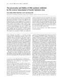
To further establish the involvement of PKG in the induction of cell growth inhibition, cells were pretreated with specific PKG inhibitors – KT5823 (3 lm) and Rp-8-
FEBS Journal 278 (2011) 3360–3369 ª 2011 The Authors Journal compilation ª 2011 FEBS
3362
F. Fallahian et al.
Cyclic GMP induced apoptosis via protein kinase G
A
B
respectively, after
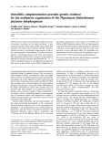
totic cells (Fig. 5). According to this method, MCF-7 and MDA-MB-468 cells were evaluated by flow cyto- metry 24 h after treatment with YC-1 and 8-br-cGMP. All data were analyzed using specific software (partec flomax). Control cells were negative for both annexin V–FITC and PI. Annexin V–FITC-positive, PI-nega- tive cells were considered to be in an early apoptotic stage, while annexin V–FITC-positive, PI-positive cells were considered to be late apoptotic or necrotic, and PI-positive cells were considered as mostly necrotic. The rate of apoptosis was calculated as the sum of early apoptotic cells and late apoptotic cells. As shown in Fig. 5, apoptotic rates ranged from 0.5% to 57.2% and from 0.53% to 53.48% in MCF-7 and MDA- MB-468 cells, respectively, after treatment with YC-1. The rates of apoptosis ranged from 0.45% to 45.51% and from 1.6% to 46.35% in MCF-7 and MDA-MB- 468 cells, treatment with 8-br- cGMP.
Involvement of caspases in apoptosis induced by YC-1- and 8-br-cGMP
The results demonstrated that treatment of MDA- MB468 cells with YC-1 and 8-br-cGMP resulted in a significant increase (P < 0.01, compared with control) in the activity of caspase-3 and caspase-9, in a time- dependent manner (Fig. 6B). Unlike the MDA-MB468 cells, the activity of caspase-3 remained unchanged but the activity of caspase-9 increased in a time-dependent manner in MCF-7 cells after treatment with YC-1 and 8-br-cGMP (Fig. 6A).
Fig. 4. Effect of KT5823 and Rp-8-pCPT-cGMP (Rp-cGMP) on YC-1- and 8-br-cGMP-induced cytotoxicity in MCF-7 (A) and MDA-MB-468 (B) cells. Cells were pretreated with the specific PKG inhibitors, KT5823 (3 lM) and Rp-8-pCPT-cGMp (20 lM), for 1 h and then trea- ted with YC-1 (40 lM) and 8-br-cGMP (50 lM) for 48 h. Each point (mean ± SD) is from three separate experiments. **P < 0.01 com- pared with the YC-1-treated group; ##P < 0.01 compared with the 8-br-cGMP-treated group.
Further confirmation for the involvement of caspas- es in the induction of apoptosis by YC-1 and 8-br- cGMP was provided by the results obtained from pre- treatment of the cells with Z-VAD-fmk (100 lm), a broad-spectrum caspase inhibitor. As shown in Fig. 7, Z-VAD-fmk significantly (P < 0.01, compared with control) inhibited YC-1 and 8-br-cGMP-induced apop- tosis in MCF-7 and MDA-MB-468 cells.
Cell cycle analysis
pCPT-cGMP (20 lm) – and then treated with YC-1 and 8-br-cGMP at their IC50 concentrations for 48 h. As shown in Fig. 4, KT5823 and Rp-8-pCPT-cGMP significantly attenuated the loss of viability caused by YC-1 and 8-br-cGMP. It is important to note that KT5823 and Rp-8-pCPT-cGMP, alone, had no effects on cell growth.
Detection of apoptosis by flow cytometry
To investigate a possible relationship between PKG activation and the induction of apoptotic cell death, fluorescein isothiocyanate (FITC)-conjugated annexin V (FL1-H) and propidium iodide (PI) (FL2-H) stain- ing (detected by flow cytometry and fluorescence microscopy) were used as criteria to distinguish apop-
Studies were also carried out to ascertain whether PKG activation was associated with a disturbance of cell cycle regulation. The cell cycle distributions of cells treated with YC-1 and 8-br-cGMP were there- fore analyzed by flow cytometry. As shown in Fig. 8, treatment of cells with YC-1 and 8-br-cGMP resulted in an increase in the percentage of cells in the sub-G1 phase. This increase was concentration dependent and consistent with the induction of apoptosis.
FEBS Journal 278 (2011) 3360–3369 ª 2011 The Authors Journal compilation ª 2011 FEBS
3363
F. Fallahian et al.
Cyclic GMP induced apoptosis via protein kinase G
C
A
)
)
Viable Early apoptosis Late apoptosis Necrosis
%
%
Viable Early apoptosis Late apoptosis Necrosis
( e t a r s i s o t p o p A
( e t a r s i s o t p o p A
YC-1 (µM)
YC-1 (µM)
D
B
)
)
Viable Early apoptosis Late apoptosis Necrosis
%
Viable Early apoptosis Late apoptosis Necrosis
%
**
**
**
**
**
**
( e t a r s i s o t p o p A
**
( e t a e r s i s o t p o p A
**
**
**
**
**
*
*
8-br-cGMP (µM)
8-br-cGMP (µM)
Fig. 5. Detection of apoptosis using flow cytometry. Flow cytometric analysis of MCF-7 cells after treatment with YC-1 (A) and 8-br-cGMP (B). MDA-MB-468 cells were also treated with YC-1 (C) and 8-br-cGMP (D) and flow cytometric analysis was carried out. The results shown represent the mean ± SD of three independent experiments. *P < 0.05; **P < 0.01 compared with the control group.
Discussion
[25]. Most previous
cGMP mediates discrete cellular functions in mamma- lian cells by interacting with target proteins, including PKG, cyclic nucleotide-gated channels and specific phosphodiesterases [8–10]. There is increasing evidence that activation of the cGMP ⁄ PKG pathway can play an important role in the inhibition of cell proliferation and induction of apoptosis in cancer cells [18–23]. Recently, a cDNA array was performed to study PKGI mRNA levels in several tumor types. It was shown that the PKG levels were dramatically reduced in tumors from liver, thyroid, lung, colon, testes and pancreas compared with their normal counterpart tis- sues [21]. The decreased expression of PKG in cancer cells is important evidence to support the anti-tumor activity of this enzyme in vivo, but the mechanism of PKG down-regulation is unknown.
Based on these findings, we designed studies to eval- uate the significance of cGMP and PKG in the regula- tion of cell-growth inhibition in the breast cancer cell lines, MCF-7 and MDA-MB-468. Elevation of cGMP
levels can be achieved by YC-1, a nitric oxide (NO)- soluble guanylyl independent and heme-dependent studies used cyclase activator chemical NO donors to raise the concentration of intracellular NO. NO interacts with the heme group of soluble guanylyl cyclase, leading to activation of the enzyme and a rise in cGMP [17,26]. However, the interpretation of results is complicated by the fact that NO can exert a range of other biological effects that are independent of cGMP [27]. Therefore, in the cur- rent research, YC-1 was used to elevate cGMP without requiring a rise in NO. Treatment of MCF-7 and MDA-MB-468 cells with YC-1 caused an increase in cGMP, confirming that the activity of the soluble form of guanylyl cyclase in these cells is increased by this compound. YC-1 also caused a time- and dose-depen- dent cell-growth inhibition in these cells. It should be noted that YC-1 may also cause cell death by cGMP- independent pathways in some cell types [28–31]; how- ever, using KT5823 and Rp-8-pCPT-cGMP (specific inhibitors of PKG), we indicated that YC-1 induced cell-growth inhibition through a cGMP-dependent
FEBS Journal 278 (2011) 3360–3369 ª 2011 The Authors Journal compilation ª 2011 FEBS
3364
F. Fallahian et al.
Cyclic GMP induced apoptosis via protein kinase G
A
A
) e m
)
Cas3-YC-1 Cas9-YC-1 Cas3-8-br-cGMP Cas9-8-br-cGMP
%
( s
i
i t r e v o e s a e r c n
s o t p o p A
i
y t i v i t c a e s a p s a C
l
d o f (
##
**
Time (h)
B
B
Cas3-YC-1
)
) e m
%
Cas9-YC-1 Cas3-8-br-cGMP Cas9-8-br-cGMP
( s
i
** **
**
**
*
*
*
s o t p o p A
i t r e v o e s a e r c n
*
i
y t i v i t c a e s a p s a C
l
d o f (
##
**
Time (h)
lane 2, YC-1 + Z-VAD-fmk;
Fig. 7. Effects of caspase inhibition on YC-1- and 8-br-cGMP- induced apoptosis in MCF-7 cells (A) and MDA-MB-468 cells (B). Lane 1, YC-1 (100 lM); lane 3, 8-br- cGMP (100 lM); lane 4, 8-br-cGMP + Z-VAD-fmk. The results repre- sent mean ± SD. **P < 0.01 compared with the YC-1-treated group; ##P < 0.01 compared with the 8-br-cGMP-treated group.
Fig. 6. Colorimetric assay of caspase-3 and caspase-9 activation after treatment with YC-1 (100 lM) and 8-br-cGMP (100 lM). In MCF-7 cells (A) the activity of caspase-9 increased in a time-depen- dent manner but the activity of caspase-3 remained unchanged. The activity of caspase-3 and caspase-9 increased in a time-depen- dent manner in MDA-MB468 cells (B). *P < 0.05; **P < 0.01 com- pared with the control group.
pathway in breast cancer cells. Moreover, employing a more potent substrate analogue as an activator of PKG, 8-br-cGMP, provided confirmation that activa- tion of the cGMP ⁄ PKG pathway might induce cell- growth inhibition in breast cancer cells. It is important to note that the expression of PKG has not been inves- tigated in MCF-7 and MDA-MB-468 cells. Accord- ingly, PKG expression was examined by RT-PCR in these cells. The results showed detectable levels of PKGIa, PKGIb and PKGII mRNA transcripts in these cell lines.
etry on a fluorescence-activated cell sorter (FACS)] was used to distinguish apoptotic cells. Analysis of treated cells by annexin-V ⁄ PI staining revealed that YC-1 and 8-br-cGMP promote a significant level of cell death via the apoptotic pathway. Furthermore, the effects of YC-1 and 8-br-cGMP on the cell-cycle profiles were evaluated using flow cytometry PI-based staining in MCF-7 and MDA-MB-468 cells. Measure- ment of the DNA content makes it possible to iden- tify apoptotic cells and to recognize the cell-cycle phase specificity. Data indicated that exposure to these compounds in both cell lines caused a significant increase in the sub-G1 phase population, which is a hallmark of apoptosis.
Apoptosis is characterized by a variety of distinct morphological and biochemical features that are used It has been to discriminate apoptotic cell death. shown that loss of phospholipid asymmetry that leads to the exposure of phosphatidylserine on the outside of the plasma membrane is an early event of apopto- sis. As annexin-V preferentially binds to the negatively charged phosphatidylserine, FITC-conjugated annexin-V (FL1-H) ⁄ PI (FL2-H) staining [detected by flow cytom-
To ascertain whether activation of the caspase fam- ily participated in the apoptosis induced by YC-1 and 8-br-cGMP, the activity of caspases was exam- ined. Our findings indicated that treatment of MDA- MB-468 cells with YC-1 and 8-br-cGMP induced a significant increase in caspase-3 and caspase-9 activ- ity. Activity of caspase-9 increased in MCF-7 cells after treatment with these compounds, while activa- tion of caspase-3 was not observed in these cells.
FEBS Journal 278 (2011) 3360–3369 ª 2011 The Authors Journal compilation ª 2011 FEBS
3365
F. Fallahian et al.
Cyclic GMP induced apoptosis via protein kinase G
Sub-G1
G0/G1
C
A
S
G2/M
)
Sub-G1
G0/G1
)
%
%
S
G2/M
( n o i t u b i r t s i d e l c y c l l e C
( n o i t u b i r t s i d e l c y c l l e C
YC-1 (µM)
YC-1 (µM)
D
B
Sub-G1 G0/G1
)
Sub-G1
G0/G1
%
)
S
G2/M
%
S
G2/M
( n o i t u b i r t s i d e l c y c l l e C
( n o i t u b i r t s i d e l c y c l l e C
8-br-cGMP (µM)
8-br-cGMP (µM)
Fig. 8. Effects of YC-1 and 8-br-cGMP on the cell-cycle distribution. MCF-7 cells were treated with YC-1 (A) and 8-br-cGMP (B) for 24 h. MDA-MB-468 cells were also treated with YC-1 (C) and 8-br-cGMP (D). The percentage of cells in each stage of the cell cycle were then evaluated by PI staining and flow cytometric analysis, as described in the Materials and methods. YC-1 and 8-br-cGMP caused an increase in the percentage of cells in the sub-G1 phase, which is consistent with the induction of apoptosis. *P < 0.05; **P < 0.01 compared with control group.
experiments using Z-VAD-fmk as inhibitor
point
to
MCF-7 cells do not express the caspase-3 protein because of the presence of a 47-bp deletion within the CASP-3 gene and our finding is exon 3 of consistent with what was reported previously [32]. a Moreover, broad-spectrum caspase the involvement of caspases in the apoptosis induced by YC-1 and 8-br-cGMP in these cells.
MCF-7 and MDA-MB-468. Our findings are consis- tent with previous evidence that displayed an increase in expression of PDE 5 and a decrease in expression of PKG Ib in breast cancer cells compared with normal mammary epithelial cells [24]. Additional studies are required to determine the precise molecular mecha- nisms by which PKG activation can inhibit growth and induce apoptosis in breast cancer cells.
Materials and methods
Chemical reagents
To examine possible differences in the growth-inhibi- tory effects of cGMP ⁄ PKG on estrogen-dependent and estrogen-independent breast cancer cells, we used MCF-7 and MDA-MB-468 [oestrogen receptor (ER)- positive and ER-negative, respectively] breast cancer cell lines. Both cell lines had the same sensitivity to the growth-inhibitory effects of cGMP; therefore, it seems that cGMP may exert its growth-inhibitory effects through an ER-independent pathway.
RPMI-1640, trypsin ⁄ EDTA, NaCl ⁄ Pi, penicillin and strepto- mycin were purchased from Gibco (Rockville, USA). YC-1 and KT5823 were purchased from Alexis Biochemicals (Lorrach, Germany). 8-br-cGMP, Rp-8-pCPT-cGMP, the annexin-V–FITC apoptosis detection kit, PI, MTT [3-(4,5- dimethylthiazol-2-yl)-2,5-diphenyltetrazolium bromide] and from Sigma-Aldrich (Munich, dimethylsulfoxide were
In conclusion, we provided evidence, for the first time, that the activation of PKG by cGMP induces growth inhibition in the human breast cancer cell lines,
FEBS Journal 278 (2011) 3360–3369 ª 2011 The Authors Journal compilation ª 2011 FEBS
3366
F. Fallahian et al.
Cyclic GMP induced apoptosis via protein kinase G
Germany). The caspase-3 and caspase-9 colorimetric assay kits and the cGMP direct immunoassay (ELISA) kit were obtained from R&D Systems Co. (Minneapolis, USA).
Cell culture
dimethylsulfoxide was added to each well. The plates were shaken for an additional 10 min and the absorbance values were read using a microplate reader (Bio-Rad, Hercules, CA, USA) at 570 nm. Stock solutions of YC-1 and KT5823 were prepared in dimethylsulfoxide and the sol- vent was added to the control cultures in all experiments. The final concentration of vehicle (dimethylsulfoxide) was 0.1%. Data were collected from several experiments and the percentage of cell-growth inhibition was determined by comparison with the dimethylsulfoxide-treated control cells.
Assay for intracellular levels of cGMP
The MCF-7 and MB-MDA-468 human breast cancer cell lines were purchased from the National Cell Bank of Iran (NCBI). The cells were grown in RPMI-1640 supplemented with 10% fetal bovine serum, 100 unitsÆmL)1 of penicillin and 100 lgÆmL)1 of streptomycin, and were maintained at 37 (cid:2)C in a humidified incubator with 5% CO2. Cells were harvested at 70–100% confluence with trypsin ⁄ EDTA and were either used fresh or were frozen on liquid nitrogen and stored at )70 (cid:2)C.
RT-PCR analysis of the expression of PKG isoforms
cGMP levels were measured using an immunoassay kit pro- vided by R&D Pharmacia Biotech (Minneapolis, USA), according to the manufacturer’s instructions. Briefly, cells were cultured in 24-well plates at a density of 3 · 105 cells per well. On the following day, the specific compound was added. After 2 h, cells were lysed by the addition of cell lysis buffer. Mixtures were placed into the wells of a 96-well plate precoated with goat antibody. Following a wash to remove excess conjugate and unbound sample, a substrate solution was added to the wells and incubated for 30 min at room temperature. Then, measurement of cGMP was carried out using a microplate reader (Bio-Rad) set to 450 nm.
Assay for apoptosis using annexin-V ⁄ PI staining
lines using Trizol Total RNA was extracted from cell reagent (Sigma-Aldrich, Munich, Germany) and RNA integrity was evaluated by agarose gel electrophoresis. cDNA was synthesized from 1 lg of total RNA, according to the manufacturer’s instructions (Fermentas, Leon-Rot, Germany) and was used as template for PCR reactions. The PCR conditions (30 cycles) were as follows: denatur- ation at 94 (cid:2)C for 60 s, annealing at 56 (cid:2)C for 60 s and extension at 72 (cid:2)C for 60 s. The PCR was performed using primers designed against sequences of the human PKG iso- forms. The primer sequences were: PKG Ia: forward, gat- caaagagctggagaag and reverse, tacaatccacaatctcctg; PKG Ib: forward, caccttgcgggatttacagt and reverse, gtagaagggca- gggtcacat; and PKG II, forward, aagcacccagatggacactc and reverse, tcagctggatgcacttgttc. Primer sequences for glyceral- dehyde-3-phosphate dehydrogenase (GAPDH), as an inter- nal control, were: forward, caaggtcatccatgacaactttg and reverse, gtccaccaccctgttgctgtag. PCR products were examined by agarose (2% w ⁄ v) gel electrophoresis and visualized by ethidium-bromide staining.
Cell viability assay with MTT reduction
Apoptotic cell death induced by YC-1 and 8-br-cGMP was quantified by flow cytometry using the Annexin-V–FITC Kit, according to the manufacturer’s protocol. In brief, the treated and untreated (control) cells, were harvested after 24 h and washed twice with cold NaCl ⁄ Pi. The cell pellets were resuspended in 500 lL of 1· binding buffer at a con- centration of 1 · 106 cellsÆmL)1. Five microlitres of annexin V–FITC and 5 lL of PI were added to the cell suspension, followed by gentle vortexing. The staining samples were incubated for 10 min at room temperature in the dark. Samples were analyzed by a FACScalibur flow cytometer (BD Biosciences, San Jose, USA) using the software sup- plied in the instrument.
Measurement of caspase activity
FEBS Journal 278 (2011) 3360–3369 ª 2011 The Authors Journal compilation ª 2011 FEBS
3367
A total of 3 · 105 cells per well were cultured overnight in 24-well plates and treated with YC-1 and 8-br-cGMP for an additional 6, 12 and 24 h. Caspase-3 and caspase-9 activity was assessed according to the manufacturer’s instructions for the Caspase Colorimetric Assay Kit (R&D systems). Briefly, cells were harvested and lysed in 50 lL of lysis buffer on ice for 10 min and then centrifuged at 10 000 g for 1 min. After centrifugation, the supernatants Cell viability was determined using the MTT assay. MCF- 7 and MDA-MB-468 cells were seeded at 5 · 103 cells per well in 5% CO2 at 37 (cid:2)C in RPMI-1640 (containing 10% serum, 100 unitsÆmL)1 of penicillin and fetal bovine 100 lgÆmL)1 of streptomycin) in 96-well plates. After over- night incubation to allow cell attachment, the RPMI-1640 in each well was replaced with media containing various concentrations of specific compound and incubated for 24, 48 and 72 h individually. Afterwards, 20 lL of MTT (5 mgÆmL)1 in NaCl ⁄ Pi) was added to each well and the cells were incubated for another 4 h at 37 (cid:2)C. The supernatants were then aspirated carefully and 200 lL of
F. Fallahian et al.
Cyclic GMP induced apoptosis via protein kinase G
5 Shimojo T, Hiroe M, Ishiyama S, Ito H, Nishikawa T & Marumo F (1999) Nitric oxide induces apoptotic death of cardiomyocytes via a cyclic-GMP-dependent pathway. Exp Cell Res 247, 38–47. were incubated with caspase-3 and caspase-9 substrate in reaction buffer. Samples were incubated in a 96-well flat- bottom microplate at 37 (cid:2)C for 1 h. The amount of p-nitroaniline released was measured using a microplate reader (Bio-Rad,) at a wavelength of 405 nm.
6 Kimberly AL, Giovanni MP, Kazerounian S & Ruiz- Stewart I (2000) Guanylyl cyclases and signaling by cyclic GMP. Pharmacol Rev 52, 375–413.
Cell cycle analysis
7 Stacey P, Rulten S, Dapling A & Phillips SC (1998) Molecular cloning and expression of human cGMP- binding cGMP-specific phosphodiesterase (PDE5). Biochem Biophys Res Commun 247, 249–254.
8 Torphy TJ (1998) Phosphodiesterase isozymes: molecu- lar targets for novel antiasthma agents. Am J Respir Crit Care Med 157, 351–370. 9 Hofmann F (2005) The biology of cyclic GMP-depen- dent protein kinases. J Biol Chem 280, 1–4. 10 Kaupp UB & Seifert R (2002) Cyclic nucleotide-gated ion channels. Physiol Rev 82, 769–824. 11 Pfeifer A, Ruth P, Dostmann W, Sausbier M, Klatt P
& Hofmann F (1999) Structure and function of cGMP- dependentprotein kinases. Rev Physiol Biochem Pharma- col 135, 105–149. PI staining was used to analyze the DNA content and the distribution of the cells in the cell cycle. After exposure of MCF-7 and MDA-MB-468 cells to each test compound for 24 h, detached and attached cells were harvested and fixed with 70% ice-cold ethanol. After fixation, the cells were washed twice with ice-cold NaCl ⁄ Pi and stained with 0.1% Triton X-100 (Sigma), 0.1% sodium citrate (Sigma), 0.1 mgÆmL)1 of RNase A (Sigma) and 0.05 mgÆmL)1 of PI (Sigma), at 37 (cid:2)C for 30 min in the dark. The DNA content of cells exposed to the test compounds was measured using a FACSCalibur flow cytometer (BD Biosciences, San Jose, USA). Apoptotic cells were considered to constitute the sub-G1 population, and the percentage of nonapoptotic cells in each phase of the cell cycle was determined. 12 Ruth P (1999) Cyclic GMP-dependent protein kinases:
Statistical analysis
understanding in vivo functions by gene targeting. Phar- macol Ther 82, 355–372.
13 Lohmann SM, Fischmeister R & Walter U (1991) Sig- nal transduction by cGMP in heart. Basic Res Cardiol 86, 503–514.
14 Lohmann SM, Vaandrager AB, Smolenski A, Walter U & De Jonge HR (1997) Distinct and specific functions of cGMP-dependent protein kinases. Trends Biochem Sci 22, 307–312. All assays were repeated at least three times. The results of IC50 quantitative studies are reported as mean ± SD. value was determined using graphpad prism5 software. All experiments were repeated at least three times. To com- pare data, One-way analysis of variance (ANOVA) with Dunnett’s post-hoc test was used. In all cases, P < 0.05 was taken as the level of significance and such differences are indicated in the figures by an asterisk.
15 Uhler MD (1993) Cloning and expression of a novel cyclic GMP-dependent protein kinase from mouse brain. J Biol Chem 268, 13586–13591.
References
1 Eigenthaler M, Lohmann SM, Walter U & Pilz RB
16 Lincoln TM, Dey N & Sellak H (2001) Signal transduc- tion in smooth muscle – Invited review: cGMP-depen- dent protein kinase signaling mechanisms in smooth muscle: from the regulation of tone to gene expression. J Appl Physiol 91, 1421–1430.
(1999) Signal transduction by cGMP-dependent protein kinases and their emerging roles in the regulation of cell adhesion and gene expression. Rev Physiol Biochem Pharmacol 135, 173–209. 2 Smolenski A, Burkhardt AM, Eigenthaler M, Butt E,
17 Kaminski A, Gao H & Morgan NG (2004) Involvement of the cGMP signalling pathway in the regulation of viability in insulin-secreting BRIN-BD11 cells. FEBS Lett 559, 118–124.
Gambaryan S, Lohmann SM & Walter U (1998) Func- tional analysis of cGMP-dependent protein kinases I and II as mediators of NO ⁄ cGMP effects. Naunyn- Schmiedebergs Arch Pharmacol 358, 134–139. 18 Browning DD (2008) Protein kinase G as a therapeutic target for the treatment of metastatic colorectal cancer. Expert Opin Biol Ther 8, 1–10.
3 Vaandrager AB & De Jonge HR (1996) Signalling by cGMP-dependent protein kinases. Mol Cell Biochem 157, 23–30. 19 Browning DD (2010) CGMP-dependent protein kinases as potential targets for colon cancer prevention and treatment. Future Med Chem 2, 65–80. 4 Loweth AC, Williams GT, Scarpello JNB & Morgan 20 Deguchi A, Thompson WJ & Weinstein IB (2004)
FEBS Journal 278 (2011) 3360–3369 ª 2011 The Authors Journal compilation ª 2011 FEBS
3368
Activation of protein kinase G is sufficient to induce apoptosis and inhibit cell migration in colon cancer cells. Cancer Res 64, 3966–3973. NG (1997) Evidence for the involvement of cGMP and protein kinase G in nitric oxide-induced apoptosis in the pancreatic B-cell line, HIT-T15. FEBS Lett 400, 285–288.
F. Fallahian et al.
Cyclic GMP induced apoptosis via protein kinase G
21 Hou Y, Gupta N, Schoenlein P, Wong E, Martindale
damage, and apoptosis in human and rat islets of Langerhans following exposure to nitric oxide, peroxynitrite, and cytokines. Nitric Oxide 2, 429–441. 28 Ferrero R & Torres M (2001) Prolonged exposure to R, Ganapathy V & Browning DD (2006) An anti-tumor role for cGMP-dependent protein kinase. Cancer Lett 240, 60–68. 22 Liu L, Li H, Underwood T, Loyd M, David M, Sperl
YC-1 induces apoptosis in adrenomedullary endothelial and chromaffin cells through a cGMP-independent mechanism. Neuropharmacology 41, 895–906. 29 Yao-Ting H, Shiow-Lin P, Jih-Hwa G, Ya-Ling C, G, Pamukcu R & Thompson WJ (2001) Cyclic GMP-dependent protein kinase activation and induc- tion by exisulind and CP461 in colon tumor cells. J Pharmacol Exp Ther 299, 583–592.
Fang-Yu L, Sheng-Chu K & Che-Ming T (2005) YC-1 suppresses constitutive nuclear factor-KB activation and induces apoptosis in human prostate cancer cells. Mol Cancer Ther 4, 1628–1635. 23 Pitari GM, Li T, Baksh RI & Waldman SA (2006) Exisulind and guanylyl cyclase C induce distinct antineoplastic signaling mechanisms in human colon cancer cells. Mol Cancer Ther 5, 1190–1196. 24 Deguchi A, Das KK, Xing SW, Oehlen B & Weinstein
30 Hsu HK, Juan SH, Ho PY, Liang YC, Lin CH, Teng CM & Lee WS (2003) YC-1 inhibits proliferation of human vascular endothelial cells through a cyclic GMP- independent pathway. Biochem Pharmacol 66, 263–271. 31 Wang SW, Pan SL, Guh JH, Chen HL, Huang DM, IB (2005) Down-regulation of the cGMP ⁄ PKG pathway in primary human colon cancers and cancer cell lines. Proc Am Assoc Cancer Res 46, 56–68.
FEBS Journal 278 (2011) 3360–3369 ª 2011 The Authors Journal compilation ª 2011 FEBS
3369
25 Wu CC, Ko FN, Kuo SC, Lee FY & Teng CM (1995) YC-1 inhibited human platelet aggregation through NO-independent activation of soluble guanylate cyclase. Br J Pharmacol 116, 1973–1978. Chang YL, Kuo SC, Lee FY & Teng CM (2005) YC-1 [3-(5_-Hydroxymethyl-2_-furyl)-1-benzyl Indazole] exhibits a novel antiproliferative effect and arrests the cell cycle in G0-G1 in human hepatocellular carcinoma cells. J Pharmacol Exp Ther 312, 917–925. 32 Janicke RU, Sprengart ML, Wati MR & Porter AG 26 Zaitsev SV, Appelskog IB, Kapelioukh IL & Yang SN (2001) Imidazoline compounds protect against IL-1– induced –cell apoptosis. Diabetes 50, 70–76. 27 Hadjivassiliou V, Green MH, James RF, Swift SM, Clayton HA & Green IC (1998) Insulin secretion, DNA (1998) Caspase-3 required for DNA fragmentation, and morphological changes associated with apoptosis. J Biol Chem 273, 9357–9360.