HUE JOURNAL OF MEDICINE AND PHARMACY ISSN 1859-3836 51
Hue Journal of Medicine and Pharmacy, Volume 14, No.2-2024
Ultrasound features and role of O-RADS classification in the diagnosis
of ovarian tumors
Nguyen Thi Trang1, Tran Van Bao1, Dang Cong Thuan1*
(1) Hue University of Medicine and Pharmacy, Hue University
Abstract
Background: Ovarian tumors are a common condition in women, with 5% - 30% cases being malignant.
Clinical symptoms are often nonspecific, causing difficulties in early diagnosis and detection. The O-RADS
classification system provides a consistent way to interpret ovarian masses on ultrasound. Aim: The aim
of this study is to (1) Describe the ultrasound characteristics of ovarian tumors according to the O-RADS
classification. (2) Investigate the signs predicting malignancy in the O-RADS 3, 4, and 5 categories. Materials
and Method: This cross-sectional study involved 188 patients who were examined and treated at the Hospital
of Hue University of Medicine and Pharmacy, diagnosed with ovarian tumors, from April 2022 to September
2023. Results: 88.8% of ovarian tumors were found to be benign (88.8%), with serous tumors being the most
common type in both benign and malignant groups. The distribution of ovarian tumors based on the O-RADS
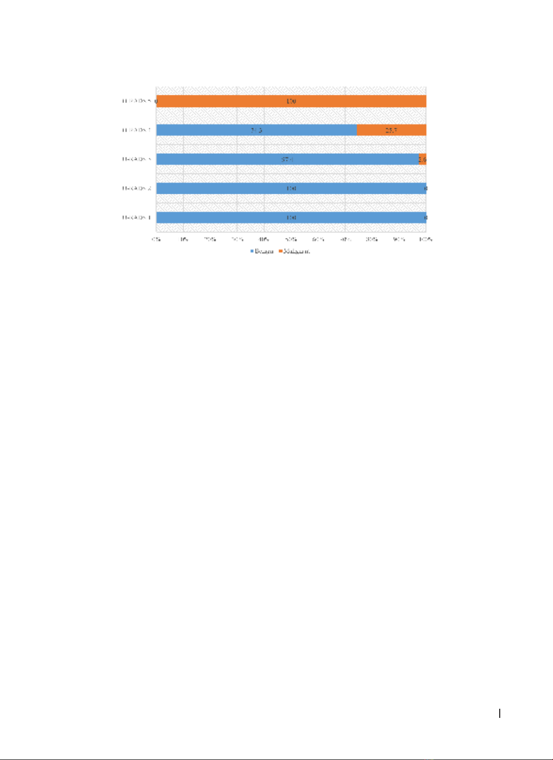
classification was as follows: O-RADS 1 (1.6%), O-RADS 2 (52.1%), O-RADS 3 (22.9%), O-RADS 4 (17.6%),
O-RADS 5 (5.9%). Most ovarian tumors were monocystic masses, without solid components (65.4%), with
diameters ranging from 50 - 100 mm (58.0%), and had smooth inner borders (79.3%). Papillary growth in
inner borders and increased vascularity in Doppler ultrasound (color score: CS = 2 - 4) were found to be
predictive factors for malignant ovarian tumors, with adjusted odds ratios (aOR) of 8.5 and 5.5, respectively.
Conclusions: Monocystic mass with solid components, multicystic mass with solid components, mass with
solid components, irregular inner borders, papillary growth in inner borders, and increased vascularity in
Doppler ultrasound (CS = 2 - 4) were identified as predictive factors for malignant ovarian tumors.
Keywords: ovary tumors, O-RADS classification, ultrasound.
Corresponding: Dang Cong Thuan, Email: dcthuan@huemed-univ.edu.vn
Recieved: 6/2/2023; Accepted: 19/2/2024; Published: 25/2/2024
DOI: 10.34071/jmp.2024.2.7
1. BACKGROUND
Ovarian tumors are the most common disease
of the ovaries with a prevalence of 5% - 30% being
malignant of cases being malignant lesions. They
often present with nonspecific clinical symptoms,
leading to challenges in early diagnosis and
detection. The disease is often detected in a late
stage [1].
There have been many scores and classification
systems introduced to improve the effect of early
diagnosis of ovarian cancer such as Schillinger,
IOTA, or GI-RADS. In 2018, the American College of
Radiology issued a consensus on using the O-RADS
classification system in the diagnosis of ovarian
tumors, providing a consistent way to interpret
ultrasound characteristics and restrict ambiguous
pictures and errors, especially in cases with potential
for malignancy, as well as proposed guidelines
for the management of risk groups. The O-RADS
classification system offers a standardized approach
to interpreting ovarian masses using ultrasound [2].
In Vietnam, there have been studies evaluating
the application of classifications and scores in
diagnosing as well as describing pathological
characteristics of ovarian tumors [3]. However, there
are still quite a few studies that fully investigate
the ultrasound image characteristics and signs for
the prediction of malignant ovarian tumors based
on the O-RADS classification when compared with
postsurgical pathological results. Therefore, we
carry out this study with 2 aims:
1. To describe the ultrasound characteristics
of ovarian tumors according to the O-RADS
classification.
2. To investigate the signs predicting malignancy
in the O-RADS 3, 4, and 5 categories.
2. MATERIALS AND METHODS
2.1. Participants
- A cross-sectional study was conducted on 188
patients who sought examination and treatment at the
Hospital of Hue University of Medicine and Pharmacy.
These patients were diagnosed with ovarian tumors
between April 2022 and September 2023.
HUE JOURNAL OF MEDICINE AND PHARMACY ISSN 1859-3836
52
Hue Journal of Medicine and Pharmacy, Volume 14, No.2-2024
2.1.1. Sample selection criteria
- Patient has ovarian tumor on gynecological
ultrasound;
- Patient has postsurgerial histopathological
results.
2.1.2. Exclusion criteria
- Patient has previously been treated for ovarian
tumors;
- Patient has cancer from other organs
metastasizes to the ovaries;
- Patient does not agree to participate in the
study.
2.1.3. Sampling method
- 188 patients satisfied the selection criteria
based on the convenience sampling method.
2.2. Study methods
2.2.1. Study design: A descriptive cross-
sectional method was applied.
2.2.2. Place and time
- The study was conducted in the Obstetrics and
Gynecology Department, Pathology Department,
and Department of Diagnostic Imaging, at Hue
University of Medicine and Pharmacy Hospital from
April 2022 to September 2023.
2.2.3. Study techniques
2.2.3.1. 2D ultrasound, Doppler ultrasound, and
histopathological techniques
- According to the instructions of the Vietnam
Ministry of Health.
2.2.3.2. Signs that need to be recorded according
to the O-RADS classification system
- Lesion category: unilocular/multilocular, solid/
without solid component
- Cystic lesions: inner margin or walls
including solid component (papillary projection
or nodule, smooth, irregular); internal content,
cystic component (anechoic fluid, hyperechoic
components)
- Solid or solid-appearing lesions: external
contour (smooth, irregular); internal contents
(acoustic shadowing)
- Maximum diameter
- Vascularity: CS = 1, CS = 2, CS = 3, CS = 4
- General and extra-ovarian findings: paraovarian
cyst, peritoneal inclusion cyst, fluid distended in
Fallopian tube, cul-de-sac fluid, ascites, peritoneal
thickening or nodules.
2.2.3.3. Histopathological classification of
ovarian tumors
- According to the classification of ovarian
tumors of World Health Organization [4].
2.2.4. Data processing and analysis
The collected data were processed according
to medical statistical algorithms, using SPSS 22.0
software. Descriptive data were shown in numbers,
percentages, mean, and standard deviation.
Algorithms were used including:
- Chi-squared test (χ2);
- Independent-samples t-test, the Wilcoxon test;
- Significance was set at p ≤ 0.05 for all tests;
- Simple linear regression and multiple linear
regression.
3. RESULTS
3.1. General characteristics
The patients were quite evenly distributed
geographically with 53.2% living in urban areas.
The group with malignant histology had an average
age of 50.62 ± 14.3, higher than the benign group
with 41.01 ± 16.5 (p = 0.012). The incidence
of general ovarian tumors and benign ovarian
tumors in premenopausal women was 2.5 times
higher than in postmenopausal women, while
malignant ovarian tumors were more common in
postmenopausal women than the premenopausal
group (p < 0.05).
3.2. Ultrasound characteristics compared with
pathological results
3.2.1. Postsurgical pathological results
The study found that the majority of ovarian tumors
were benign, accounting for 88.8% of cases, with
the highest proportion being serous cystadenoma,
followed by dermoid cysts, mature teratomas,
and endometriosis. Malignant tumors accounted
for 11.2% with serous carcinoma being the most
common, followed by mucinous carcinoma.
HUE JOURNAL OF MEDICINE AND PHARMACY ISSN 1859-3836 53
Hue Journal of Medicine and Pharmacy, Volume 14, No.2-2024
3.2.2. Distribution of ovarian tumors according to O-RADS classification compared with pathology
results
Chart 1. Distribution of ovarian tumors according to O-RADS classification
compared with pathology results
Based on the O-RADS classification, the
distribution of ovarian tumors is as follows: O-RADS
1 (1.6%), O-RADS 2 (52.1%), O-RADS 3 (22.9%),
O-RADS 4 (17.6%), O-RADS 5 (5.9%). 100% O-RADS
1 and O-RADS 2 ovarian tumors were benign. All
tumors classified as O-RADS 5 were malignant.
3.2.3. Major ultrasound features of ovarian
tumors
66.0% of ovarian tumors were unilocular without
solid components and the majority of these tumors
were benign (123/124 patients). Solid or solid-
appearing tumors were only found in 5.9% of cases
but the malignant rate was 2 times higher than
benign tumors in this group.
The average diameter was 84.4 ± 46.2 mm. In
particular, the malignant group was 129.5 ± 68.6
mm higher than the benign group was 78.8 ± 39.4
mm. Most ovarian tumors in general and benign
tumors had a diameter of 5 - 10 cm, 56.4%, and
58.7% respectively. While malignant tumors were
mainly 10 cm (57.1%). This difference is statistically
significant (p < 0.05).
86.2% of tumors without solid components
are benign, and only 4/148 cases are malignant.
In contrast, tumors with solid components are up
to 81% malignant. The difference was statistically
significant with p < 0.005.
The majority of ovarian tumors in general and
benign tumors in particular had regular internal
margins with 81.4%. 100% of solid tumors with
irregular outer margins were malignant tumors.
100% of malignant tumors didn’t have acoustic
shadowing.
46.8% of ovarian tumors was an anechoic fluid
and/or with little echogenicity inside. Hypoechoic
ovarian tumors accounted for the lowest rate of
4.3%.
The majority of benign tumors didn’t have
vascular proliferation (84%). Ovarian tumors with
increased vascularity (CS = 2 - 4) had a significantly
higher malignant risk than those without increased
vascularity (p < 0.001).
3.2.4. Extra-ovarian findings
In most cases, no cul-de-sac fluid, peritoneal
thickening, or nodules was present. 87.5% of tumors
accompanied by peritoneal fluid were malignant,
and 4/4 of cases with thickened peritoneal or
nodules were malignant. This difference was
statistically significant p < 0.05.
3.3. Signs predicting malignancy in the O-RADS
3, 4, and 5 categories
We conducted data selection for patients in
O-RADS 3,4,5 groups and regression analysis based
on 84 patients in O-RADS 3,4,5 groups.
HUE JOURNAL OF MEDICINE AND PHARMACY ISSN 1859-3836
54
Hue Journal of Medicine and Pharmacy, Volume 14, No.2-2024
3.3.1. Simple linear regression of signs predicting malignancy
Table 1. Simple linear regression of signs predicting malignancy
Ultrasound characteristics OR (CI 95%) p
Multilocular 0.6 (0.2 - 1.6) 0.3
Unilocular cyst with solid component(s) 12 (1.2 - 133.7) 0.03
Multilocular cyst without solid component(s) 2.7 (0.2 - 28.4) 0.4
Multilocular cyst with solid component(s) 11.4 (1.8 - 120.3) 0.04
Solid tumor 35 (3.3 - 368.6) 0.03
Solid appearing 8.5 (2.5 - 28.4) 0.001
Maximum diameter 5 - 10 cm 1.4 (0.1 - 14.3) 0.7
Maximum diameter ≥ 10 cm 1.3 (0.1 - 13.1) 0.8
Papillary projection of the inner margin 16.3 (3.1 - 87.1) 0.001
Irregular inner margin 7.5 (1.1 - 52.4) 0.04
Vascularity CS = 2 - 4 20.1 (5.6 - 71.4) < 0.001
Cul-de-sac fluid N/A P > 0.05
Peritoneal thickening, or nodules N/A P > 0.05
Unilocular cyst with solid component(s), multilocular cyst with solid component(s), solid tumor, solid
appearance, papillary projection of the inner margin, irregular inner margin, and vascularity CS=2-4 were
independent risk factors of malignant ovarian tumors.
3.3.2. Multiple linear regression of signs predicting malignancy
We conducted multiple linear regression analyses based on characteristics with OR > 1 and p < 0.05.
Including solid appearance, papillary projection of the inner margin, irregular inner margin, and vascularity
CS = 2 - 4.
Table 2. Multiple linear regression of signs predicting malignancy
Ultrasound characteristics OR CI 95% p
Solid appearing 0.9 0.1 - 6.9 0.7
Papillary projection of the inner margin 8.5 1.2 - 60.4 0.03
Irregular inner margin 3.5 0.3 - 42.8 0.3
Vascularity CS=2-4 5.5 1 - 29.2 0.04
There was a statistically significant association between findings: papillary projection of the inner margin
and vascularity CS=2-4 with the risk of malignant ovarian tumors.
3.3.3. The value of signs predicting malignancy
Table 3. The value of signs predicting malignancy
Ultrasound
characteristics
Sensitivity
(%)
Specificity
(%)
Positive
predictive value
Negative
predictive value
Papillary projection
of inner margin 64.3 81.4 45.0 90.6
Vascularity CS=2-4 80.9 82.5 60.2 92.8
Vascularity CS=2-4 had quite high sensitivity and specificity, while papillary projection of the inner margin
hadn’t high sensitivity in predicting malignant ovarian tumors.
4. DISCUSSION
4.1. General characteristics
Our study is similar to some other studies that
found that the average age of the malignant group
was higher than those of the benign group [5, 6,
7]. The incidence of ovarian tumors in general and
benign tumors in premenopausal women was 2.5
times higher than in postmenopausal women,
while malignant tumors were more common in
postmenopausal women (p < 0.05). Other authors
HUE JOURNAL OF MEDICINE AND PHARMACY ISSN 1859-3836 55
Hue Journal of Medicine and Pharmacy, Volume 14, No.2-2024
also have similar results [3, 5]. It can be explained
since the symptoms of ovarian tumors in the early
stages are often vague and difficult to detect. It
is not until the late stages of the disease that the
clinical symptoms are clear and easy to detect [8].
4.2. Ultrasound characteristics compared
with pathological results
4.2.1. Postsurgical pathological results
In our study, 88.8% of cases were benign
tumors, with the highest proportion being serous
cystadenoma (40.4%), followed by dermoid cysts,
mature teratomas, and endometriosis. Malignant
tumors accounted for 11.2% with serous carcinoma
being the most common, followed by mucinous
carcinoma. Studies by Vo Thi Quynh Nhu (2022) and
Xie T (2022) also showed similar results. We saw
that there is diversity in the histopathological results
of ovarian tumors; the difference in rates may be
due to the location of each study [6, 9].
4.2.2. O-RADS classification
In general, the results of our study and those
of the authors were different due to many reasons
such as sample size or study location, but there
was a tendency for a malignancy rate of > 75% in
the O-RADS 5 group; 20 - 79% in O-RADS 4 group;
1 - 16% in O-RADS 3 and 0 - 1% in O-RADS 2 group.
According to a publication by the US, the O-RADS
2 group was almost certainly benign (< 1% risk of
malignancy); O-RADS 3, low risk of malignancy (1%
to < 10%); O-RADS 4, intermediate risk of malignancy
(10% to < 50%); O-RADS 5, high risk of malignancy (≥
50%). Therefore, our results were appropriate [2].
4.2.3. Major ultrasound features of ovarian
tumors
In our study, the majority of benign ovarian
tumors were unilocular without solid components
(73.7%), solid or nearly solid tumors accounted for a
high proportion in the malignant group (33.3%). The
percentage of malignant tumors containing solid
components was 81.0% while most benign tumors
did not contain solid components with 86.2% (p <
0.001). Other studies also agreed that the tumors
without solid components tended to be benign,
and tumors with solid components tended to be
malignant [3, 7].
The average diameter of malignant tumors was
129.5 ± 68.6 mm, higher than that of benign tumors,
which was 78.8 ± 39.4 mm. Our study had a higher
rate of unilocular tumors than multilocular tumors
in both benign and malignant groups. The difference
was not statistically significant (p > 0.05), similar to
Tran Doan Tu (2020) [5]. However, Vo Thi Quynh
Nhu found that 75.4% of malignant tumors were
multilocular lesions [6].
Most benign tumors had regular inner margins
(87.1%). While the majority of malignant tumors
had a papillary projection (64.3%) or irregular inner
margins (21.4%). There were 11 solid lesions we
recorded, 4/11 had regular outer margins and all of
them were benign tumors, 7/11 cases had irregular
outer margins and all of them were malignant. This
difference was statistically significant with p < 0.05.
The papillary projection or irregular inner margins
had a higher risk of being malignant than the regular
inner margins without papillary [10].
Acoustic shadowings were not recorded in
malignant lesions and were recorded in 35/188
benign lesions. Some authors have observed that
adding acoustic shadowing to the O-RADS system
improved the area under the curve (AUC) to 0.94 (p =
0.01), similar to the assessment of other neoplasms
in the ANDEX model (AUC = 0.95, p = 0.35) [11].
Since 1989, T. Bourne has begun to describe
perfusion imaging and concluded that vascular flow
imaging on transvaginal ultrasound can be used
for screening purposes. Avoid missing potentially
malignant ovarian masses in the early stages [12]. In
our study, 92.2% of benign ovarian tumors did not
have increased vascularity, and 81% of malignant
ovarian tumors had mild, moderate to strong
vascular proliferation. The difference is statistically
significant p < 0.001.
4.2.4. Extra-ovarian findings
Recording extra-ovarian lesions such as fluid
retention and peritoneal inclusion cysts didn’t
increase the risk score for O-RADS, but other
features such as free peritoneal fluid, peritoneal
thickening, or nodules might increase the category
to O-RADS 5, so they play an important role in
detecting malignant ovarian tumors [2]. In our
study, 100% of patients with peritoneal thickening
or nodules were malignant and 33.3% of malignant
tumors had the presence of peritoneal fluid. This
rate is equivalent to Vo Thi Quynh Nhu’s study
(2022) [6].
4.3. Signs predicting malignancy in the O-RADS
3, 4, and 5 categories
Simple linear regression analysis of ultrasound
characteristics in 84 patients in the O-RADS 3,4,5
group, we found an association between some
factors and the risk of malignant ovarian tumors.
4.3.1. Lesion category
In our study, unilocular cysts with solid
component (OR = 12.0; CI 1.2 - 133.7; p < 0.001),