JOURNAL OF 108 - CLINICAL MEDICINE AND PHARMACY Vol. 19 - Dec./2024 DOI: https://doi.org/10.52389/ydls.v19ita.2510
54
A patient with life-threatening hemoptysis from a
ruptured pulmonary artery aneurysm was successfully
treated with endovascular coil interventions and a
pulmonary lobectomy
Do Thanh Hoa, Le Duc Duan anh Le Xuan Duong*
108 Military Central Hospital
Summary
Life-threatening hemoptysis due to ruptured pulmonary artery aneurysm is a rare emergency with a
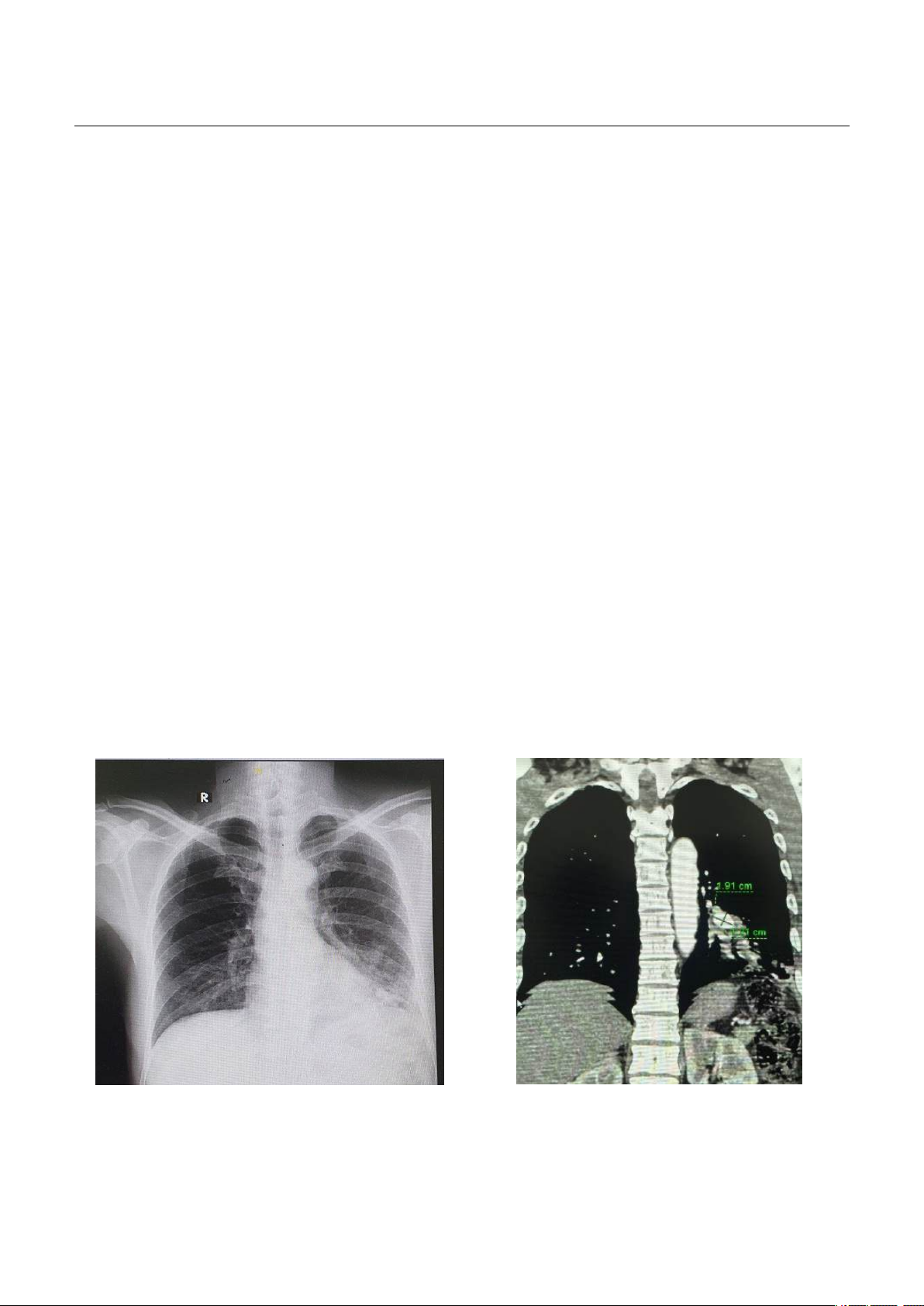
high mortality rate if not treated promptly. Computed Tomography Angiography (CTA) is an important
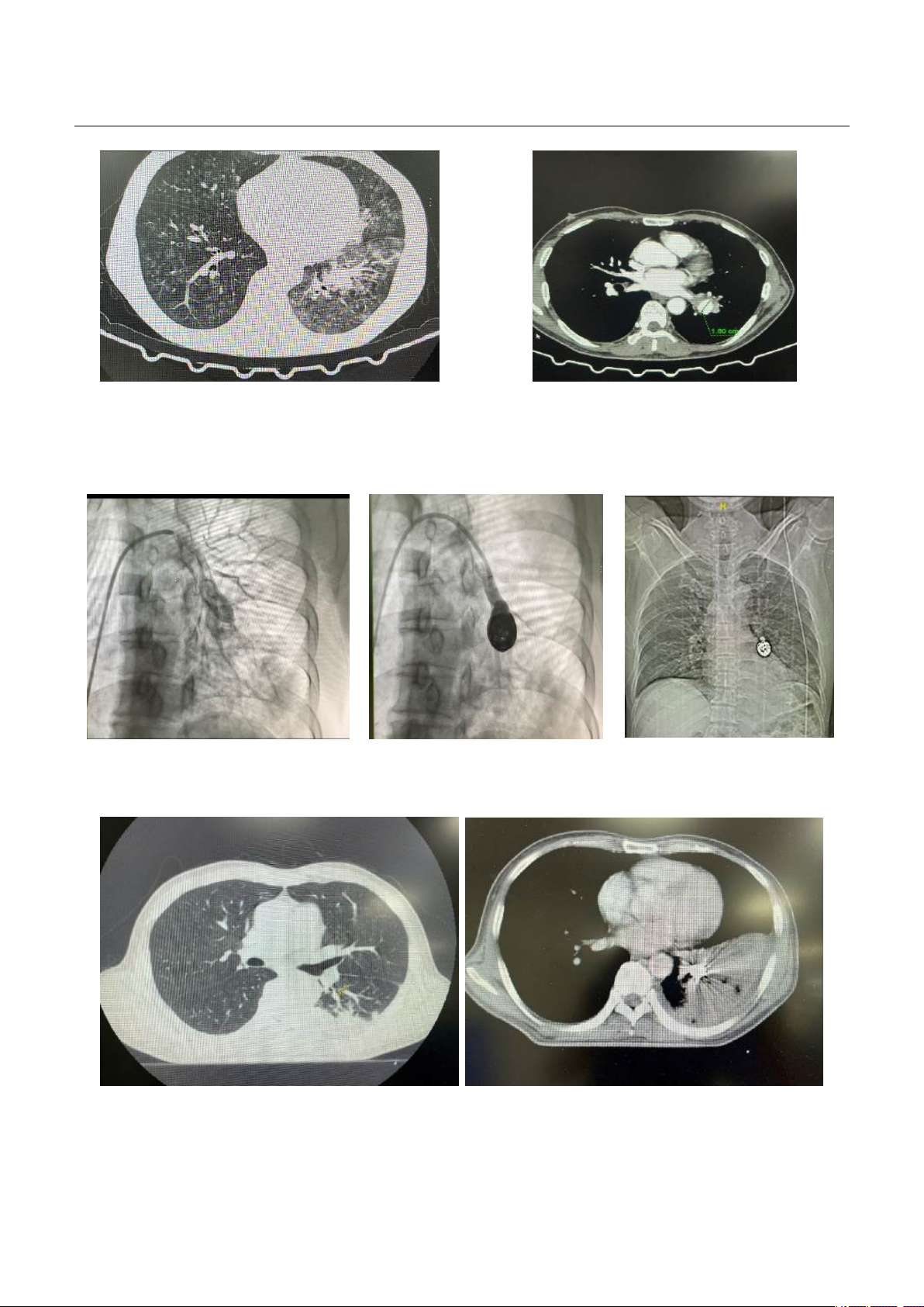
diagnostic method to detect causes of hemoptysis, especially severe hemoptysis. Endovascular coil
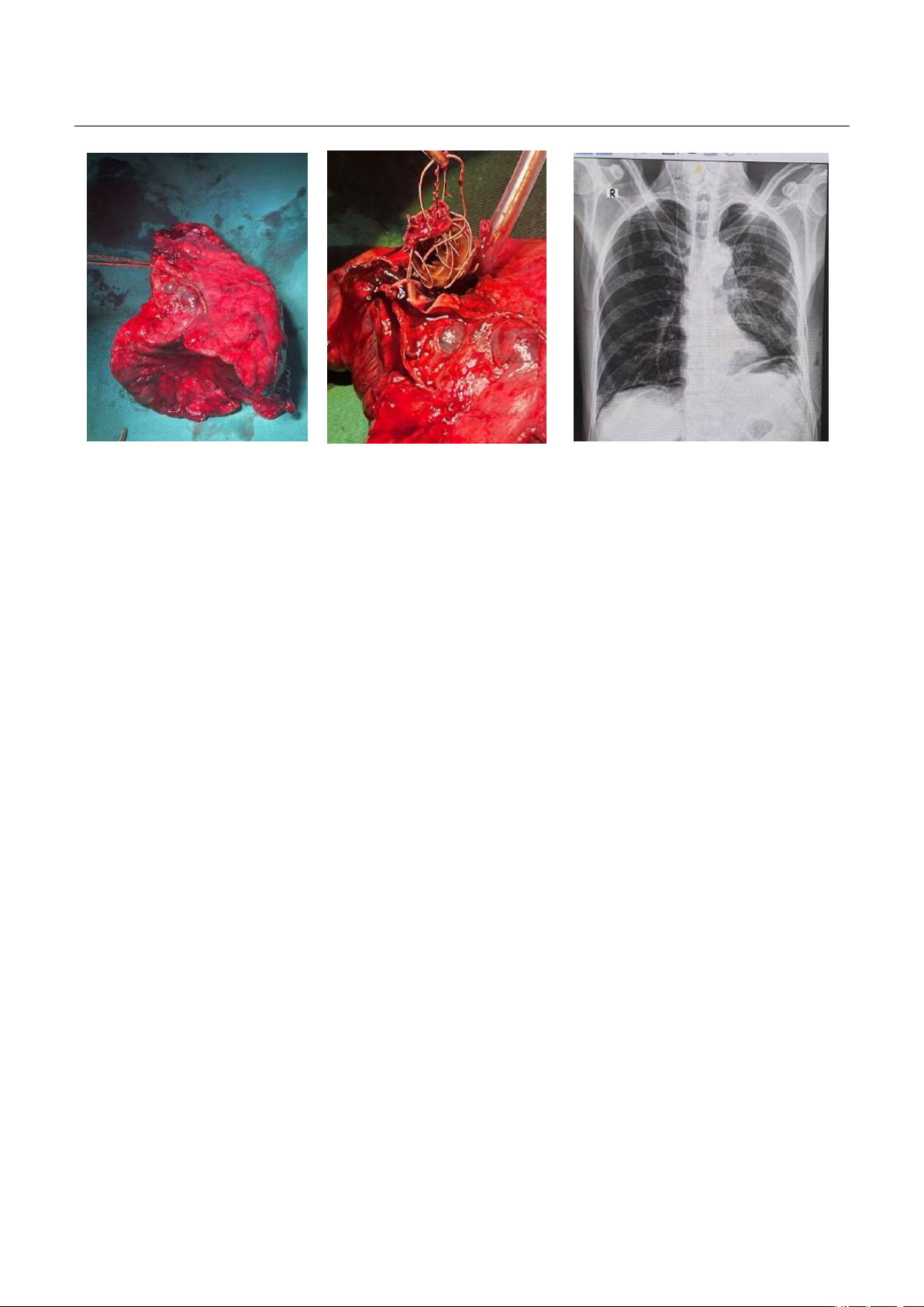
interventions and surgery are the cornerstones of management for pulmonary aneurysms and cessation
of hemoptysis.
Keywords: Life-threatening hemoptysis, pulmonary artery aneurysm, emergency endovascular
coiling, pulmonary lobectomy.
I. BACKGROUND
Life-threatening hemoptysis is coughing up
blood regardless of the amount of blood that
causes hemodynamic disturbances or respiratory
failure leading to death if left untreated1, there is no
consensus on the amount of blood coughed up, but
the authors suggest that coughing up at a rate of
more than 100ml/hour or over 500ml/24 hours
poses a life-threatening risk2. About 5-14% of
patients who cough up blood will have life-
threatening hemoptysis. The mortality rate from life-
threatening hemoptysis is reported to be around 9-
38%. About 90% of hemoptysis is from bronchial
artery circulation due to high pressure. There are
also other vascular sources from non-bronchial
artery circulation such as: aorta, intercostal artery,
subclavicular artery. In which the source from the
pulmonary artery accounts for only about 5% of the
total number of cases2. Pulmonary artery aneurysm
Received: 16 October 2024, Accepted: 30 November 2024
*Corresponding author: duongicu108@gmail.com -
108 Military Central Hospital
rupture is one of the very rare causes of hemoptysis.
The prevalace according to a previous study based
on an analysis of 109,571 autopsies found 8 cases of
pulmonary aneurysms3, that is equivalent to a rate of
about 0.0073%. The cause and pathogenesis are not
completely clear. Pulmonary artery aneurysms are
usually asymptomatic, some may manifest initially
with hemoptysis. Massive hemoptysis and life-
threatening hemoptysis are common symptoms,
accounting for 20-60% of pulmonary aneurysm
ruptures4. Currently, recommendations for surgery
and definitive treatment of pulmonary aneurysms
are not yet available. When there is a life-
threatening hemoptysis, emergency endovascular
intervention and surgery are important measures to
save the patient's life. Below we report a clinical case
of life-threatening hemoptysis caused by ruptured
pulmonary aneurysm treated with endovascular
interventions with coils and pulmonary lobectomy.
II. CASE PRESENTATION
49 year-old male patient with a history of type 2
diabetes mellitus treated with metformin
1700mg/day and had no any previous lung disease
JOURNAL OF 108 - CLINICAL MEDICINE AND PHARMACY Vol. 19 - Dec./2024 DOI: https://doi.org/10.52389/ydls.v19ita.2510
55
and heart disease. Two weeks before admitted to
the hospital, the patient had hemoptysis, about 2ml
each time, 3-5 times a day and mild fever. The
patient went to a local hospital and took a chest X-
ray, revealed thick interstitial tissue and ground
glass opacity in the base of the left lung. The initial
diagnosis was pneumonia, which was treated with
antibiotics and hemostatic medications. However,
the hemoptysis continued.
After that, the patient suddenly coughed up a
large amount of blood of about 200ml, then, he was
admitted to the Emergency Department of 108
Military Central Hospital in a state of alertness, stable
hemodynamic and no respiratory failure. The patient
underwent airway control and took a pulmonary
artery computed tomography scan, revealed a left
lower lobe pulmonary aneurysm measuring 1.91 x
1.21 x 1.80cm, with ground glass opacity at base of
the 2 lungs. The patient was quickly transferred to
endovacular coilings.
During the procedure, the patient continued to
cough up large amounts of blood again for
approximate 300ml, causing respiratory failure. The
patient was intubated to control airway and the
procedure continued. After the coiling intervention
the patient stopped hemoptysis. After the patient
was stable, he underwent a flexible bronchoscopy,
the results showed no abnormalities in the airways,
only little residual blood in the lower left lobe and
the lumen was pushed inward corresponding to the
coil placement. Patient had negative bacterial
culture, negative microscopy of bronchial lavage
fluid for AFB and QuantiFERON TB. Sars-CoV-2 and
influenza test were negative. Blood clotting tests
and echocardiogram were normal. Finally, the
patient was diagnosed with life-threatening
hemoptysis caused by an aneurysm of left branch of
pulmonary artery.
4 days after the procedure, the patient coughed
up blood again with the amount of about 150ml.
The patient was re-evaluated by pulmonary artery
CT scan immediately, suspected another small
pulmonary aneurysm on the periphery of the left
lung and had an extensive consolidation in lower
lobe of the left lung due to blood clot. Therefore, the
patient underwent surgery to remove the lower lobe
of the left lung to solve both radically residual
pulmonary aneurysm and large consolidation. After
surgery, the patient had hemoptysis cessated for 7
days and had no complication from surgery and was
discharged from the hospital.
Figure 1a Figure 1b
JOURNAL OF 108 - CLINICAL MEDICINE AND PHARMACY Vol. 19 - Dec./2024 DOI: https://doi.org/10.52389/ydls.v19ita.2510
56
Figure 1c Figure 1d
Figure 1. 1a: Left basal alveolar opacity on posteroanterior chest X-ray; 1b: Left lower lobe pulmonary artery
aneurysm on coronal plane thoracic CT-scan. 1c: Mass in left lower lobe and ground-glass opacites in lower lobes
of lung on chest-CT scan; 1d: left lower lobe pulmonary artery aneurysm on axial plane chest CT-scan.
Figure 2a Figure 2b Figure 2c
Figure 2. Endovascular intervention with coil into aneurysm. 2a: DSA image before the coiling; 2b: DSA image
after coiling. 2c: Chest X-ray image after intervention (DSA: Digital Subtraction Angiography).
Figure 3a Figure 3b
Figure 3. 3a: A suspicious small pulmonary aneurysm on the periphery of the left lung after being revaluated by
chest-CT scan. 3b: Extensive consolidation in the lower lobe of left lung due to blood clot.
JOURNAL OF 108 - CLINICAL MEDICINE AND PHARMACY Vol. 19 - Dec./2024 DOI: https://doi.org/10.52389/ydls.v19ita.2510
57
Figure 4a Figure 4b Figure 4c
Figure 4. 4a. A picture of the lower lobe of the left lung that has been surgically removed;
4b. The image of the coil was removed. 4c. Chest X-ray after removal of the left lower lobe of lung.
III. DISCUSSION
Pulmonary aneurysm is defined by pulmonary
artery dilation at least 1.5 times the normal size and
damage to all 3 layers of the walls of blood vessels.
Distinguishing from pulmonary pseudo-aneurysms
when damage to less than 3 layers of artery walls5.
About 89% of the aneurysm occurs in the
pulmonary artery trunk, only 8% of the damage in
the right pulmonary artery branch, and 3% of the
damage in the left branch6. Our clinical case has a
left lower lobe pulmonary aneurysm.
Etiology and classification: Pulmonary artery
aneurysms can be divided into 2 main groups: Those
with high endovascular pressure and those with low
endovascular pressure6.
High pressure pulmonary artery aneurysms
include:
Pulmonary artery aneurysms with pulmonary
hypertension associated with heart disease, most
commonly due to ductus arteriosus, interventricular
septal abnormalities, and atrial septal
abnormalities7. Generally these abnormalities are
caused by left-right shunt formation that increases
pressure on the vascular wall and forms an
aneurysm8.
Pulmonary artery aneurysms with idiopathic
pulmonary hypertension and pulmonary artery
aneurysms with pulmonary hypertension due to
other causes (such as chronic pulmonary arterial
thrombosis) 6.
Low pressure pulmonary artery aneurysms
include:
Due to infections: Untreated syphilis and
tuberculosis were often associated with pulmonary
artery aneurysms. Pulmonary artery aneurysms in
syphilis are usually in large pulmonary arteries. In
tuberculosis, it usually forms in the lung
parenchyma, called Rasmussen aneurysm5. Today,
pyogenic bacteria such as Staphylococcus increase
the risk of pseudo-aneurysms.
Vasculitis: Behcet is an unexplained systemic
vasculitis, characterized by damage to the mucosa
and skin, presenting in the eyes, and systemic
vasculitis of both arteries and veins of all sizes.
Pulmonary artery aneurysms are feature of this
disease9. Hughes–Stovin syndrome, this is a very rare
vasculitis that affects mainly large blood vessels,
consisting of 3 main stages: stage I is
thrombophlebitis. Stage II is the formation of
multiple pulmonary artery aneurysms and stage III is
the rupture of the aneurysms causing massive
hemoptysis10.
JOURNAL OF 108 - CLINICAL MEDICINE AND PHARMACY Vol. 19 - Dec./2024 DOI: https://doi.org/10.52389/ydls.v19ita.2510
58
Connective tissue diseases include Marfan
syndrome and Loeys–Dietz syndrome (or
osteoarthritis-aneurysm syndrome).
Pulmonary artery dilation after pulmonary
artery valve stenosis.
Idiopathic pulmonary artery aneurysms: Being a
rare form, this is the final diagnosis after excluding
possible causes.
Patients we report had a normal
echocardiogram should be in the low-pressure
pulmonary artery aneurysm group. There were no
signs of infection, tests for tuberculosis were
negative. There were no other systemic
manifestations of vasculitis or connective tissue
disease. So, there was a high probability of an
idiopathic aneurysm.
About clinical manifestations: Usually no clinical
symptoms or nonspecific clinical symptoms, which
could be seen in many different diseases such as:
Symptoms caused by aneurysms compressing
the surrounding structure such as bronchial
compression causing shortness of breath, cough,
post-obstructive pneumonia, bronchiectasis.
Hemoptysis due to rupture of an aneurysm:
Amount of blood of 100-1000ml per day are
considered massive1. There are many causes of
hemoptysis such as bronchiectasis, tuberculosis,
fungal infections, lung cancer. In which, vascular
abnormalities are one of the possible causes of life-
threatening hemoptysis. However, hemoptysis
caused by vascular abnormalities accounts for only
about 0.2% of all causes of hemoptysis13. Our clinical
case had life-threatening hemoptysis from a
ruptured pulmonary artery aneurysm.
Symptoms of aneurysm dissection: This is a rare
symptom that can cause chest pain, cardiogenic
shock and sudden death.
Symptoms of the cause of the aneurysm:
depends on each cause.
Diagnosis: Diagnosis of pulmonary artery
aneurysm by pulmonary angiography, however this
is an invasive technique and only lumen of
aneurysm is identified. Currently, pulmonary artery
CT scans have helped confirm the diagnosis, assess
thrombosis and other abnormalities of the walls of
blood vessels.
Treatment: There are currently no clear
recommendations for the treatment of pulmonary
artery aneurysm, treatment methods may include:
Conservative treatment can be applied to
asymptomatic cases, the absence of significant
pulmonary hypertension and stable size. Some
authors suggest conservation for cases of aneurysm
diameter < 6cm12. Conservative treatment is the
medical treatment of underlying causes such as
immunosuppressants for vasculitis, anti-tuberculosis
drugs, antibiotics for syphilis.
Endovascular intervention: Various types of
intervention materials such as coils, glues, covered
stents, detachable balloons, vascular plug and PDA
closure devices, depending on the anatomical
position and characteristics of the aneurysm, there
are many different endovascular embolization
procedures13. Treatment of endovascular
intervention is one of the fairly new approaches,
especially applied to small peripheral branches. The
advantage of the aneurysm coiling is that it can
retain many peripheral pulmonary artery branches
and preserve lung function in the distal segment of
the aneurysm14. Our patients had an aneurysm in
the left pulmonary artery branch, so initial treatment
option with the aneurysm coiling was reasonable
and feasible. According to the medical literature, the
initial success rate was 95%. However, placing coils
within an aneurysm carries a potential risk of
rupture. Care must be taken to avoid overpacking or
advancing the catheter and coils through the
aneurysm wall14. In our case, the picture after
surgery showed, coiling still worked well.
Surgical treatment remains the foundational
treatment for pulmonary artery aneurysms, and
there is insufficient evidence of a pulmonary trunk
aneurysm diameter threshold for surgery7. Surgery is
usually performed in cases of large aneurysms (>
6cm), which are particularly symptomatic in patients
with signs of right ventricular dysfunction and
pulmonary hypertension12. In cases of focal
pulmonary artery dilation, the aneurysm may be
surgically removed. In cases of diffuse dilation, the
entire trunk of the pulmonary artery is replaced by
an artificial or allogeneic artery segment. However,
peripheral pulmonary aneurysms are more difficult