JOURNAL OF MILITARY PHARMACO-MEDICINE N04 - 2025
22
RESULTS OF ALZHEIMER’S DISEASE ANIMAL MODEL INDUCTION
BASED ON THE INTRAHIPPOCAMPAL INJECTION OF AMYLOID
Β-PEPTIDE (1-42) AT VIETNAM MILITARY MEDICAL UNIVERSITY
Nguyen Ha Hoa1*, Do Duc Thuan1, Do Xuan Hai2
Abstract
Objectives: To evaluate some behaviors and histological results of the
hippocampus in a rat model of Alzheimer's disease (AD). Methods: A
longitudinal, descriptive study was conducted on 21 Wistar rats. Intrahippocampal
amyloid beta (Aβ) injection was performed, followed by behavioral testing and
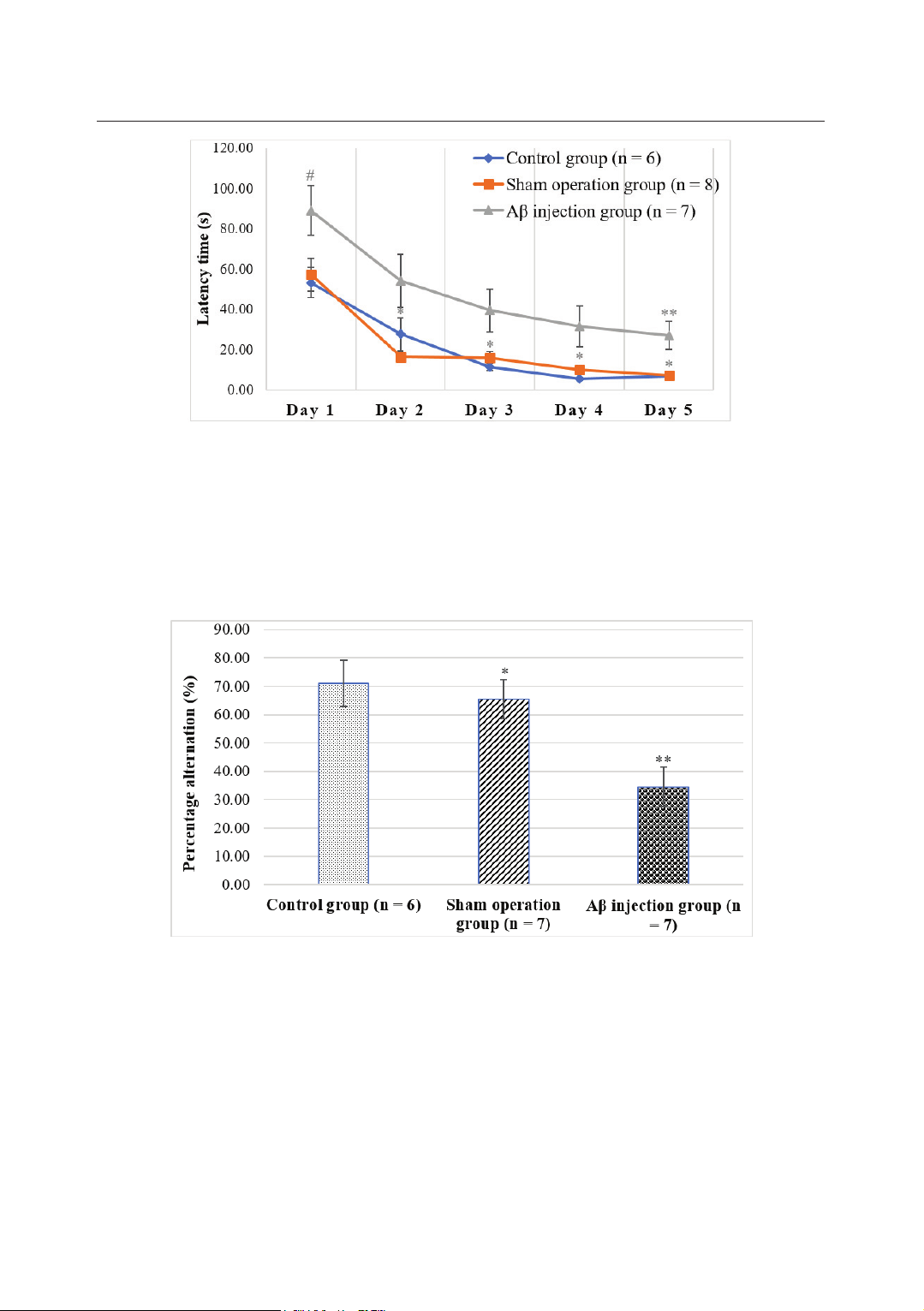
analysis and hippocampal histological examination. Results: Rats in the AD model
showed learning impairment with a reduced average latency time (52.09 ± 10.67s)
and reduced latency time on the 5th day (27.08 ± 6.88s) during the learning phase
of the Morris water maze (MWM) test. Memory impairment was indicated by a
decreased percentage of spontaneous alternations (34.45 ± 7.03%) in the Y-maze
test, reduced time spent (14.84 ± 4.61s), and shorter distance traveled (2.26 ±
0.82m) in the target quadrant during the probe trial of the MWM test.
Neurodegeneration in the hippocampus was observed, with an increased
degeneration score (1.83 ± 0.31). Conclusion: Intrahippocampal Aβ injection is
an effective method for inducing the AD model in rats, characterized by learning
and memory impairments as well as neurodegeneration in experimental animals.
Keywords: Alzheimer's disease model; Hippocampus; Stereotaxic surgery.
INTRODUCTION
Alzheimer's disease is a progressive,
age-related degenerative brain disorder
with a multifactorial and heterogeneous
etiology. Among many hypotheses
proposed for the pathogenesis of AD,
the amyloid hypothesis is the most
widely accepted pathological mechanism.
Extracellular amyloid plaques, intracellular
neurofibrillary tangles, neuronal
degeneration, and consequent memory
impairment are hallmark features of AD [1].
1Department of Neurology, Vietnam Military Medical University
2Practical and Experimental Surgery Department, Vietnam Military Medical University
*Corresponding author: Nguyen Ha Hoa (bsnavyhh@gmail.com)
Date received: 08/01/2025
Date accepted: 27/02/2025
http://doi.org/10.56535/jmpm.v50i4.1180
JOURNAL OF MILITARY PHARMACO-MEDICINE N04 - 2025
23
AD models serve as valuable tools
for replicating pathological changes,
deciphering the disease's pathogenesis,
and testing novel therapeutic approaches
[1]. Many AD models have been
developed on a global scale, with the rat
intrahippocampal Aβ injection model
being demonstrated as one of the most
representative and reliable AD animal
models [2]. However, this specific AD
model has not yet been reported in
Vietnam. Therefore, this study aims to:
Evaluate behaviors and histological
outcomes of AD rat model induction via
intrahippocampal Aβ injection.
MATERIALS AND METHODS
1. Subjects
Including 21 adult healthy Wistar
rats experimented in strict compliance
with The Animal Center Guidelines for
Care and Use of Laboratory Animals at
Vietnam Military Medical University.
* Location and time: The study was
conducted at the Practical and Experimental
Surgery Department from September
2022 to July 2024.
2. Methods
* Study design: A longitudinal,
descriptive study.
* Sample size: The sample size was
determined using the formula:
n = DF/k + 1
n: Sample size of each group; DF:
Degree of freedom with a value of 10 or 20;
k: Number of comparison groups. With
k = 2, the sample size of each group is:
6 n 11 [3]
* Research procedure:
21 rats were divided into 3 groups:
The control group (n = 6), the sham
operation (SO) group (n = 8), and the
Aβ injection (AB) group (n = 7).
Drug: Aβ1-42 (ab120959) was purchased
from Abcam and was dissolved in PBS
(2 μg/μL).
Surgery: Rats were anesthetized with
intraperitoneal ketamine and xylazine,
and their heads were shaved and fixed
onto the stereotaxic apparatus (Japan).
Injection points (coordinates: AP =
-4mm, ML = ± 2.2mm) were marked in
stereotaxic frame. The hole was drilled
into the skull using an electric driller
(Komax drill, Germany). The Hamilton
needle (Hamilton 10μL pump, USA)
was lowered into the hippocampus at a
depth of DV = -3.2mm. For the AB
group, 2.5μL of Aβ (2 μg/μL) was
injected bilaterally. In the SO group, the
needle was inserted without injecting
any substance. After the injection, the
needle was carefully withdrawn, the
skull opening sealed with composite
solder, and the skin sutured [2].
Behavioral test:
+ Y-maze test: The Y-maze test was
performed according to the procedures
JOURNAL OF MILITARY PHARMACO-MEDICINE N04 - 2025
24
described in a previous study (Prieur E
et al., 2019). The Y-maze is a three-arm
maze with equal angles between all
arms of 75cm in length and 15cm in
width, with 15cm-high walls. The maze
floor and the walls were constructed
using black-painted wood. Rats were
initially placed within one arm, and the
sequence and number of arm entries
were recorded over an 8-minute period
for each rat and monitored using a video
tracking system (ANY-maze, Stoelting,
USA) [9].
+ MWM test: The MWM test was
performed according to the procedures
described in a previous study (Vorhees
CV et al., 2006) [4]. The MWM is a
black circular pool (150cm in diameter
and 60cm in height). The circular pool
was filled with water at a temperature of
21°C ± 1°C. The pool was divided into
four equal quadrants. A transparent
platform (10cm in diameter and 28cm
in height) was centered in one of the
four quadrants of the pool and
submerged 2cm below the water's
surface so that it was invisible at the
surface. The water maze experiment
was performed on 6 days. On the 5th of
learning, the rat received swimming
training for 120 seconds in the presence
of the platform. The rat underwent a
daily session of four training trials each,
with an inter-trial interval of 2 minutes.
In each training trial, rats were placed in
water facing the wall of the pool in a
randomly selected pool quadrant. Once
the rat located the platform, it was
allowed to remain on it for 10 seconds.
If the mouse failed to locate the
platform within 120 seconds, it was
placed on it for 10 seconds and then
removed. On the 6th day, in the probe
trial, the platform was removed, and
rats were tested for memory retrieval by
swimming for 60 seconds. The trajectory
of each rat while swimming was
monitored using a video tracking
system (ANY-maze, Stoelting, USA).
Histological analysis: At the end of
the experimental phase, the hippocampus
of 6 rats of each group (AB and SO) was
extracted and fixed in 10% formalin,
then paraffin block was cast and cut into
3 - 4μm slices and stained with
Hematoxylin-Eosin (HE). The specimens
were observed under an optical microscope.
* Research variables and indicators:
Behavioral assessment indicators:
Determined by Anymaze software:
+ Number of alternating movements
is the number of times the rat successfully
enters three consecutive wings (ABC,
ACB, BCA, etc.) and percent alternation
[9]. % Alternation = (Number of
alternating movements)/(Total number
of times entering the wings -2) x 100%.
JOURNAL OF MILITARY PHARMACO-MEDICINE N04 - 2025
25
+ The average escape latency (s) on
each day of learning. The swimming
time (s) and distance (m) at the target
quadrant in the probe trial [4, 5].
Pathological indicators:
Neurodegeneration is determined by
cell damage in the hippocampus on HE
staining, which includes cytoplasm
eosinophilic, vacuole, dispersed
chromatin, and loss of nuclear
membrane integrity. Each damage is
given 1 score; the neurodegenerative
score is the sum of the scores of the
lesions [6].
* Data collection and processing
methods: Behavioral data were extracted
from the Excel files of Anymaze software.
Statistical analysis was performed using
SPSS software. Quantitative variables
were expressed as Mean ± SEM.
Alternation (%) in the Y-maze test, the
time and distance of swimming in the
target quadrant in the MWM probe trial
were analyzed using one-way ANOVA,
the Tukey's post-hoc test in case of
multiple comparisons. Escape latencies
and swimming distance in the training
trials in the MWM test were analyzed
using repeated-measures two-way
ANOVA and Bonferroni adjustment for
multiple comparisons. Neurodegenerative
scores were analyzed using the Independent-
Sample T-test. The differences were
considered statistically significant with
p < 0.05.
3. Ethics
The research was conducted
according to Decision No. 3424/QĐ-
HVQY dated September 19, 2022. The
Department of Practical and Experimental
Surgery granted permission for the use
and publication of the research data. The
authors declare to have no conflicts of
interest in the research.
RESULTS
1. Learning performance of AD
model in rats
Repeated measure two-way ANOVA
test showed that there was a difference
in latency time between groups [(F(2,
10) = 14.07; p = 0.001]. Bonferroni
adjustment for multiple comparisons
showed that the latency time of the AB
group (52.09 ± 10.67s) was higher than
that of the SO group (22.84 ± 2.62s) and
the control group (20.94 ± 1.93s). On
the 5th day of the learning phase, the
latency time of the AB group (27.08 ±
6.88s) was higher than that of the SO
group (7.18 ± 0.88s) and the control
group (6.73 ± 0.83s); the difference was
statistically significant with p < 0.05.
There was no difference in latency time
between the SO group and the control
group during learning days with p > 0.05.
JOURNAL OF MILITARY PHARMACO-MEDICINE N04 - 2025
26
Figure 1. The swimming time to the platform of rats in the MWM.
(**: Compare between the AB group and SO group, control group (** p < 0.05);
*: Compare between the SO and control group (* p > 0.05). #: Compare between
three group (# p > 0.05))
2. Memory impairment of AD model in rats
Figure 2. Percentage alternation of rats in Y-maze.
(**: Compare between the AB group and SO group, control group (** p < 0.05);
*: Compares between the SO and control group (* p > 0.05))
One-way ANOVA test showed that there were significant differences in %
alternation between the three groups [F(2, 19) = 7.36; p = 0.005], Tukey’s post-
hoc test showed that % alternation in the AB group (34.45 ± 7.03%) was lower
than that in the SO group (65.44 ± 6.84%) and control group (71.09 ± 8.19%); the