
64
Journal of Medicine and Pharmacy, Volume 12, No.07/2022
Corresponding author: Nguyen Hoang Bach. Email: nhbach@huemed-univ.edu.vn
Recieved: 12/10/2022; Accepted: 15/11/2022; Published: 30/12/2022
Identification of bacterial pathogens from clinical samples using 16S
rRNA sequencing
Nguyen Hoang Bach1*, Mai Thi Thao Nhi2, Ung Thi Thuy1, Nguyen Thi Khanh Linh1
(1) Department of Microbiology, University of Medicine and Pharmacy, Hue University, Vietnam
(2) Department of Basic Medical Sciences, Phan Chau Trinh University, Quang Nam, Vietnam
Abstract
Introduction: Bacterial infections have a substantial impact on global health and can become serious if
misdiagnosed with several diseases related to the central nervous, cardiovascular, and respiratory systems.
The prognosis in patients with infectious disease strongly depends on early diagnosis and appropriate
antibiotic therapy. We aimed to compare the accuracy of genus and species-level identification bacteria
using biochemical testing and 16S rRNA sequence analysis. Material and methods: 50 clinical samples were
isolated and identified the pathogenic bacteria by routine laboratory methods. In parallel, DNA was extracted
from isolate’s colonies and amplified the 16S rRNA gene by using specific primers. The PCR products were
evaluated by agarose gel electrophoresis and direct sequencing by the Sanger method. The sequence data
were manipulated by Geneious Prime software. The sequence data matching the Prokaryotic 16S Ribosomal
RNA database with a similarity score of ≥ 98% were selected. Results: Total of 50 clinical samples were isolated
and identified the pathogenic bacteria with common biochemical test and API® Microbial Identification. The
sequencing data showed that almost species identified by 16S rRNA sequencing matched the biochemical test
method. There are 3 species (6%) were identified as different species with the routine methods. Conclusions:
16S rRNA gene sequencing is more sensitive, easier to manage, more accurate and especially for bacteria that
are difficult to identify. 16S rRNA sequencing is considered an effective method to early identify pathogens in
clinical samples, and this technique is increasingly being used in microbiology laboratories.
Keywords: 16S rRNA gene, Sanger sequencing, bacterial identification, misdiagnosed.
1. INTRODUCTION
Bacterial infections have a substantial impact
on global health and can become serious if
misdiagnosed with several diseases related to the
central nervous, cardiovascular, and respiratory
systems. These contribute to increased morbidity
and mortality rates, especially in immunodeficiency
patients. The prognosis in patients with infectious
disease strongly depends on early diagnosis and
appropriate antibiotic therapy [1,2]. Therefore, rapid
and sensitive identification of pathogenic bacteria is
essential for initiating timely and effective antibiotic
treatment and preventing disease spread [3].
Cultivation and phenotypic identification methods
(culture-dependent methods) for determining
antimicrobial resistance remain the gold standard
approach in clinical microbiology. However, the
sensitivity of culture methods is influenced by
patient characteristics, laboratory practices, and
the spectrum of bacterial pathogens. These are also
time-consuming, taking at least 24 - 48h to complete
which leads to delayed appropriate treatment
in critically ill patients. Such delays may worsen
the patients’conditions and increase mortality. In
addition, it is challenging to identify by culture
with fastidious, slowly growing microorganisms or
antibiotic exposure prior to sample collection and
generally fails to differentiate between species
of the genus. The other methods for microbial
identification in the laboratory is the genotypic
identification - molecular diagnostic method [4].
Molecular approaches have been offered
as an alternative or complement to phenotypic
methods. Typically, conserved sequences within
phylogenetically informative genetic targets, such
as the 16S rRNA coding gene, are used for bacterial
genotypic identification [5,6]. In this study, we
report a comparison of two bacterial identification
methods which rely on phenotypic/biochemical
tests and 16S rRNA gene sequence analysis. The
ability of these two methods to accurately identify
50 clinical isolates at levels of specificity: genus and
species, was examined.
2. MATERIALS AND METHODS
2. 1. Materials
Forty clinical samples were collected from Dec
2020 to April 2021 at Hospital of Hue University
DOI: 10.34071/jmp.2022.7.9

65
Journal of Medicine and Pharmacy, Volume 12, No.07/2022
of Medicine and Pharmacy. All samples were
transported to the Microbiology Department of
Hospital of Hue University of Medicine and Pharmacy
within 2 hours after collection for microbiological
analysis.
Ten QA-QC samples from OUCRU (Oxford
University Clinical Research Unit - Vietnam) were
performed the species identification as control
group.
2.2. Methods
Research method: cross-sectional study.
Isolation and phenotypic identification
The samples were processed for bacterial
isolation and identification by routine microbiological
methods such as culture and biochemical tests
following the guideline of the Ministry of Health
- Vietnam. These strains were isolated and
phenotypically identified by means of the API® 20
E for Gram-negative bacilli; API® 20 NE for Gram-
negative non-Enterobacteriaceae; API 20 strep for
Streptococci and API® Coryne for Corynebacteria.
The isolates were stored in Brain Heart Infusion
Broth (E&O Laboratories, Bonnybridge, Scotland)
with 20% of sterile glycerol in a cryovial at -80oC for
long-term storage.
DNA extraction
The colonies were picked up from the primary
plate of each isolate and resuspended in 200 uL 1×
TE buffer. The samples were centrifuged at 15,000 ×
g for 15 min. The supernatant was eliminated, and
the pellet was resuspended in molecular biology-
grade water (Eppendorf, Hamburg, Germany), then
centrifuged at 15,000 × g for 10 min. The supernatant
was eliminated, and the pellet was resuspended in
40 μL of molecular biology-grade water, subjected to
boiling at 100°C in a water bath for 10 mins, cooled
on ice, and centrifuged at 15,000 × g for 10 s. The
supernatant was transferred to a new tube before it
was stored at -20°C [7 - 9].
PCR amplification of 16S rRNA gene
The 16S rRNA gene has been a mainstay of
sequence-based bacterial analysis until today. The
gene is large enough, with sufficient interspecific
polymorphisms of 16S rRNA gene. Conventional
PCR was performed by using forward primers 5’-
AGAGTTTGATCMTGGCTCAG-3’ and reverse primer
5’-TACGGYTACCTTGTTACGACTT -3 located at
position 27 and 1492 respectively , which specifically
targets approximately 1500 bp of the 16S rRNA gene
[10]. A total of 50 ng genomic DNA, 0.5 μM for each
primer, and 12.5 µL MyTaq Mix 2× Bioline (Meridian
Bioscience International Limited) were combined in
a 25 μL total volume reaction. The PCR amplification
was profiled as follows: initial denaturation at 95oC
for 5 minutes, followed by 30 cycles of 94oC for 30
seconds, 60oC for 30 seconds, 72oC for 90 seconds,
then 72oC for 7 minutes in Veriti™ 96-Well Thermal
Cycler (Applied Biosystems, USA). 4μL of PCR
products were separated by electrophoresis on
1% agarose gel with 1× GelRed and digitized with
GelDocs XR (Biorad, CA, USA).
16S ribosomal RNA gene profile analysis
10 ng of PCR products having 16S rRNA
amplicon and 0.32 µM of each primer were used
for direct sequencing. To sequence both strands,
two primers were run for each isolate. Forward and
reverse sequences were assembled into consensus
sequences using Geneious Prime v2020.0.3 to
get the consensus 16S rRNA sequences, primers
were trimmed manually, and ambiguous bases
were resolved based on visual inspection of the
chromatograms. Consensus sequences were
taxonomically classified via Geneious Prime BLAST
Plugin. The sequence data matching the partial
sequence of the Prokaryotic 16S Ribosomal RNA
databases with a similarity score of ≥ 98% were
selected.
3. RESULTS
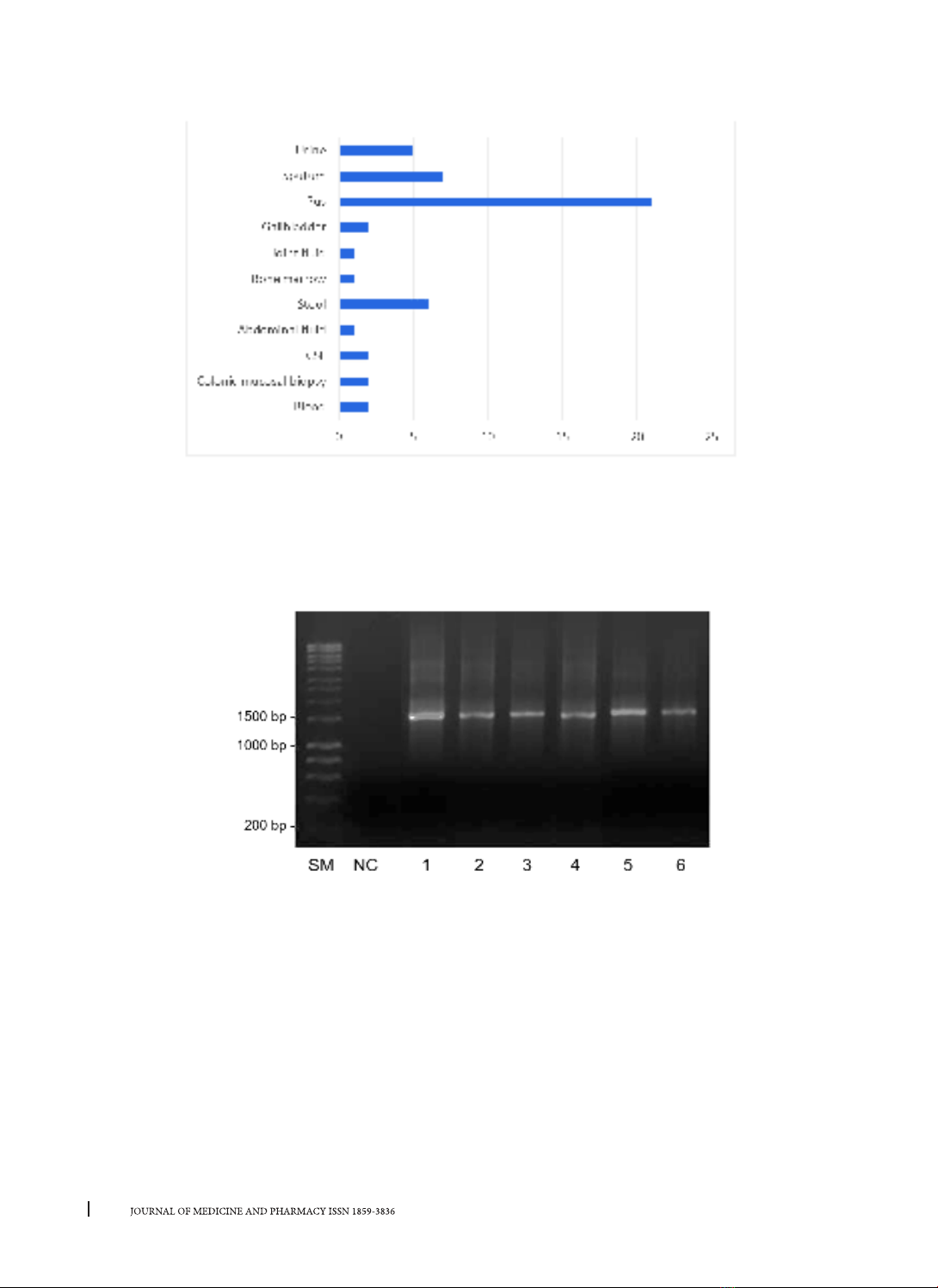
Fifty clinical samples composed of 11 types
of clinical samples were isolated the pathogenic
bacterial by the biochemical tests-based method
(Fig.1). There are 35 different strains with 21 samples
identified as more than one strain, 6 samples were
identified as Stenotrophomonas maltophilia (12%),
3 were identified as Klebsiella pneumonia (6%),
4 were identified as Morganella morganii (8%), 2
were identified as Serratia odorifera (4%), 2 were
identified as coagulase-negative Staphylococci
(CoNS) (4%), 4 were identified as Klebsiella oxytoca
(4%), and 29 samples were identified in single
species (Table 1).
66
Journal of Medicine and Pharmacy, Volume 12, No.07/2022
Fig.1. Type of clinical sample and the number of isolates of each type of clinical sample.
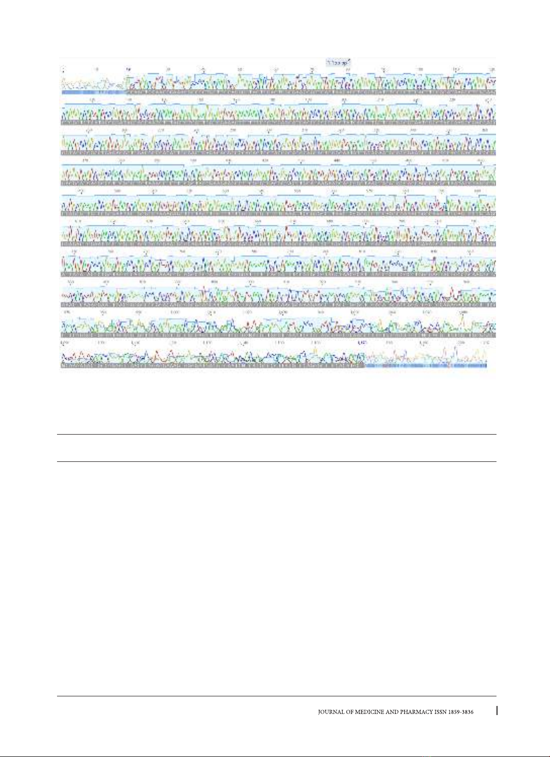
Amplification of 16S rRNA gene
A sequence including the near full-length of the 16S rRNA gene was obtained from PCR reactions with
27F and 1492R universal primers in all of the samples. Amplification of the 16S rRNA gene was confirmed by
gel electrophoresis. The expected size of approximately 1500 bp was amplified (Fig. 2). The specific primers
worked correctly in all samples. The remaining PCR product was sequenced in a total of 50 samples.
Fig.2. Amplicon of 16S rRNA gene on 1% agarose gel. SM: 1kb plus DNA ladder (ThermoFisher, USA); NC:
non-template control; lane 1-6: amplicons of 16S rRNA gene
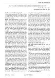
Sequencing and species classification
Fifty PCR products were sequenced and the data were manipulated with Geneious Prime software. The
primers were trimmed manually, and ambiguous bases were resolved based on visual inspection of the
chromatograms (Fig.3). The forward and reverse sequences were assembled into consensus of 16S rRNA
gene for each isolate. Consensus sequences were taxonomically classified via Geneious Prime BLAST Plugin.
All 50 sequence data were matching the partial sequence of the prokaryotic 16S ribosomal RNA databases
with a similarity score of ≥ 98-99%. The species classification was shown in the right column of table 1. There
are 3 samples in which the species taxonomy is different from the conventional microbiology method (grey
row with underlined taxonomy name) (Table 1): Burkholderia pseudomallei, Escherichia coli, Streptococcus
constellatus (Fig.4).
67
Journal of Medicine and Pharmacy, Volume 12, No.07/2022
Fig 3. Raw data of forward sequence by the Sanger sequencing method (chain-termination). The good
quality sequenced bases are more than 1100bp for each strain.
Table 1. Result of bacterial identification by biochemical tests-based method versus 16S rRNA gene
sequencing method
Sample
ID Biochemical test methods Specimens 16S rRNA sequencing
1Morganella morganii Pus Morganella morganii
2Stenotrophomonas maltophilia Pus Stenotrophomonas maltophilia
3Providencia stuartii Urine Providencia stuartii
4Stenotrophomonas maltophilia Pus Stenotrophomonas maltophilia
5Providencia rettgeri Urine Providencia rettgeri
6Morganella morganii Sputum Morganella morganii
7Chryseobacterium
eningosepticum
Urine Chryseobacteriumm
eningosepticum
8Morganella morganii Pus Morganella morganii
9Stenotrophomonas maltophilia Pus Stenotrophomonas maltophilia
10 Klebsiella oxytoca Colonic mucosal
biopsy
Klebsiella oxytoca
11 Stenotrophomonas maltophilia Blood Stenotrophomonas maltophilia
12 Stenotrophomonas maltophilia Stool Stenotrophomonas maltophilia
13 Alcaligenes spp. Pus Alcaligenes spp.
14 Plesiomonas shigelloides Gallbladder Plesiomonas shigelloides

68
Journal of Medicine and Pharmacy, Volume 12, No.07/2022
15 Serratia marcescens Pus Serratia marcescens
16 Aeromonas salmonicida Sputum Burkholderia pseudomallei*
Accession: OP890627
17 Streptococcus anginosus Pus Streptococcus anginosus
18 Klebsiella pneumoniae Urine Klebsiella pneumoniae
19 Burkholderia pseudomallei Blood Burkholderia pseudomallei
Accession: OQ076306
20 Staphylococcus aureus Sputum Staphylococcus aureus
21 Streptococcus constellatus Pus Streptococcus constellatus
22 Coagulase-negative Staphylococci Pus Staphylococcus intermedius
23 Aeromonas hydrophila Abdominal fluid Aeromonas hydrophila
24 Serratia odorifera Bone narrow Escherichia coli*
25 Serratia odorifera Pus Serratia odorifera
26 Coagulase-negative Staphylococci Pus Staphylococcus haemolyticus
27 Stenotrophomonas maltophilia Pus Stenotrophomonas maltophilia
28 Elizabethkingia meningoseptica Sputum Elizabethkingia meningoseptica
29
Chromobacterium
violaceum Joint fluid Chromobacterium violaceum
30 Klebsiella pneumoniae Pus Klebsiella pneumoniae
31 Klebsiella oxytoca Sputum Klebsiella oxytoca
32 Klebsiella pneumoniae CSF Klebsiella pneumoniae
33 Gemella morbillorum Pus Gemella morbillorum
34 Kluyvera intermedia Gallbladder Kluyvera intermedia
35 Chryseobacterium indologenes Pus Chryseobacterium indologenes
36 Streptococcus agalactiae Pus Streptococcus agalactiae
37 Aerococcus urinae Pus Aerococcus urinae
38 Leuconostoc spp. Pus Streptococcus constellatus*
39 Aerococcus viridan Pus Aerococcus viridan
40 Achromobacter xylosoxidans Pus Achromobacter xylosoxidans
41 Morganella morganii Sputum Morganella morganii
42 Klebsiella oxytoca Sputum Klebsiella oxytoca
43 Stenotrophomonas maltophilia Pus Stenotrophomonas maltophilia
44 Citrobacter koseri CSF Citrobacter koseri
45 Enterococcus spp. Stool Enterococcus spp.
46 Enterococcus durans Stool Enterococcus durans
47 Salmonella spp. Stool Salmonella spp.
48 Enterococcus faecium Urine Enterococcus faecium
49 Enterococcus faecalis Stool Enterococcus faecalis
50 Enterococcus group D Stool Enterococcus group D
* Identification by 16S rRNA sequencing are different from biochemical tests-based method.


![Phương pháp ELISA: [Thêm từ mô tả/định tính để tăng CTR]](https://cdn.tailieu.vn/images/document/thumbnail/2014/20140329/phamtrong91/135x160/8501396068823.jpg)


![Giáo trình Lý thuyết Vi sinh 2 (Cao đẳng Y tế Thanh Hoá): Kỹ thuật xét nghiệm [Mới nhất]](https://cdn.tailieu.vn/images/document/thumbnail/2025/20251231/gaupanda090/135x160/18981772781252.jpg)


