MINIREVIEW
Hyaluronan matrices in pathobiological processes
Aimin Wang
1
, Carol de la Motte
2
, Mark Lauer
1
and Vincent Hascall
1
1 Department of Biomedical Engineering, The Cleveland Clinic, Cleveland, OH, USA
2 Department of Pathobiology, The Cleveland Clinic, Cleveland, OH, USA
Mechanism of hyaluronan synthesis
Hyaluronan (HA) is a glycosaminoglycan that is syn-
thesized by a distinctly different mechanism from the
other glycosaminoglycans (chondroitin sulfate, heparan
sulfate, keratan sulfate). A diagram showing the mech-
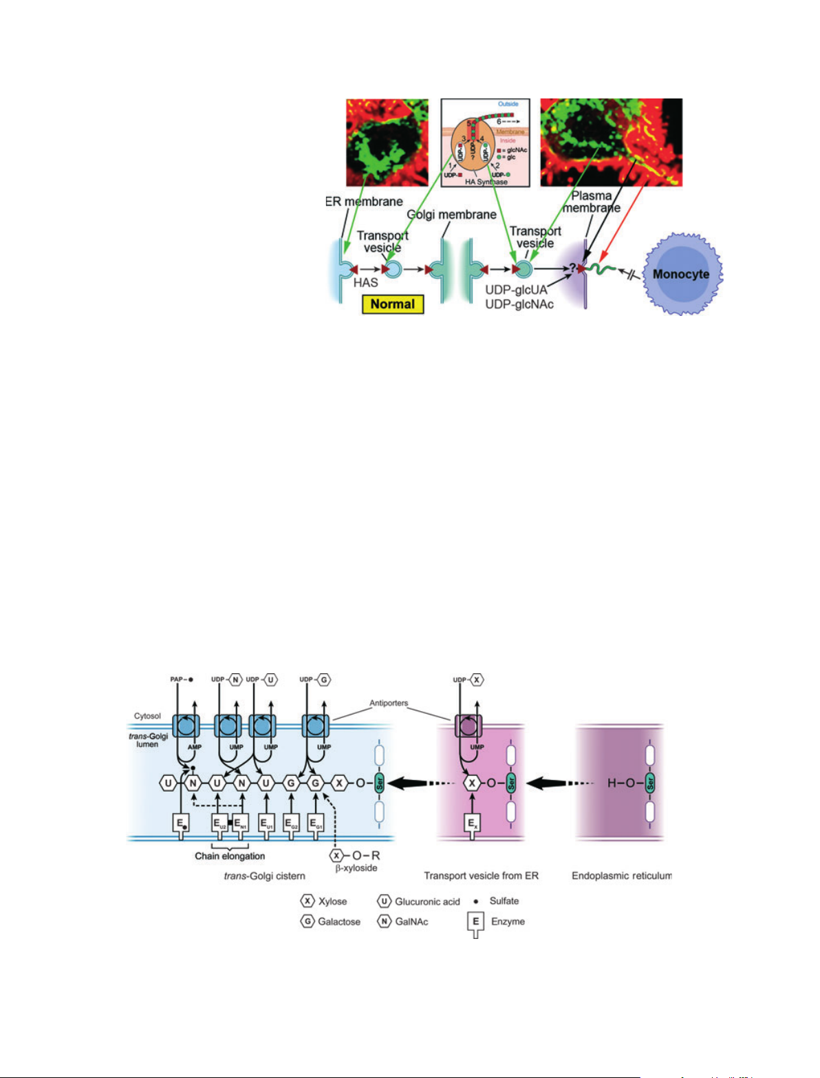
anism of HA synthesis is given in Fig. 1. Hyaluronan
synthase (HAS) enzymes are synthesized in the endo-
plasmic reticulum (ER) in an inactive form and must
be transported in vesicles to and through the Golgi for
insertion into the plasma membrane. After the enzyme
has been activated, it utilizes the cytosolic substrates,
UDP-glucuronate (UDP-glcUA) and UDP-N-acetyl-
glucosamine (UDP-glcNAc), and adds them alternately
to the reducing end of the chain with release of the
anchoring UDP. The elongating chain is extruded into
the extracellular compartment. Confocal microscopy
images of live cells that were transfected with green
fluorescent protein (GFP)-HAS3 are shown in Fig. 1
[1]. The localization of the enzyme (green) in perinucle-
ar regions (ER Golgi) and in transport vesicles is
apparent. The active enzyme in the plasma membrane
(yellow) extrudes HA into the normal extracellular
fuzzy coats (red) with which monocytes do not interact
[2] (see accompanying article by Tammi et al. [3]).
This mechanism of HA synthesis has several unique
features [4]: (a) the extruded chain is not modified by
the addition of sulfoesters or epimerases that modify
other glycosaminoglycans; (b) the final chain can be
extremely large, > 10 million Da; (c) a core protein is
Keywords
autophagy; CD44; diabetes; diabetic
nephropathy; endoplasmic reticulum stress;
golgi; hyaluronan; hyaluronan synthase
proteoglycan synthesis; inflammation
Correspondence
A. Wang, Department of Biomedical
Engineering ND20, Lerner Research
Institute, The Cleveland Clinic, 9500 Euclid
Ave., Cleveland, OH 44195, USA
Fax: 216 444 9198
Tel: 216 445 3237
E-mail: wanga@ccf.org
(Received 1 November 2010, revised 9
February 2011, accepted 25 February
2011)
doi:10.1111/j.1742-4658.2011.08069.x
Hyaluronan matrices are ubiquitous in normal and pathological biological
processes. This remarkable diversity is related to their unique mechanism
of synthesis by hyaluronan synthases. These enzymes are normally acti-
vated in the plasma membrane and utilize cytosolic substrates directly to
form these large polyanionic glycosaminoglycans, which are extruded
directly into the extracellular space. The extracellular matrices that are
formed interact with cell surface receptors, notably CD44, that often dic-
tate the biological processes, as described in the accompanying minireviews
of this series. This article focuses on the discovery in recent studies that
many cell stress responses initiate the synthesis of a monocyte-adhesive
hyaluronan extracellular matrix, which forms a central focus for subse-
quent inflammatory processes that are modulated by the dialogue between
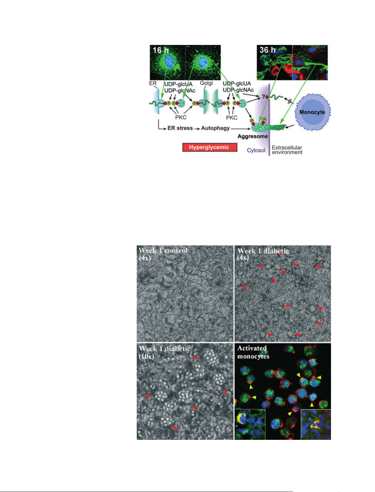
the matrix and the inflammatory cells. The mechanisms involve active hyal-
uronan synthases at the cell membrane when cell stresses occur at physio-
logical levels of glucose. However, dividing cells at hyperglycemic levels of
glucose initiate the synthesis of hyaluronan in intracellular compartments,
which induces endoplasmic reticulum stress and autophagy, processes that
probably contribute greatly to diabetic pathologies.
Abbreviations
CD44, cluster of differentiation 44; ER, endoplasmic reticulum; galNAc, N-acetylgalactosamine; GFP, green fluorescent protein; glcUA,
glucuronate; glcNAc, N-acetylglucosamine; HA, hyaluronan; HAS, hyaluronan synthase; PKC, protein kinase C.
1412 FEBS Journal 278 (2011) 1412–1418 ª2011 The Authors Journal compilation ª2011 FEBS
not required, unlike all proteoglycans; (d) the rate of
synthesis can be modulated as a function of the con-
centrations of the cytosolic UDP-sugar substrates; (e)
it is energetically efficient; UDP-glcUA is synthesized
by two oxidation steps from UDP-glucose yielding two
molecules of NADPH. It is also important to prevent
the activation of HAS enzymes in intracellular com-
partments, which causes pathological consequences as
described below.
In contrast with HA, all other glycosaminoglycans
are synthesized on core proteins inside the Golgi to
form the large family of proteoglycans (Fig. 2). The
UDP-sugar and phosphoadenosinephosphosulfate sub-
strates are synthesized in the cytoplasm and shuttled
into the Golgi by antiporters that remove a down-
stream product (UMP, AMP) for each substrate,
which is used to synthesize the oligosaccharide attach-
ment region, to add the alternating sugar residues onto
the nonreducing end of the growing chain and to add
sulfoesters. This antiporter mechanism controls the
concentrations of UDP-sugar substrates in the Golgi
according to the rate of glycosaminoglycan synthesis
on the proteoglycans, and is therefore independent of
the changes in the UDP-sugar concentrations in the
cytosol.
Monocyte-adhesive HA matrices
synthesized by stressed cells in normal
glucose
Biology has taken advantage of the unique mechanism
of HA synthesis to produce normal pericellular glycoc-
alyces on most cells and to contribute to normal extra-
cellular matrices. Notably, in cartilage, HA anchors
the aggrecan proteoglycan aggregates, and this
HA–aggrecan complex provides the tissue with its abil-
Fig. 1. Model for the normal transport of
hyaluronan synthase (HAS) from the endo-
plasmic reticulum (ER) to the plasma mem-
brane, where it is activated to synthesize
and extrude hyaluronan. The confocal micro-
graphs show live cells that were transfected
with GFP-Has3 (green) and stained for hyal-
uronan (red). They demonstrate ER Golgi
localization (left), transport vesicles (right),
active HAS in plasma membranes (yellow)
and extracellular hyaluronan (red). Micro-
graphs provided by Kirsi Rilla (see the article
by Tammi et al. [3] in this series).
Fig. 2. Model for the biosynthesis of proteoglycans (see text for details). ER, endoplasmic reticulum; PAP, phosphoadenosinephosphosulfate.
A. Wang et al. Hyaluronan matrices in pathobiological processes
FEBS Journal 278 (2011) 1412–1418 ª2011 The Authors Journal compilation ª2011 FEBS 1413
ity to respond to compressive loads. However, biology
has also utilized the synthesis of HA to form abnormal
matrices when cells are stressed by a variety of condi-
tions. This was initially shown in a study with cultures
of smooth muscle cells isolated from normal human
colons [5,6]. Cultures stressed by viral infection or by
treatment with poly(I:C), which initiates responses sim-
ilar to viral infection, synthesized an extensive HA
matrix with structural information that was recognized
by monocytes macrophages, which bind at 4 C and
rapidly phagocytose the matrix at a physiological tem-
perature of 37 C (Fig. 3) [6]. An increasing number of
studies have now demonstrated that the same or simi-
lar monocyte-adhesive HA matrices are synthesized in
response to a variety of stresses in cell models both
in vitro and in vivo. For example, the section (Fig. 4)
from a biopsy taken from an asthmatic patient during
an inflammatory response shows an extensive patho-
logical HA matrix (green) with embedded inflamma-
tory cells exhibiting capped CD44 (red). Other
examples include responses to ER stress at physiologi-
cally normal levels of glucose [7], wound healing
[8–10], idiopathic pulmonary hypertension [11], airway
smooth muscle cells in vitro and airway interstitial cells
in mouse asthma models [12–14], adipocytes in adipose
tissue in a diabetic mouse model [15] and renal tubular
endothelial stress [16–18]. Further, removal of this
monocyte-adhesive matrix by inflammatory cells is
essential and requires the cell surface HA receptor,
CD44. This was demonstrated by showing that the
lungs of CD44 null mice subjected to noxious bleomy-
cin inhalation synthesized and continuously accumu-
lated HA matrix which could not be removed by the
influx of monocytes and macrophages [19], and most
of the animals died. In contrast, irradiated CD44 null
mice repopulated with normal bone marrow aspirates
were able to generate normal monocytes and macro-
phages that were able to remove this matrix, with
subsequent survival and restoration of normal lung
function after bleomycin treatment. (For a further
insight into the roles of HA interactions with CD44
and its variants, and their importance in malignancy,
see the accompanying article by Misra et al. [20].)
Monocyte-adhesive HA matrices
synthesized by dividing cells in
hyperglycemic glucose
More recently, a unique activation of HASs in intra-
cellular compartments has been identified in cells stim-
ulated to divide in hyperglycemic medium (25 mm
glucose), typical of uncontrolled diabetes [21,22].
Mesangial cells isolated from rat kidneys were growth
arrested and then stimulated to divide in hyperglyce-
mic medium. This initiated a protein kinase C (PKC)
response, which led to the activation of HASs in intra-
cellular compartments, including, most probably, the
Fig. 3. U937 monocytic cells, using the
receptor CD44 (red), bind to hyaluronan
cable structures (green) on the surface of
poly(I:C)-stimulated cultures of intestinal
smooth muscle cells at 4 C (left panel) [6].
When the cultures are warmed (37 C for
30 min), the monocytic cells relocate, or
‘cap’, CD44 to one pole and internalize
hyaluronan as shown in the enlarged inset.
The left panel is reprinted from ref. [6] with
permission from the American Society for
Investigative Pathology.
Fig. 4. A section from a lung biopsy taken from a patient with an
asthmatic flare stained for hyaluronan (green), CD44 (red) and
nuclei (blue).
Hyaluronan matrices in pathobiological processes A. Wang et al.
1414 FEBS Journal 278 (2011) 1412–1418 ª2011 The Authors Journal compilation ª2011 FEBS
ER, Golgi and transport vesicles. This is shown in the
confocal micrographs of cells permeabilized at 16 h
after the initiation of cell division in hyperglycemic
medium and stained for HA (Fig. 5, left images). The
resulting ER stress in this model initiated an autopha-
gic response near the end of cell division, which
involved a large upregulation of cyclin D3 and the for-
mation of intracellular aggresomes that co-stained for
HA, cyclin D3 and microtubule protein 9 light chain 3,
a marker for autophagy [22,23]. This was followed by
the formation of an extensive monocyte-adhesive HA
matrix between and through neighboring cells after
completion of the cell cycle, as shown in the confocal
images of cultures 36 h after stimulation to divide in
hyperglycemic medium (Fig. 5, right images). The inhi-
bition of protein kinase C or the treatment of the cells
Fig. 5. Model for the intracellular activation
of hyaluronan synthases in cells that divide
in hyperglycemic medium (25 mMglucose).
The images on the left are mesangial
cells stimulated to divide in hyperglycemic
medium, permeabilized at 16 h and stained
for hyaluronan (green). Intracellular hyaluro-
nan is observed in endoplasmic reticulum
(ER) Golgi regions and in transport vesicles
[21]. The images on the right show permea-
bilized cells (left) and nonpermeabilized cells
(right) stained for hyaluronan (green),
cyclin D3 (red) and nuclei (blue) 36 h after
stimulation to divide in hyperglycemic
medium [21]. PKC, protein kinase C.
Fig. 6. Adhesion of U937 monocytes to kid-
ney sections from a control and a streptozo-
tocin-induced diabetic rat, 1 week after the
induction of hyperglycemia. An enlargement
of the diabetic kidney section (bottom
left) shows clusters of monocytes over
glomeruli. The adhesion was performed at
4C. When a section from the diabetic
kidney was warmed to 37 C, most of the
monocytes detached. They were then
spread on a slide and stained for hyaluronan
(green), CD44 (red) and nuclei (blue) (bottom
right). Examples of capped CD44 are appar-
ent (arrowheads). The insets in this panel
show macrophages in glomeruli in sections
that co-stain for CD44 and hyaluronan
(yellow), providing evidence for monocyte
macrophage activity in the glomeruli.
A. Wang et al. Hyaluronan matrices in pathobiological processes
FEBS Journal 278 (2011) 1412–1418 ª2011 The Authors Journal compilation ª2011 FEBS 1415
with cyclin D3 siRNA prior to stimulation to divide
prevented these responses. This mechanism occurs
within the first week in vivo after the initiation of
hyperglycemia in streptozotocin-treated rats [21,22].
Confocal analyses showed the presence of an abnormal
HA matrix with embedded macrophages in sections
from diabetic rat kidneys after 1 week [21,22]. Further,
Fig. 6 shows that U937 monocytes adhere to glomeruli
in such sections at 4 C, and that they phagocytose
HA out of the section when warmed to 37 C.
Intracellular HA: a new frontier in
diabetes
A previous review has suggested the possibility that
intracellular HA may be a new frontier for inflamma-
tory pathologies [24]. An important experiment which
formed the basis for this possibility showed that divid-
ing aortic smooth muscle cells accumulate intracellular
HA during the cell cycle, which is considered to be a
potentially normal process [25]. However, the medium
used in these experiments was hyperglycemic (25 mm
glucose) which, according to our results with mesangial
cells, would have activated HASs within the dividing
cells. In an unrelated earlier study, scratch wounds of
endothelial cell cultures demonstrated that monocytes
adhered to the migrating and dividing cells at the
edges of the scratch wounds, but did not adhere to the
adjacent nondividing cells [26]. These experiments were
also performed in medium that contained a higher
than normal glucose level (15 mm), which is above the
levels shown to trigger HA synthesis within dividing
mesangial cells [21]. This suggests that monocyte adhe-
sion is most probably the result of the formation of a
monocyte-adhesive HA matrix by the dividing cells. In
a third case, 3T3-L1 cells, an accepted model for
adipogenesis, were routinely stimulated to divide in a
standard hyperglycemic (25 mmglucose) medium
before stimulating their adipogenic responses. After
cell division, the medium became extraordinarily
viscous as a result of the synthesis of HA [27]. Figure 7
shows that, under the same conditions, adipogenic
3T3-L1 cells undergo autophagy (cyclin D3-stained ag-
gresomes [22]) and produce an extensive HA matrix
that is monocyte adhesive. These three culture models
with distinctly different cell types indicate the likeli-
hood that an intracellular HA stress response that
drives autophagy and the formation of a monocyte-
adhesive HA matrix will occur in most, if not all, cells
stimulated to divide in hyperglycemic medium. Investi-
gators should be aware of the glucose levels in experi-
mental medium, as commonly used hyperglycemic
media may induce intracellular HA responses in divid-
ing cells in culture, which may confound the interpre-
tation of the results.
Cytosolic UDP-sugar concentrations increase in cells
in response to hyperglycemic conditions [28–30]. This
led us to ask whether the intracellular HA synthesis
response could be inhibited if the concentrations
of UDP-sugars were diminished. As shown in Fig. 2,
xylosides, which enter cells, enter the Golgi compart-
ment and bypass the need for a core protein to stimu-
late chondroitin sulfate synthesis. The capacity of cells
to synthesize chondroitin sulfate is usually much
greater than the rate required to complete the proteo-
glycans. For example, 4-methylumbelliferol-xyloside
increases chondroitin sulfate synthesis in airway
smooth muscle cell cultures by eight- to ten-fold [31].
To accommodate this rate of synthesis, the antiporters
must increase the entry of UDP-glcUA (a substrate for
Fig. 7. 3T3-L1 cells dividing in hyperglycemic medium undergo
autophagy and synthesize an extensive monoctye-adhesive matrix.
3T3-L1 cells were stimulated to divide in hyperglycemic medium
(25 mMglucose), routinely used to promote adipogenesis in this
model. At 48 h, a permeabilized culture (top panel) was stained for
hyaluronan (green), cyclin D3 (red) and nuclei (blue). The presence
of hyaluronan cables (green) and cyclin D3-stained aggresomes
(red) indicates that the cells underwent autophagy and cyclin D3-
mediated formation of a hyaluronan matrix. The bottom left panel
shows extensive U937 monocyte adhesion to an identically treated
culture, which was lost when the culture was treated with Strepto-
myces hyaluronidase (selective for hyaluronan) (bottom right panel).
Hyaluronan matrices in pathobiological processes A. Wang et al.
1416 FEBS Journal 278 (2011) 1412–1418 ª2011 The Authors Journal compilation ª2011 FEBS