
Open Access
Available online http://ccforum.com/content/12/2/R42
Page 1 of 12
(page number not for citation purposes)
Vol 12 No 2
Research
Circulating plasma factors induce tubular and glomerular
alterations in septic burns patients
Filippo Mariano1*, Vincenzo Cantaluppi2*, Maurizio Stella3, Giuseppe Mauriello Romanazzi2,
Barbara Assenzio4, Monica Cairo3, Luigi Biancone2, Giorgio Triolo1, V Marco Ranieri4 and
Giovanni Camussi2
1Dipartimento di Area Medica, Unita' di Nefrologia e Dialisi, Ospedale CTO, Via G. Zuretti 29, Torino, 10126, Italy
2Dipartimento di Medicina Interna, Centro Interdipartimentale di Biotecnologie Molecolari e Centro Ricerca Medicina Sperimentale (CeRMS),
Universita' di Torino, Corso Dogliotti, 14, Torino, 10126, Italy
3Dipartimento di Chirurgia Plastica, Centro Grandi Ustionati, Ospedale CTO, Via G. Zuretti 29, Torino, 10126, Italy
4Dipartimento di Anestesiologia e Rianimazione, Università di Torino, Ospedale S Giovanni Battista-Molinette, Corso Dogliotti 14, Torino, 10126, Italy
* Contributed equally
Corresponding author: Giovanni Camussi, giovanni.camussi@unito.it
Received: 7 Dec 2007 Revisions requested: 9 Jan 2008 Revisions received: 8 Feb 2008 Accepted: 25 Mar 2008 Published: 25 Mar 2008
Critical Care 2008, 12:R42 (doi:10.1186/cc6848)
This article is online at: http://ccforum.com/content/12/2/R42
© 2008 Mariano et al.; licensee BioMed Central Ltd.
This is an open access article distributed under the terms of the Creative Commons Attribution License (http://creativecommons.org/licenses/by/2.0),
which permits unrestricted use, distribution, and reproduction in any medium, provided the original work is properly cited.
Abstract
Background Severe burn is a systemic illness often
complicated by sepsis. Kidney is one of the organs invariably
affected, and proteinuria is a constant clinical finding. We
studied the relationships between proteinuria and patient
outcome, severity of renal dysfunction and systemic
inflammatory state in burns patients who developed sepsis-
associated acute renal failure (ARF). We then tested the
hypothesis that plasma in these patients induces apoptosis and
functional alterations that could account for proteinuria and
severity of renal dysfunction in tubular cells and podocytes.
Methods We studied the correlation between proteinuria and
indexes of systemic inflammation or renal function prospectively
in 19 severe burns patients with septic shock and ARF, and we
evaluated the effect of plasma on apoptosis, polarity and
functional alterations in cultured human tubular cells and
podocytes. As controls, we collected plasma from 10 burns
patients with septic shock but without ARF, 10 burns patients
with septic shock and ARF, 10 non-burns patients with septic
shock without ARF, 10 chronic uremic patients and 10 healthy
volunteers.
Results Septic burns patients with ARF presented a severe
proteinuria that correlated to outcome, glomerular (creatinine/
urea clearance) and tubular (fractional excretion of sodium and
potassium) functional impairment and systemic inflammation
(white blood cell (WBC) and platelet counts). Plasma from
these patients induced a pro-apoptotic effect in tubular cells and
podocytes that correlated with the extent of proteinuria. Plasma-
induced apoptosis was significantly higher in septic severe
burns patients with ARF with respect to those without ARF or
with septic shock without burns. Moreover, plasma from septic
burns patients induced an alteration of polarity in tubular cells,
as well as reduced expression of the tight junction protein ZO-1
and of the endocytic receptor megalin. In podocytes, plasma
from septic burns patients increased permeability to albumin
and decreased the expression of the slit diaphragm protein
nephrin.
Conclusion Plasma from burns patients with sepsis-associated
ARF contains factors that affect the function and survival of
tubular cells and podocytes. These factors are likely to be
involved in the pathogenesis of acute tubular injury and
proteinuria, which is a negative prognostic factor and an index of
renal involvement in the systemic inflammatory reaction.
Introduction
Acute renal failure (ARF) is frequently associated with a sys-
temic inflammatory response due to sepsis [1]. Circulating fac-
tors have been proposed as responsible mediators for the
systemic micro-vascular injury occurring in these patients [2].
During sepsis, bacterial endo- or exotoxins can act on
ADQI = Acute Dialysis Quality Initiative; ARF = acute renal failure; BCrC = blood creatinine clerance; BUC = blood urea clearance; FeNa = fractional
sodium excretion; FeK = fractional potassium excretion; LPS = lipopolysaccharide; Pto/Cro = proteinuria/creatininuria ratio; RRT = renal replacement
therapy; TER = trans-epithelial resistance; WBC = white blood cells.

Critical Care Vol 12 No 2 Mariano et al.
Page 2 of 12
(page number not for citation purposes)
glomerular podocytes and tubular epithelium, stimulating syn-
thesis of cytokines and other inflammatory mediators [3-6]. In
patients with sepsis-associated ARF, a marked dissociation
between the degree of tubular necrosis and the renal dysfunc-
tion has been described [7]. By contrast, clinical and experi-
mental data suggest that apoptosis plays an important role in
sepsis-induced ARF [4-9].
In severe burns patients, sepsis almost always develops, lasts
for several weeks and is frequently associated with ARF [10-
19]. A constant feature of burn-associated kidney injury is pro-
teinuria, which starts in the first days post-injury and increases
over time. Proteinuria is a consequence of increased glomeru-
lar permeability and of decreased tubular re-absorption of fil-
tered proteins [13,20,21].
Immediately after burn injury, onset of proteinuria can depend
on filtered breakdown proteins derived from massive tissue
destruction or on renal involvement as a consequence of the
increased systemic capillary permeability. Subsequently, pro-
teinuria reflects the involvement of the kidney in the septic
process [13,20-22].
In the present study, we investigated whether circulating fac-
tors present in the plasma of septic severe burns patients
could induce tubular and glomerular alterations that could
account for proteinuria and ARF.
Materials and methods
Patients
From January 2003 to December 2005, from 258 patients
admitted to the Burn Center (Dipartimento di Chirurgia Plas-
tica, Centro Grandi Ustionati, Ospedale CTO, Via G. Zuretti
29, Torino, 10126, Italy) (mean (standard error (SE) burned
surface area 24.5 ± 1.3%, range 1–98%, mortality rate
19.4%), 19 patients with severe burns and septic shock who
developed ARF (8–10 days after burn injury) were enrolled in
a prospective study ("burns septic ARF" group). Demographic
and clinical data (Table 1) for these 19 patients were recorded
and blood and urine biochemical parameters were analyzed.
Plasma samples collected at the time of ARF onset before the
start of renal replacement therapy (RRT) were used for labora-
tory studies. Informed consent was obtained according to the
Declaration of Helsinki and study approval was obtained by
the Center for Molecular Biotechnology Institutional Review
Board, University of Torino.
All 19 patients fulfilled the criteria for septic shock [23] and
ARF according to Acute Dialysis Quality Initiative – Risk, Injury,
Failure, Loss, and End-stage kidney disease (ADQI-RIFLE)
classification [24]. Patients with chronic cardiovascular sys-
tem failure (New York Heart Association (NYHA) class III),
chronic respiratory failure (chronic hypoxia, hypercapnia),
chronic liver failure (biopsy-confirmed cirrhosis or portal hyper-
tension), neoplastic diseases, collagenopathies, insulin-
Table 1
Demographic and clinical characteristics
Septic burns patients with ARF
(n = 19) Septic burns patients
(n = 10) Septic ARF patients
(n = 10) Septic patients
(n = 10)
Female (n) 7 (36.8%) 2 (20%) 4 (40%) 3 (30%)
Age (years) 50.4 ± 4.6 55.5 ± 6.0 62.4 ± 4.4 58.2 ± 3.8
Burned surface area (%) 51.3 ± 5.4 44.9 ± 6.2 - -
SOFA score (at ARF onset) 11.8 ± 0.4 - 11.7 ± 1.1 -
Non-survivors (n) 10 (52.6%) 8 (80%) 8 (80%) 5 (50%)
Renal replacement therapy (n) 15 (78.9%) - 10 (100%) -
Duration of renal replacement therapy (days) 22.4 ± 2.6 - 12.9 ± 2.7 -
Hemoculture* n (all patients)
Staphylococcus aureus 12
Acinetobacter baumannii 11
Pseudomonas aeruginosa 9
Candida albicans 4
Escherichia coli 1
Klebsiella pneumoniae 1
*All patients had positive blood cultures. Some patients were simultaneously positive for more than one pathogen strain.
Data shows mean ± standard error (SE) unless otherwise stated.
ARF, acute renal failure; SOFA, Sequential Organ Failure Assessment.

Available online http://ccforum.com/content/12/2/R42
Page 3 of 12
(page number not for citation purposes)
dependent diabetes mellitus and known aortic aneurysm were
excluded from the study.
As controls, we studied 10 burns patients with septic shock
without ARF ("burns septic" group), 10 septic shock patients
with ARF ("septic ARF" group), 10 patients with septic shock
without ARF ("septic" group), 10 stable uremic patients ("ure-
mic" group) and 10 healthy volunteers ("healthy" group) (Table
1).
Systemic treatment of burns patients was based on current
guidelines and consisted of prompt hydro-electrolytic replace-
ment, ventilatory support when necessary, pain control and
surgical treatment by multiple stage skin excisions and subse-
quent reconstruction by means of autografts or donor allo-
grafts assured by the Regional Skin Bank. Septic events
mainly caused by multiresistant bacterial strains were treated
according to current guidelines with antibiotic regimens fol-
lowing blood cultures and sensitivity tests. When glycopep-
tides or aminoglycosydes were used, the administered dose
was calculated on creatinine clearance and adjusted following
the results of drug blood levels [10].
Biochemical parameters
The clinical observation of the burns septic ARF group was
divided in two periods; the first from burn injury to the onset of
ARF (pre-ARF period) and the second from the onset of ARF
to functional recovery or to exitus (ARF period). Recovery from
ARF was defined as blood creatinine improvement back up to
starting values. The mean times of observation in pre-ARF and
ARF periods were 18.9 ± 3.6 and 19.8 ± 3.8 days, respec-
tively (mean ± SE).
Biochemical evaluation included blood creatinine clearance
(BCrC) and blood urea clearance (BUC) measured by 24-h
diuresis, fractional excretion of sodium (FeNa) and potassium
(FeK), and hemochromocytometer examination.
Proteinuria was determined in an automated analyzer by ben-
zethonium chloride dye binding method (Roche Modular Sys-
tem, Roche Diagnostics GmbH, Mannheim, Germany) [25].
Tumor necrosis factor (TNF)α concentrations in plasma were
measured by Bio-Plex cytokine assay (Biorad, Hercules, CA,
USA).
Laboratory studies
Cells
Podocytes and proximal tubular epithelial primary cultures
were obtained from normal cortex fragments of surgically
removed kidneys. Immortalized tubular cells and podocyte
lines were generated by infection with a hybrid Adeno5/SV40
virus as previously described [26,27].
Viability and apoptosis assays
Cellular viability was studied by using the XTT-based colori-
metric method (Sigma, St Louis, MO, USA).
In all groups studied, apoptosis was evaluated by terminal uri-
dine deoxynucleotidyl transferase dUTP nick-end labeling
(TUNEL) assay (ApopTag, Oncor, Gaithersburg, MD, USA).
Moreover, in selected experiments tubular apoptosis was con-
firmed by identification of intranucleosomal DNA fragmenta-
tion after 1% agarose gel electrophoresis (BioVision Research
Products, Mountain View, CA, USA). The activities of cas-
pases 3, 8 and 9 were assessed by a colorimetric assay
(Chemicon International, Temecula, CA, USA).
Polarity assay
Transepithelial electrical resistance (TER), an indicator of cell
polarity, was measured by using an epithelial volt-ohm meter
(EVOM, World Precision Instruments, Inc., Sarasota, FL, USA)
after incubation of tubular cells or podocytes with different
stimuli. All measures were normalized for the area of the mem-
brane used in the experimental procedure and expressed as
ohm/cm2.
Permeability assay
Permeability was evaluated by diffusion of Trypan blue-albu-
min complexes across podocyte confluent monolayers cul-
tured on Transwell (Greiner Bio-One, Frickenhausen,
Germany) under conditions of continuous slight agitation. Aliq-
uots of medium from the upper and the lower wells were trans-
ferred to a 96-well plate and analyzed at the 590 nm
wavelength (Model 680 Spectrophotometer, Biorad, Her-
cules, CA). Results are given as percentage of increase of
albumin diffusion in comparison to vehicle alone [28].
Gene array
The Human GEarray for the study of apoptosis markers
(Superarray Inc., Bethesda, MD, USA) was performed and
analyzed according to the manufacturer's instructionsto char-
acterize the expression profile of tubular cells incubated in
presence of different plasma samples. These data are freely
available from the ArrayExpress databank of the European Bio-
informatics Institute (experiment name: Human Apoptosis –
Burn; ArrayExpress accession: E-MEXP-1510).
Immunofluorescence, fluorescence-activated cell sorting
(FACS) and Western blot analysis
For immunofluorescence studies, antibodies directed against
Fas (CD95), CD40 (Upstate, Charlottesville, VA, USA), meg-
alin, ZO-1, E-cadherin and pan-cytokeratins (Santa Cruz Bio-
tech, Santa Cruz, CA, USA) were used with tubular cells. Fas
and CD40 expression were also evaluated by FACS analysis
(Becton Dickinson, Mountain View, CA, USA). Antibodies
directed against nephrin (Progen, Heidelberg, Germany), nes-
tin (Santa Cruz) and ZO-1 were used with podocytes. Specific
Alexa Fluor-conjugated antibodies were used as secondary

Critical Care Vol 12 No 2 Mariano et al.
Page 4 of 12
(page number not for citation purposes)
antibodies (Invitrogen, Carlsbad, CA, USA). Fluorescein iso-
thiocyanate (FITC)-conjugated phalloidin (Sigma) was used to
evaluate cytoskeleton actin distribution on tubular cells and
podocytes by ultraviolet (UV) light microscopy.
For Western blot analysis, cell lysates were separated by
SDS-PAGE and immunoblotted with anti-human Bax, anti-
human Bcl-2, anti-human megalin (tubular cells) or anti-human
nephrin (podocytes) antibodies.
Statistical analysis
Descriptive statistics, Student t test, linear regression analysis
with curves of minimal square and analysis of variance
(ANOVA) with Dunnet or Newman-Keuls multi-comparison
test (Statistica 6.1, StaSoft Inc, Tulsa, OK, USA) were per-
formed. Values were expressed as mean ± SE. p Values <
0.05 were considered statistically significant.
Results
Proteinuria was related to patient outcome and systemic
inflammation
Clinical characteristics of the 19 burns septic ARF group
patients with positive blood cultures are given in Table 1. The
burns septic ARF patients presented marked and persistent
proteinuria (mean ± SE value during 8-week observation
period of 2,074.8 ± 113.4 mg/day). When proteinuria was
expressed as proteinuria/creatininuria ratio (Pto/Cro), a con-
tinuous increase of protein excretion was observed over the
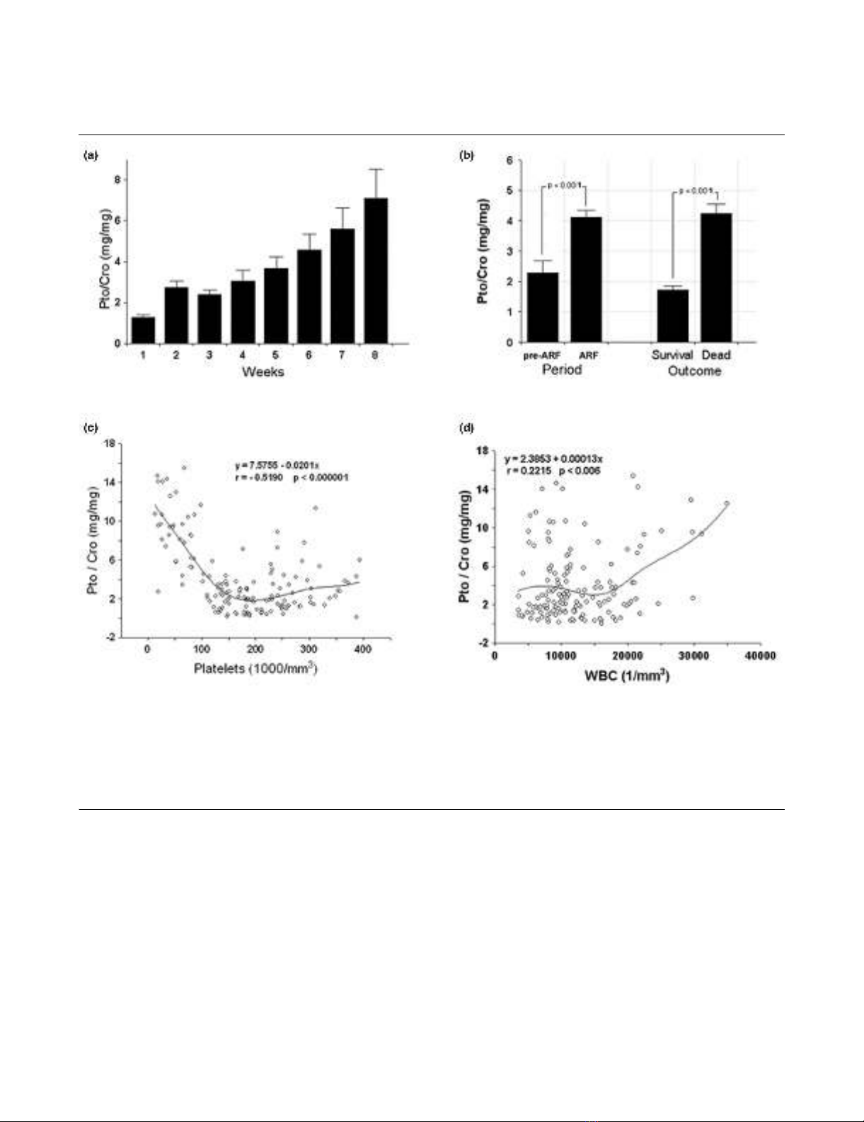
time (Figure 1A). In addition, Pto/Cro was significantly higher
in the ARF period than in the pre-ARF period, and in non-sur-
vivors compared to survivors (Figure 1B).
In the ARF period Pto/Cro significantly correlated in a negative
manner with platelet counts (Figure 1C) and in a positive man-
ner with white blood cell (WBC) counts (Figure 1D). By con-
trast, in the pre-ARF period no significant correlations were
observed between Pto/Cro and platelet (y = 1.9501 +
0.00125, r 0.10041, p 0.27312) or WBC counts (y = 1.8256
+ 0.00004, r 0.10481, p 0.25258).
Proteinuria correlated with loss of glomerular and
tubular functions
During the ARF period, Pto/Cro negatively correlated both
with decreased BCrC and BUC (Figure 2A,B) and positively
correlated both with FeNa and FeK (Figure 2C,D). The best lin-
ear correlation was observed between Pto/Cro and FeK, with
a Pearson coefficient > 0.6 (Figure 2D). By contrast, in the
pre-ARF period no significant correlations were observed
between Pto/Cro and BCrC, BUC, FeNa and FeK.
Burns septic ARF group plasma exerted a cytotoxic
effect on tubular cells and on podocytes
Incubation with increasing doses of burns septic ARF group
plasma (range 1–10%) induced a marked reduction of viability
of tubular cells (Figure 3A) and of podocytes (data not shown).
The cytotoxic effect was evident from 12 h, increased over
time and peaked at 48–72 h (data not shown). No significant
cytotoxic effect was observed after addition of control healthy
group plasma.
Burns septic ARF group plasma induced tubular cell
apoptosis correlated with proteinuria
Consistent with the cytotoxicity data, we observed a signifi-
cant increase of apoptotic tubular cells detected by TUNEL
assay after incubation with lipopolysaccharide (LPS) (positive
control), septic, septic ARF, burns septic and burns septic
ARF group plasma (Figure 3B). The maximal effect was
observed with burns septic ARF group plasma (Figure 3B),
and a similar pro-apoptotic effect was also observed on podo-
cytes (data not shown). Apoptosis was confirmed by detection
of DNA fragmentation (Figure 3B, inset). Since burns patients
were treated with potentially nephrotoxic antibiotics, we used
healthy plasma as control in absence or presence of the same
concentrations of vancomycin (10 μg/ml) and gentamicin (2
μg/ml) detected in the plasma of treated patients. Addition of
antibiotics did not induce a significant increase of tubular
apoptosis (Figure 3B). Furthermore, when we tested plasma
from stable chronic uremic patients, no significant effects
were observed (Figure 3B). The rate of tubular apoptosis
induced by burns septic ARF group plasma significantly corre-
lated with Pto/Cro ratio (Figure 3C).
The apoptotic effect of burns septic ARF group plasma could
be only partially accounted to the presence of LPS. Indeed,
pre-treatment of plasma with polymyxin B significantly
reduced, but did not suppress this effect (Figure 3D), suggest-
ing the presence in plasma of harmful mediator/s other than
LPS. Indeed, the level of TNF-α (81.3 ± 9.4 pg/ml) in these
plasma samples was significantly higher than in healthy con-
trols (5.9 ± 0.8 pg/ml). In addition, pre-incubation of tubular
cells with burns septic ARF group plasma and subsequent
addition of LPS resulted in a significant worsening of tubular
apoptosis in comparison to stimulation with LPS alone (Figure
3D).
The activity of caspases 3, 8 and 9 significantly increased in
tubular cells after incubation with burns septic ARF group
plasma (Figure 4A). Via FACS (Figure 4B–D) and immunoflu-
orescence analysis (Figure 4B–D, insets), we observed a
slight basal expression of Fas (Figure 4B). Fas expression did
not change in the presence of healthy plasma (Figure 4C),
whereas it was markedly up-regulated with burns septic ARF
group plasma (Figure 4D).
In addition, burns septic ARF group plasma, but not control
healthy plasma, induced the up-regulation of Bax and the
down-regulation of Bcl-2 (Figure 4E), proteins known to mod-
ulate the mitochondrial apoptotic pathway. Gene array analy-
sis demonstrated that burns septic ARF group plasma
induced an up-regulation of several pro-apoptotic genes such
Available online http://ccforum.com/content/12/2/R42
Page 5 of 12
(page number not for citation purposes)
as Fas, Fas-Ligand (Fas-L), Bax and Bak and of positive regu-
lators of apoptosis (Abl1, Gadd45a). The negative regulators
of apoptosis, Birc5 and Bnip3, were down-regulated. In addi-
tion, the CD40 gene and its transduction factor TRAF-3, as
well as the nuclear factor (NF)-kB activator Ripk2 gene, were
up-regulated, suggesting an inflammatory activation of tubular
cells (Figure 4F). Up-regulation of CD40 was confirmed by
FACS analysis (Figure 4G). This is consistent with previous
observations indicating that CD40 is over-expressed in renal
tubular epithelial cells during inflammatory injury [29].
Burns septic ARF group plasma altered cytoskeleton
distribution and megalin expression in tubular cells
Burns septic ARF group plasma induced a marked alteration
of distribution of cytoskeleton actin fibers in tubular cells. In
comparison to control healthy plasma (Figure 5A), burns sep-
tic ARF group plasma promoted the formation of "heaps"
(Figure 5B) composed of tubular cells grouped in clusters, an
effect likewise ascribed to apoptosis.
We then tested whether burns septic ARF group plasma
could induce early alterations in tubular cells not accountable
to apoptosis such as down-regulation of megalin, an endocytic
receptor involved in re-absorption of filtered proteins. Burns
Figure 1
Proteinuria correlates with patient outcome and with markers of systemic inflammationProteinuria correlates with patient outcome and with markers of systemic inflammation. (a) Proteinuria expressed as proteinuria/creatininuria ratio
(Pto/Cro, mg/mg) in the weeks following patient admission. Data are given as weekly average of daily values. Only non-oliguric patients were
included and the number of patients for each week was: week 1, n = 19; week 2, n = 19; week 3, n = 17; week 4, n = 15; week 5, n = 10; week 6,
n = 7; week 7, n = 6; week 8, n = 5. (b) Overall mean proteinuria in pre-acute renal failure (ARF) vs ARF periods and in deceased vs surviving
patients. (c, d) Relationship between Pto/Cro and indexes of systemic inflammatory state in ARF period. Pto/Cro negatively correlated with platelet
count (c) and positively with white blood cell (WBC) count (d). Student t test and linear regression analysis were performed where appropriate.
Data for different parameters are also shown as minimal square fitting curves.


![Báo cáo seminar chuyên ngành Công nghệ hóa học và thực phẩm [Mới nhất]](https://cdn.tailieu.vn/images/document/thumbnail/2025/20250711/hienkelvinzoi@gmail.com/135x160/47051752458701.jpg)


%20--%3e%3cdefs%3e%3cstyle%3e%20.st0%20{%20fill:%20%23fff;%20}%20.st1%20{%20fill:%20%237800fa;%20}%20%3c/style%3e%3c/defs%3e%3cpath%20class='st1'%20d='M117.78,12.18H43.11c2.9,3.47,4.65,7.94,4.65,12.82,0,5.6-2.3,10.66-6.01,14.29h76.02l7.22-13.56-7.22-13.56Z'/%3e%3cg%3e%3cpath%20class='st0'%20d='M53.58,26.17h-.59v-1.46h.59v-4.96h2.83c1.78,0,2.67.94,2.67,2.82v5.76c0,1.87-.89,2.81-2.67,2.81h-2.83v-4.96ZM55.36,21.37v3.34h1.1v1.46h-1.1v3.34h1.01c.61,0,.91-.37.91-1.1v-5.93c0-.74-.3-1.1-.91-1.1h-1.01Z'/%3e%3cpath%20class='st0'%20d='M65.99,31.14h-1.8l-.31-2.07h-2.19l-.31,2.07h-1.64l1.82-11.39h2.62l1.82,11.39ZM65.28,18.04c-.25.46-.51.77-.75.94-.21.15-.47.22-.79.22-.26,0-.57-.07-.92-.22l-.38-.15c-.14-.05-.26-.07-.37-.07-.3,0-.53.18-.71.54l-.91-.68c.25-.46.51-.77.75-.94.21-.14.48-.21.79-.21.26,0,.57.07.92.21l.38.15c.14.05.26.07.37.07.3,0,.53-.18.71-.54l.91.68ZM61.91,27.52h1.73l-.87-5.76-.87,5.76Z'/%3e%3cpath%20class='st0'%20d='M74.53,26.89v1.52c0,1.91-.89,2.86-2.67,2.86s-2.67-.95-2.67-2.86v-5.93c0-1.91.89-2.86,2.67-2.86s2.67.95,2.67,2.86v1.11h-1.69v-1.22c0-.75-.31-1.12-.93-1.12s-.93.37-.93,1.12v6.15c0,.74.31,1.11.93,1.11s.93-.37.93-1.11v-1.63h1.69Z'/%3e%3cpath%20class='st0'%20d='M81.4,31.14h-1.8l-.31-2.07h-2.19l-.31,2.07h-1.64l1.82-11.39h2.62l1.82,11.39ZM75.9,19.2l1.52-1.91h1.71l1.51,1.91h-1.61l-.76-.95-.75.95h-1.61ZM77.32,27.52h1.73l-.87-5.76-.87,5.76ZM83.1,15.99l-1.76,1.91h-1.26l1.17-1.91h1.86Z'/%3e%3cpath%20class='st0'%20d='M84.86,19.75c1.78,0,2.67.94,2.67,2.82v1.48c0,1.87-.89,2.81-2.67,2.81h-.85v4.28h-1.79v-11.39h2.64ZM84.01,21.37v3.86h.85c.58,0,.87-.36.87-1.08v-1.71c0-.71-.29-1.07-.87-1.07h-.85Z'/%3e%3cpath%20class='st0'%20d='M93.51,19.75c1.78,0,2.67.94,2.67,2.82v1.48c0,1.87-.89,2.81-2.67,2.81h-.85v4.28h-1.79v-11.39h2.64ZM92.66,21.37v3.86h.85c.58,0,.87-.36.87-1.08v-1.71c0-.71-.29-1.07-.87-1.07h-.85Z'/%3e%3cpath%20class='st0'%20d='M98.8,31.14h-1.79v-11.39h1.79v4.88h2.03v-4.88h1.83v11.39h-1.83v-4.88h-2.03v4.88Z'/%3e%3cpath%20class='st0'%20d='M105.36,24.55h2.46v1.62h-2.46v3.34h3.09v1.63h-4.88v-11.39h4.88v1.63h-3.09v3.18ZM108.17,17.29l-1.76,1.91h-1.26l1.17-1.91h1.86Z'/%3e%3cpath%20class='st0'%20d='M112.2,19.75c1.78,0,2.67.94,2.67,2.82v1.48c0,1.87-.89,2.81-2.67,2.81h-.85v4.28h-1.79v-11.39h2.64ZM111.35,21.37v3.86h.85c.58,0,.87-.36.87-1.08v-1.71c0-.71-.29-1.07-.87-1.07h-.85Z'/%3e%3c/g%3e%3ccircle%20class='st1'%20cx='25'%20cy='25'%20r='20'/%3e%3cpath%20class='st0'%20d='M32.78,19.27c2.92,0,4.43,2.55,5.28,5.33l.71,2.17c.14.38-.33.75-.71.75h-5.61c.19-.33.24-.71.09-1.08l-.75-2.45c-.43-1.32-.99-2.64-1.79-3.77.75-.57,1.65-.94,2.78-.94h0ZM25,18.38c3.25,0,4.9,2.78,5.89,5.89l.76,2.45c.14.42-.33.8-.8.8h-11.69c-.42,0-.94-.38-.8-.8l.75-2.45c.99-3.11,2.64-5.89,5.89-5.89h0ZM25,11.35c1.74,0,3.11,1.37,3.11,3.11s-1.37,3.11-3.11,3.11-3.11-1.41-3.11-3.11,1.41-3.11,3.11-3.11h0ZM17.27,19.27c1.08,0,1.98.38,2.73.94-.8,1.13-1.37,2.45-1.74,3.77l-.8,2.45c-.14.38-.05.75.09,1.08h-5.56c-.42,0-.9-.38-.75-.75l.71-2.17c.9-2.78,2.41-5.33,5.33-5.33h0ZM17.27,12.91c1.51,0,2.78,1.27,2.78,2.83s-1.27,2.83-2.78,2.83-2.83-1.27-2.83-2.83,1.27-2.83,2.83-2.83h0ZM32.78,12.91c1.56,0,2.78,1.27,2.78,2.83s-1.23,2.83-2.78,2.83-2.83-1.27-2.83-2.83,1.27-2.83,2.83-2.83h0ZM27.07,28.56v.09c0,.57-.24,1.08-.61,1.46h0v.05c-.38.33-.9.57-1.46.57s-1.08-.24-1.46-.61h0c-.38-.38-.61-.9-.61-1.46v-.09h1.41v.09c0,.19.05.38.19.47v.05c.09.09.28.19.47.19s.38-.09.47-.19v-.05c.14-.09.24-.28.24-.47t-.05-.09h1.41ZM30.99,28.56v.09c0,1.65-.66,3.16-1.74,4.24-1.08,1.08-2.59,1.79-4.24,1.79s-3.16-.71-4.24-1.79l-.05-.05c-1.04-1.08-1.7-2.55-1.7-4.2v-.09h1.41v.09c0,1.27.47,2.4,1.27,3.25h.05c.85.85,1.98,1.37,3.25,1.37s2.4-.52,3.25-1.37c.85-.8,1.37-1.98,1.37-3.25v-.09h1.37ZM34.99,28.56v.09c0,2.78-1.13,5.28-2.92,7.07-1.79,1.79-4.29,2.92-7.07,2.92s-5.23-1.13-7.07-2.92c-1.79-1.79-2.92-4.29-2.92-7.07v-.09h1.41v.09c0,2.4.94,4.53,2.5,6.08,1.56,1.56,3.72,2.5,6.08,2.5s4.52-.94,6.08-2.5c1.56-1.56,2.5-3.68,2.5-6.08v-.09h1.41Z'/%3e%3c/svg%3e)