
CAS E REP O R T Open Access
Total aortic arch replacement under intermittent
pressure-augmented retrograde cerebral
perfusion
Hiroshi Kubota
1*
, Kunihiko Tonari
1
, Hidehito Endo
1
, Hiroshi Tsuchiya
1
, Hideaki Yoshino
2
, Kenichi Sudo
1
Abstract
Kitahori, Kawata, Takamoto et al. described the effectiveness of a novel protocol for retrograde cerebral perfusion
that included intermittent pressure augmentation for brain protection in a canine model. Based on their report, we
applied this novel technique clinically. Although the duration of circulatory arrest with retrograde cerebral perfu-
sion was long, the patient recovered consciousness soon after the operation and had no neurological deficit. Near-
infrared oximetry showed recovery of intracranial blood oxygen saturation every time the pressure was augmented.
Background
To prolong the safe limits of conventional retrograde
cerebral perfusion (RCP), Kitahori, Kawata, Takamoto
et al. assessed a novel protocol, intermittent pressure-
augmented retrograde cerebral perfusion (IPA-RCP), in
acaninemodel[1-3].Thisnew protocol was clinically
applied to a 51 year-old-male with a diagnosis of acute
aortic dissection. Near infrared oximetry showed recov-
ery of intracranial blood oxygen saturation during the
pressure augmentation. Although duration of RCP was
long, the patient recovered consciousness 30 min after
the operation free of any neurological deficit after total
arch replacement.
Case presentation
On July 24, 2006, a 51 year-old-male with a diagnosis of
acute aortic dissection (DeBakey I, Stanford A) was
transferred to our hospital from a nearby hospital, and
emergency operation was performed the same day. The
pericardium was opened through a median sternotomy
and a cardiopulmonary bypass was established by can-
nulations the inferior and superior venae cavae and the
right femoral artery. Circulatory arrest with retrograde
cerebral perfusion was commenced when the patient’s
tympanic temperature reached to 18.0°C. A large longi-
tudinal intimal tear was present in the greater curvature
of the aortic arch, and it ended just proximal to the left
subclavian artery. The aorta was transected between the
left common carotid artery and the left subclavian
artery. The aorta was reinforced with two Teflon felt
strips, and a four-branch 24-mm graft was anastomosed.
After anastomosis of the left common carotid artery, the
graft was clamped, and antegrade perfusion via a side
branch and rewarming were started. The brachiocepha-
lic artery was then anastomosed and perfused. Finally,
the proximal anastomosis was performed, and the aortic
clamp was released. Weaning from the cardiopulmonary
bypass was achieved smoothly.
Retrograde cerebral perfusion
Conventional retrograde cerebral perfusion (RCP) with
15 mmHg of superior vena cava pressure was performed
first, and 30 min later, when the anesthesiologist alert
that near-infrared oximetry showed a low value under
50%, we converted to the intermittent pressure augmen-
ted retrograde cerebral perfusion (IPA-RCP) method
with superior vena cava pressure increased to 45
mmHg. The intervals and durations of the augmenta-
tions were irregular, because when the backflow from
the cervical branch disturbed the anastomosis, the pres-
sure decreased expediently. The maximum duration of
augmentation was limited to 30 sec. The circulatory
arrest time, conventional RCP time, IPA-RCP time were
85 min, 30 min, and 55 min, respectively, and a total of
10 augmentations were performed. Intracranial regional
* Correspondence: kub@ks.kyorin-u.ac.jp
1
Department of Cardiovascular Surgery, Kyorin University, Tokyo, Japan
Full list of author information is available at the end of the article
Kubota et al.Journal of Cardiothoracic Surgery 2010, 5:97
http://www.cardiothoracicsurgery.org/content/5/1/97
© 2010 Kubota et al; licensee BioMed Central Ltd. This is an Open Access article distributed under the terms of the Creative Commons
Attribution License (http://creativecommons.org/licenses/by/2.0), which permits unrestricted use, distribution, and reproduction in
any medium, provided the original work is properly cited.
oxygen saturation (rSO
2
) was measured with a TOS-96
brain oximeter (TOSTEC Co., Ltd. Tokyo, Japan).
Results
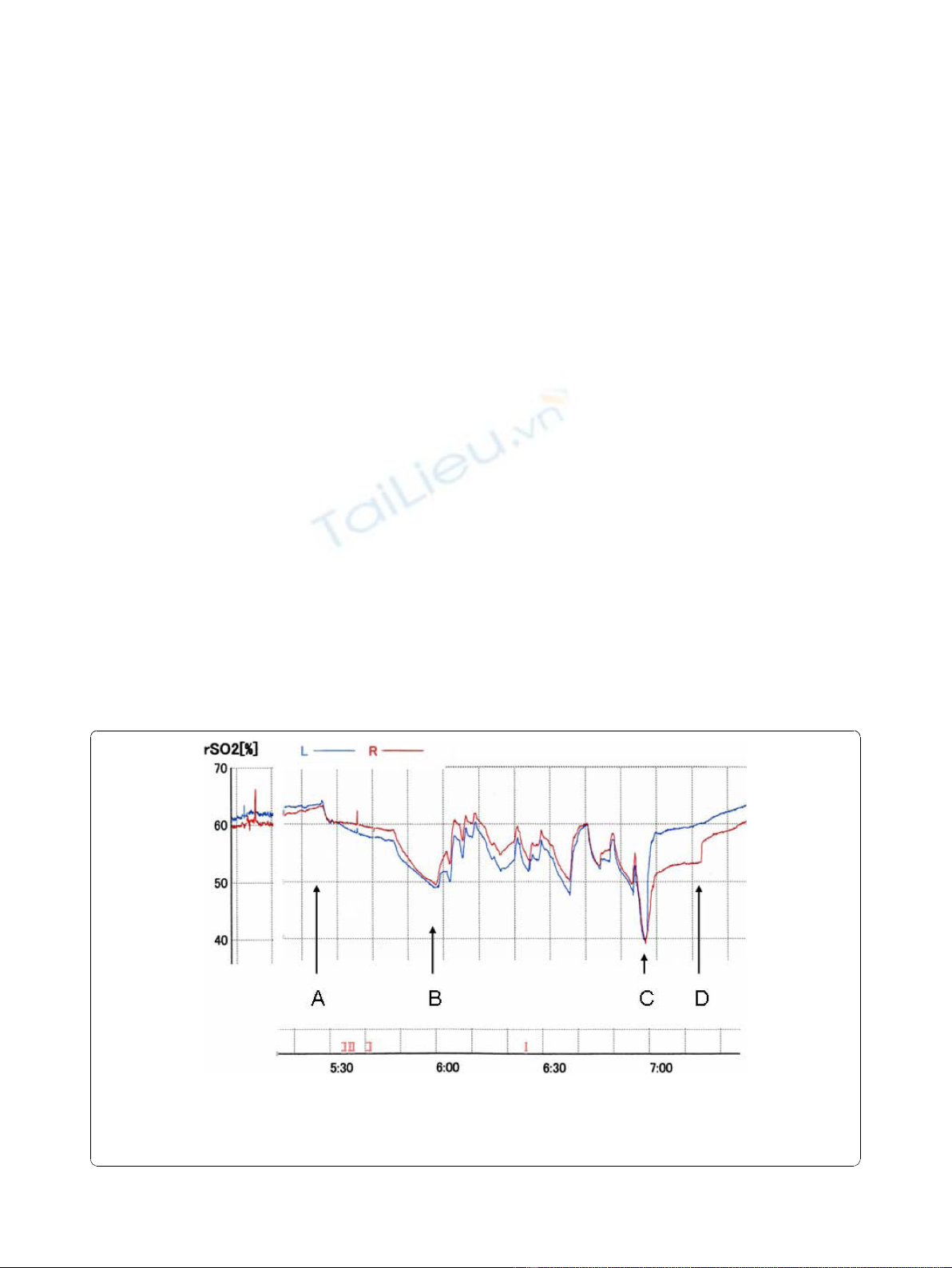
Prior to the anesthesia, the rSO
2
was 61% (Left) and
60% (Right). At the beginning of the cardiopulmonary
bypass, the rSO
2
was 55% (Left) and 56% (Right). At
profound hypothermia, the rSO
2
was 64% (Left) and
63% (Right), it gradually decreased to 49% (Left) and
50% (Right). After commencing the IPA-RCP, the rSO2
rose to around 60% at every augmentation, but it
decreased when the augmentation ceased. Just after the
resuming antegrade perfusion via a side branch of the
graft, the rSO
2
decreased to 40%, then recovered
smoothly (Figure 1)
.
The rSO
2
on the right side recov-
eredinastepwisemanner.Thepatientrecoveredcon-
sciousness 30 min after the operation free of any
neurological deficit and the postoperative course was
uneventful.
Conclusions
RCP by augmentation of CVP to 15 to 20 mmHg is rou-
tinely used in our institute for the additional brain pro-
tection during deep hypothermic circulatory arrest
because much evidence has been accumulated to sug-
gest an increased risk of perfusion-induced brain injury
associated with RCP, especially when continuously high
RCP pressures are used [4]. However, there is a safety
limit of the deep hypothermic circulatory arrest duration
because it cannot open all intracranial vessels but par-
tially. To overcome this drawback, Kitahori, Kawata,
Takamoto et al. developed a new intermittent pressure
augmentation method in which CVP is intermittently
increased to 45 mmHg [1-3]. They used a canine model,
and showed that the retinal vessels were effectively
dilated at an augmented pressure of 45 mm Hg (arteries,
107% + 3% of control veins, 114% + 3% of control),
whereas when antegrade selective cerebral perfusion was
used, the retinal vessels were smaller than the corre-
sponding preoperative vessels. They concluded that the
intermittent pressure augmentation allows an adequate
blood supply without injuring the brain and provides
adequate neuroprotection equivalent to that provided by
antegrade cerebral perfusion. In the canine model, they
administered the RCP through the maxillary vein to
overcome the drawbacks of jugular vein valves to reach
directly the cranial veins. In the majority of humans, as
de Brux et al. described, the jugular vein had competent
valvesanditishypothesizedthattheRCPgainsthe
brain through a collateral network of veins (azygos,
intercostal, medullary and vertebral veins). The useful-
ness of higher perfusion pressure could be either to dis-
tendthevalvesormoreprobablytoincreasethe
pressure in the collateral vein network to improve cere-
bral oxygenation [5]. Thus, the clinical effectiveness of
the IPA-RCP through a cannulae inserted to the SVC is
unknown field. We examined the effect of the IPA-RCP
by measuring rSO
2
which represents the brain blood
Figure 1 rSO
2
during deep hypothermic circulatory arrest.L:leftrSO
2
,R:rightrSO
2
. Initial 30 min of conventional retrograde cerebral
perfusion (RCP), rSO
2
gradually declined. When intermittent-pressure-augmented (45 mmHg) retrograde cerebral perfusion (IPA-RCP) was
induced, rSO
2
rose. The maximum duration of pressure augmentation was limited to 30 sec. A total of 10 augmentations at irregular intervals
were tried. A. Start of deep hypothermic circulatory arrest and conventional RCP. B. Start of IPA-RCP. C. Final dip: Start of the antegrade perfusion
to the left common carotid artery, and the left subclavian artery via graft branch. D. Start of antegrade perfusion via the brachiocephalic artery.
Kubota et al.Journal of Cardiothoracic Surgery 2010, 5:97
http://www.cardiothoracicsurgery.org/content/5/1/97
Page 2 of 4

perfusion. Although only the anterior part of the brain
rSO
2
is assessed by a TOS-96 brain oximeter, because
most attenuation of near-infrared light in human cere-
bral tissues is due to absorption by deoxyhemoglobin
and oxyhemoglobin, brain tissue is suitable for determi-
nation of rSO2. Only determination of rSO2 is an easily
available method to assess the real-time adequacy of
cerebral perfusion during deep hypothermic time-
restricted aortic arch surgery [6].
At first, we planned to perform the operation on our
patient using conventional RCP. However, because the
rSO2 declined to 49%, the duration of circulatory arrest
time was expected to exceed 60 min due to the fragile
aortic wall to reinforce and deep distal anastomosis, we
applied the intermittent pressure augmentation techni-
que for the first time. According to the original report,
the central venous pressure was controlled at 15 mm
Hg and it was augmented to 45 mm Hg quickly and
then decreased again to the baseline level of 15 mmHg
as soon as it reached 45 mm Hg every 30 seconds.
However, the same protocol is difficult to apply clini-
cally because backflow from the three arch vessels
increased and disturbed the anastomosis when CVP was
augmented. CVP was decreased to 15 mmHg expedi-
ently. Although the optimal duration of pressure aug-
mentation during deep hypothermic circulatory arrest in
clinical settings is unknown, to prevent the brain edema,
the maximum duration of pressure augmentation that
we set was 30 sec.
Along with every pressure augmentation, rSO2
showed immediate recovery up to 60% and it decreased
when the augmentation ceased. The essential effect of
IPA-RCP may not only be a temporary increase in rSO2
but elevation of the declining curve during RCP. Our
preliminary randomized comparative study in clinical
aortic arch replacement cases of IPA-RCP (n = 10) and
standard RCP (n = 10) showed that the interval from
the end of the operation to full awakeness of the IPA-
RCP group was 85 ± 64 min. in contrast with 310 ± 282
min. in RCP group (p < 0.05) accompanying with the
rSO2 decline ratio 60 min after the initiation of the
IPA-RCP group was 13.1 ± 3.7% in contrast with 24.5 ±
13.1% in RCP group (p < 0.05). There was no significant
difference of the used amount of the anesthetic agent. It
may support the “bottom raising effect”of this new
protocol.
Just after the resumption of antegrade perfusion, the
rSO
2
decreased to 40%, but then recovered smoothly.
We named this phenomenon the “final dip”.Whenwe
use RCP, the final dip always appears just after the
resumption of antegrade perfusion. This phenomenon
may represent wash out of deoxygenated blood that
remained and did not circulate in the brain despite the
performance of retrograde cerebral perfusion. The
stepwise recovery of the rSO
2
of the right side may
mean that the resumption of antegrade perfusion via the
left arch branches was insufficient to wash out the
remaining blood in our patient. In conclusion, this novel
protocol may have some advantages over conventional
RCP. Because it is difficult to verify the efficacy of IPA-
RCP by quantitative analysis, accumulation and analysis
of data e.g. measurement of the concentration of Tau
proteins in the CSF, comparison of the pre- and post-
operative cognitive function, measurement of the dia-
meters of the retinal vessels during IPA-RCP may
demonstrate the advantages of this new method of brain
protection [7].
Acknowledgements
We would like to gratefully acknowledge the outstanding original idea of
the IPA-RCP protocol, laboratory investigation, and cooperation given to us
by all the cardiac surgeons at the Mitsui Memorial Hospital: S Takamaoto, T
Miyairi, Columbia University Medical Center: H Takayama, and Tokyo
University Hospital: M Kawata, T Taketani, K Kitahori, K Nawata, T Morota, N
Motomura, M Ono.
Author details
1
Department of Cardiovascular Surgery, Kyorin University, Tokyo, Japan.
2
Department of Cardiology, Kyorin University, Tokyo, Japan.
Authors’contributions
HK, KT, HE, HT conceived of the study, and participated in its design and
coordination. HY and SK participated in the sequence alignment. All authors
read and approved the final manuscript.
Competing interests
The authors declare that they have no competing interests.
Received: 7 June 2010 Accepted: 2 November 2010
Published: 2 November 2010
References
1. Kitahori K, Takamoto S, Takayama H, Suematsu Y, Ono M, Motomura N,
Morota T, Takeuchi K: A novel protocol of retrograde cerebral perfusion
with intermittent pressure augmentation for brain protection. J Thorac
Cardiovasc Surg 2005, 130:363-370.
2. Kawata M, Takamoto S, Kitahori K, Tsukihara H, Morota T, Ono M,
Motomura N, Murakami A, Suematsu Y: Intermittent pressure
augmentation during retrograde cerebral perfusion under moderate
hypothermia provides adequate neuroprotection: An experimental
study. J Thorac Cardiovasc Surg 2006, 132:80-88.
3. Kawata M, Sekino M, Takamoto S, Ueno S, Yamaguchi S, Kitahori K,
Tsukihara H, Suematsu Y, Ono M, Motomura N, Morota T, Murakami A:
Retrograde cerebral perfusion with intermittent pressure augmentation
provides adequate neuroprotection: diffusion- and perfusion-weighted
magnetic resonance imaging study in an experimental canine model. J
Thorac Cardiovasc Surg 2006, 134:933-40.
4. Usui A, Oohara K, Liu TL, Murase M, Tanaka M, Takeuchi E, Abe T:
Determination of optimum retrograde cerebral perfusion conditions. J
Thorac Cardiovasc Surg 1994, 107:300-8.
5. De Brux JL, Subayi JP, Pegis JD, Pillet J: Retrograde cerebral perfusion:
anatomic study of the distribution of blood to the brain. Ann Thorac Surg
1995, 60:1294-8.
6. Ogino H, Ueda Y, Sugita T, Morioka K, Sakakibara Y, Matsubayashi K,
Nomoto T: Monitoring of regional cerebral oxygenation by near-infrared
spectroscopy during continuous retrograde cerebral perfusion for aortic
surgery. Eur J Cardiothorac Surg 1998, 14:415-8.
7. Kubota H, Takamoto S, Yoshino H, Kitahori K, Kawata M, Tonari K, Endo H,
Tsuchiya H, Inaba Y, Takahashi Y, Sudo K: Clinical Application of
Kubota et al.Journal of Cardiothoracic Surgery 2010, 5:97
http://www.cardiothoracicsurgery.org/content/5/1/97
Page 3 of 4

Intermittent Pressure-Augmented Retrograde Cerebral Perfusion. Ann
Thorac Surg 2010, 90:1340-3.
doi:10.1186/1749-8090-5-97
Cite this article as: Kubota et al.: Total aortic arch replacement under
intermittent pressure-augmented retrograde cerebral perfusion. Journal
of Cardiothoracic Surgery 2010 5:97.
Submit your next manuscript to BioMed Central
and take full advantage of:
• Convenient online submission
• Thorough peer review
• No space constraints or color figure charges
• Immediate publication on acceptance
• Inclusion in PubMed, CAS, Scopus and Google Scholar
• Research which is freely available for redistribution
Submit your manuscript at
www.biomedcentral.com/submit
Kubota et al.Journal of Cardiothoracic Surgery 2010, 5:97
http://www.cardiothoracicsurgery.org/content/5/1/97
Page 4 of 4


![Báo cáo seminar chuyên ngành Công nghệ hóa học và thực phẩm [Mới nhất]](https://cdn.tailieu.vn/images/document/thumbnail/2025/20250711/hienkelvinzoi@gmail.com/135x160/47051752458701.jpg)


