JOURNAL OF MILITARY PHARMACO-MEDICINE N04 - 2025
229
RESULTS OF BILIARY DILATION AND STONE REMOVAL
VIA FLEXIBLE CHOLANGIOSCOPY FOR THE TREATMENT
OF PRIMARY BILIARY STRICTURES AND STONES
AT MILITARY HOSPITAL 103
Do Son Hai1*, Nguyen Thi Dieu Lien2, Tong Tho Thang1
Nguyen Quang Nam1, Nguyen Anh Tuan3
Abstract
Objectives: To evaluate the results of biliary dilation and stone removal via
flexible cholangioscopy for the treatment of primary biliary strictures (BS) and
stones. Methods: A prospective, descriptive, uncontrolled study was conducted on
62 patients with primary BS and stones treated by biliary dilation and stone removal
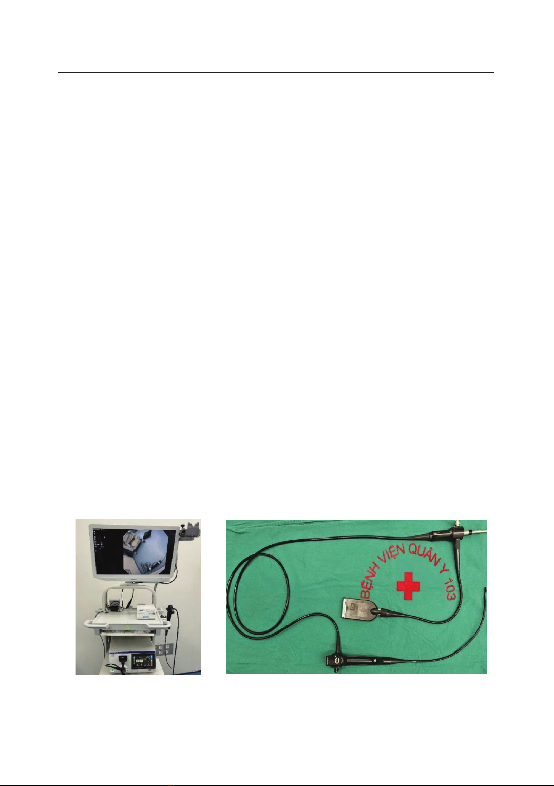
via flexible cholangioscopy at Abdominal Surgery Centre, Military Hospital 103,
from July 2021 to July 2024. Results: The mean age was 60.1 ± 14.1; the female/male
ratio was 1.69/1. 75.8% of patients had a history of biliary stones. Most patients
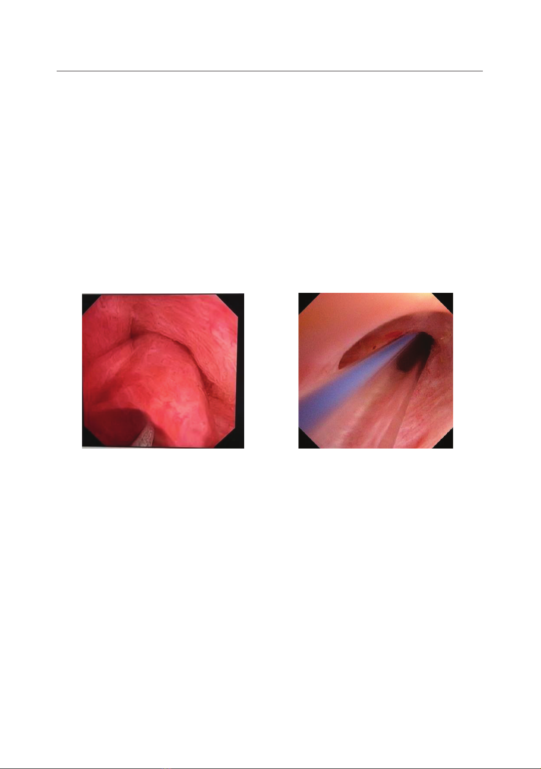
had multiple stones (79%), including choledocholithiasis and hepatolithiasis. BS
were mostly in one location (90.3%), intrahepatic strictures (88.7%), and were all
benign. The mean length and diameter of the strictures were 3.96 ± 2.9mm and
3.6 ± 0.7mm, respectively. Surgical methods were choledochotomy with
intraoperative cholangioscopy (90.3%) and percutaneous cholangioscopy (9.7%).
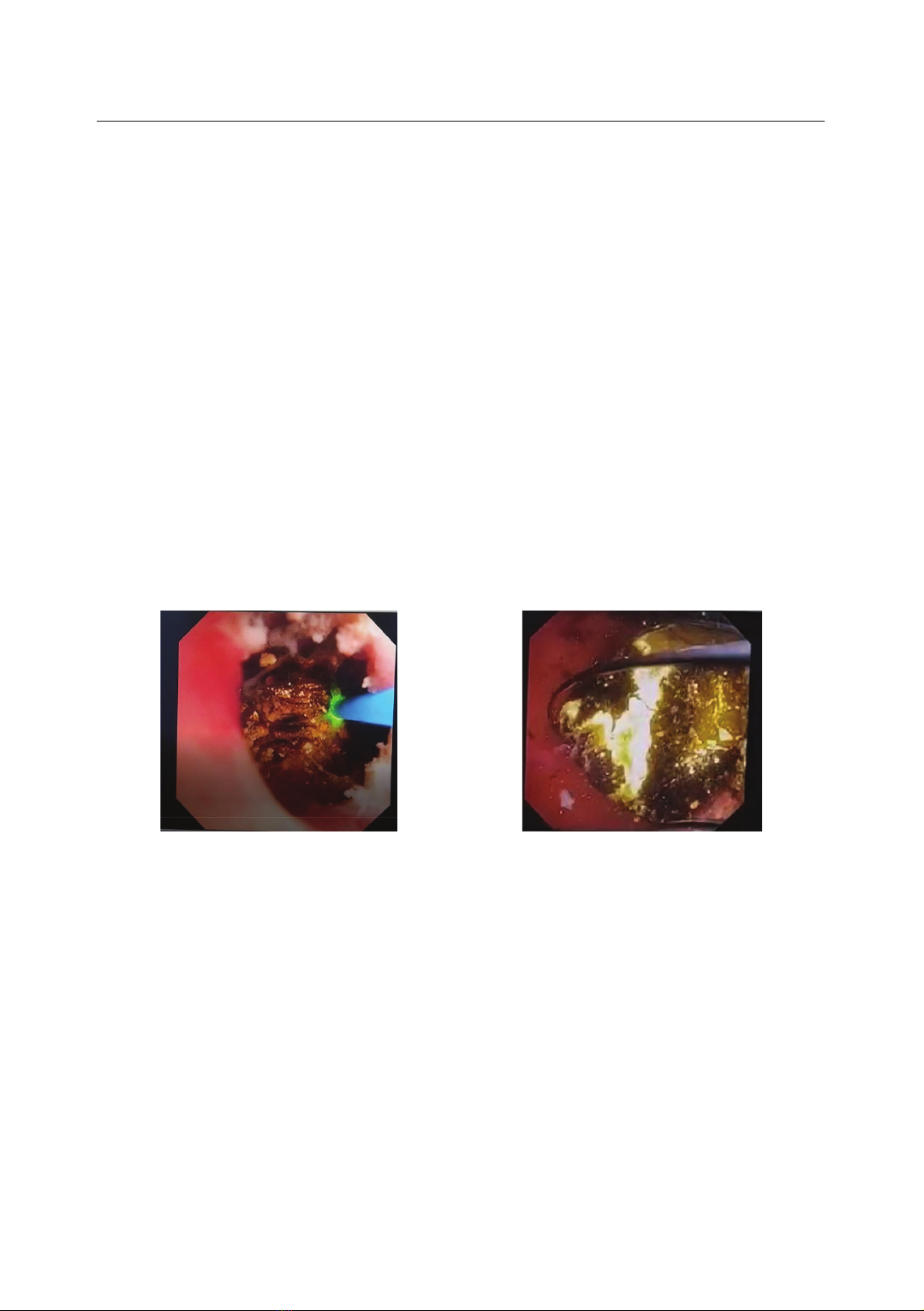
Stone removal was performed using baskets, electrohydraulic, and/or laser
lithotripsy. BS was performed using balloon dilation; then, biliary-cutaneous stents
were placed in 64.5% of cases at risk of recoil. Intraoperative complications
accounted for 16.1%; postoperative complications accounted for 12.9%. The rate
of stone clearance and successful stricture dilation after surgery was 83.9% and
87.1%. Rechecked at 1 month, 3 months, and 6 months after operation, the ratio of
recurrent stones and BS was 0%, 0%, 5.8% and 1.9%, 7.4%, 11.1%, respectively.
Conclusion: Stone removal and stricture dilation by flexible cholangioscopy is a
safe and effective method for treating primary BS and stones.
Keywords: Flexible cholangioscopy; Biliary stricture; Primary bile duct stone;
Biliary balloon dilator; Laser lithotripsy; Electrohydraulic lithotripsy.
1Abdominal Surgery Centre, Military Hospital 103, Vietnam Military Medical University
2HaDong General Hospital
3Abdominal Surgery Centre, 108 Military Central Hospital
*Corresponding author: Do Son Hai (dosonhai@vmmu.edu.vn)
Date received: 06/01/2025
Date accepted: 20/02/2025
http://doi.org/10.56535/jmpm.v50i4.1175
JOURNAL OF MILITARY PHARMACO-MEDICINE N04 - 2025
230
INTRODUCTION
Biliary stricture is a common disease
in East Asia, Southeast Asia, and Vietnam.
According to statistics, the rate of BS in
patients with primary bile duct stones
can reach 45.6 - 70% [1]. This disease
greatly affects the treatment results of
biliary stones and is the main cause of
stone recurrence, requiring patients to
undergo multiple surgeries.
In 2022, IHPBA (International
Hepatobiliary-Pancreatic Association)
defined: "BS is a localized reduction in
the diameter of the bile duct compared
to the adjacent biliary ducts, accompanied
by the dilation above the stricture" [2].
In patients with both BS and stones,
whether BS is the cause or consequence
of stones has not been proven. They
combine each other to create a
pathological spiral. Although BS has
always been considered a challenging
problem, the diagnosis and treatment of
BS were rarely mentioned in the past
due to difficulties in directly investigated
imaging. Until Shore first performed
intraoperative flexible cholangioscopy
in 1970, BS had gradually been mentioned.
Since then, flexible cholangioscopy
has become an effective method for
diagnosing and treating BS by its ability
to show clear images. Based on the
cholangioscope, Lee SK proposed a
classification of BS that has been widely
applied nowadays [3]. In 2023, the ACG
(American College of Gastroenterology)
issued recommendations on the technique
of cholangioscopy, BS dilation, and
biliary stenting [4].
Up to now, very few Vietnamese
authors have published researches that
deeply evaluate the treatment results
of BS. At Military Hospital 103,
cholangioscopy has been performed
since 2008 to treat stones. In recent
years, we have developed a technique
for dilating the BS with balloon
dilation. The research problem was
posed with the question: What is the
effectiveness of the technique of stone
removal combined with biliary dilation
in the treatment of primary BS and
stones? Because of that, we conducted
this research with the aim to: Evaluate
the results of biliary dilation and stone
removal via flexible cholangioscopy for
the treatment of primary BS and stones.
MATERIALS AND METHODS
1. Subjects
Including 62 patients with primary
BS and stones treated by biliary dilation
and stone removal via flexible
JOURNAL OF MILITARY PHARMACO-MEDICINE N04 - 2025
231
cholangioscopy at Abdominal Surgery
Centre, Military Hospital 103 from July
2021 to July 2024.
* Inclusion criteria: Patients were
diagnosed with primary BS and stones,
treated by stone removal and stricture
dilation via flexible cholangioscopy.
* Exclusion criteria: Patients had
tumors or cancers in the bile duct, liver,
pancreas’s head or Vater’s ampulla;
patients had ASA score > 3 (classification
of patient's health status before surgery
according to the American Society of
Anesthesiologists).
2. Methods
* Study design: A prospective,
descriptive, uncontrolled study.
* Research process:
Patients were clinically examined and
had para-clinical tests for preoperative
diagnosis. If patients had primary BS
and stones, met the selection criteria,
and did not fall under the exclusion
criteria, surgery was performed.
Surgical methods:
- If the patient had not previously
undergone surgery, choledochotomy with
intraoperative flexible cholangioscopy
was performed to remove stones and
dilate the stricture.
- If the patient had undergone
percutaneous biliary drainage,
percutaneous flexible cholangioscopy
via the tunnel was performed to remove
stones and dilate the stricture.
* Stone removal and biliary dilation
via flexible cholangioscopy technique:
We used a cholangioscopic system
with a Japanese Olympus CHF-V2 5mm
diameter flexible cholangioscope, 2mm
diameter instrument channel combined
with irrigation way, 2-directions adjustable
controller (up 160° - down 130°).
Figure 1. Cholangioscopic system and Olympus CHF-V2 flexible cholangioscope.
JOURNAL OF MILITARY PHARMACO-MEDICINE N04 - 2025
232
The surgeon took the cholangioscope
with the right hand and held the
controller with the left hand. Then, the
cholangioscope was inserted into the
bile duct. A continuously irrigated
stream of NaCl 0.9% was used to dilate
the bile duct and create a clean
environment during cholangioscopy.
The common bile duct was investigated
first, then stones were removed to check
the sphincter of Oddi and enterohepatic
circulation. After that, the cholangioscope
was controlled up to the common
hepatic duct and the intrahepatic bile
ducts sequentially. Within intrahepatic
bile ducts, cholangioscopy was performed
in order from the right hepatic duct,
right anterior section, segments 5, 8,
right posterior section, segments 6, 7, to
the left hepatic duct, segments 2, 3, 4,
and 1.
In this research, we performed stone
removal using baskets, laser, and
electrohydraulic lithotripsy and pumped
small fragments outside or into the
duodenum through the Oddi’s sphincter.
Stone removal was performed from near
to far, to make a good enterohepatic
circulation.
Figure 2. Stone removal with laser lithotripsy and basket.
After removing as many stones as
possible, the bile ducts were examined
for BS. When BS was found, its
characteristics were evaluated, and the
stricture was immediately biopsied with
biopsy forceps. If the biopsy result was
benign, the biliary dilation would be
performed. In this research, we applied
the balloon dilation method.
- Balloon dilation technique: Using a
specialized 3-stage Biliary Balloon
Dilation, 0.2 x 290cm, Model BD-410X
Olympus, Japan. The dilation technique
was applied according to the
recommendations of Nunes T (2021)
[5]. Based on the diameter of the BS,
the appropriate type of dilation balloon
was chosen, with a diameter 0.5 - 1mm
JOURNAL OF MILITARY PHARMACO-MEDICINE N04 - 2025
233
larger than the BS. The uninflated
dilation balloon was inserted via the
cholangioscope's instrument channel
through the stricture position. Then, the
balloon was slowly inflated with
increasing pressure from 1 - 20atm,
depending on the desired dilation size,
and was held for about 2 minutes. This
process was performed under the
observation of the flexible cholangioscope
to directly assess the result. The biliary
dilation would be performed many
times until the cholangioscope could go
through the stricture. If the dilation was
successful, we would continue to
remove stones behind the stricture.
After that, the indication for placing a
biliary stent was considered. If the
dilation was unsuccessful, consider
other methods such as liver resection,
biliary-enteric anastomosis, or conservative
procedure.
Figure 3. BS’s balloon dilation and stent placement.
- Biliary stent placement: ACG clinical
guideline 2023 recommended that if the
BS was membranous, biliary stent
placement would not be needed because
it usually did not recoil after dilation. In
other cases, the biliary stent should be
placed [4]. In this study, we used a 16Fr
biliary-cutaneous stent made of latex
(T-tube drainage) or silicon.
- Patients who had completed stone
clearance and BS dilation would be
re-examined after 1, 3, and 6 months for
re-evaluation. They would be clinically
and para-clinically examined with
ultrasound and magnetic resonance
cholangio-pancreatography to check and
compare with their surgery’s results.
* Data analysis: The information
was recorded and arranged in detail
according to the research medical
record form. The research data was
processed using SPSS 20.0 software.