HUE JOURNAL OF MEDICINE AND PHARMACY ISSN 3030-4318; eISSN: 3030-4326HUE JOURNAL OF MEDICINE AND PHARMACY ISSN 3030-4318; eISSN: 3030-4326
122 123
Hue Journal of Medicine and Pharmacy, Volume 15, No.2/2025 Hue Journal of Medicine and Pharmacy, Volume 15, No.2/2025
Bioactive constituents of Goniothalamus tamirensis essential oil and
its anticancer activity
Nguyen Khanh Thuy Linh*, Doan Quoc Tuan, Nguyen Duy Co, Le Duong Yen Nhi
University of Medicine and Pharmacy, Hue University
Abstract
Background and objectives: Cancer is a major challenge, affecting health, society, and the economy.
Natural therapies may help reduce side effects of conventional treatment. The present study aimed to
determine phytochemical characterization and evaluation of in vitro anticancer activity of the essential oil
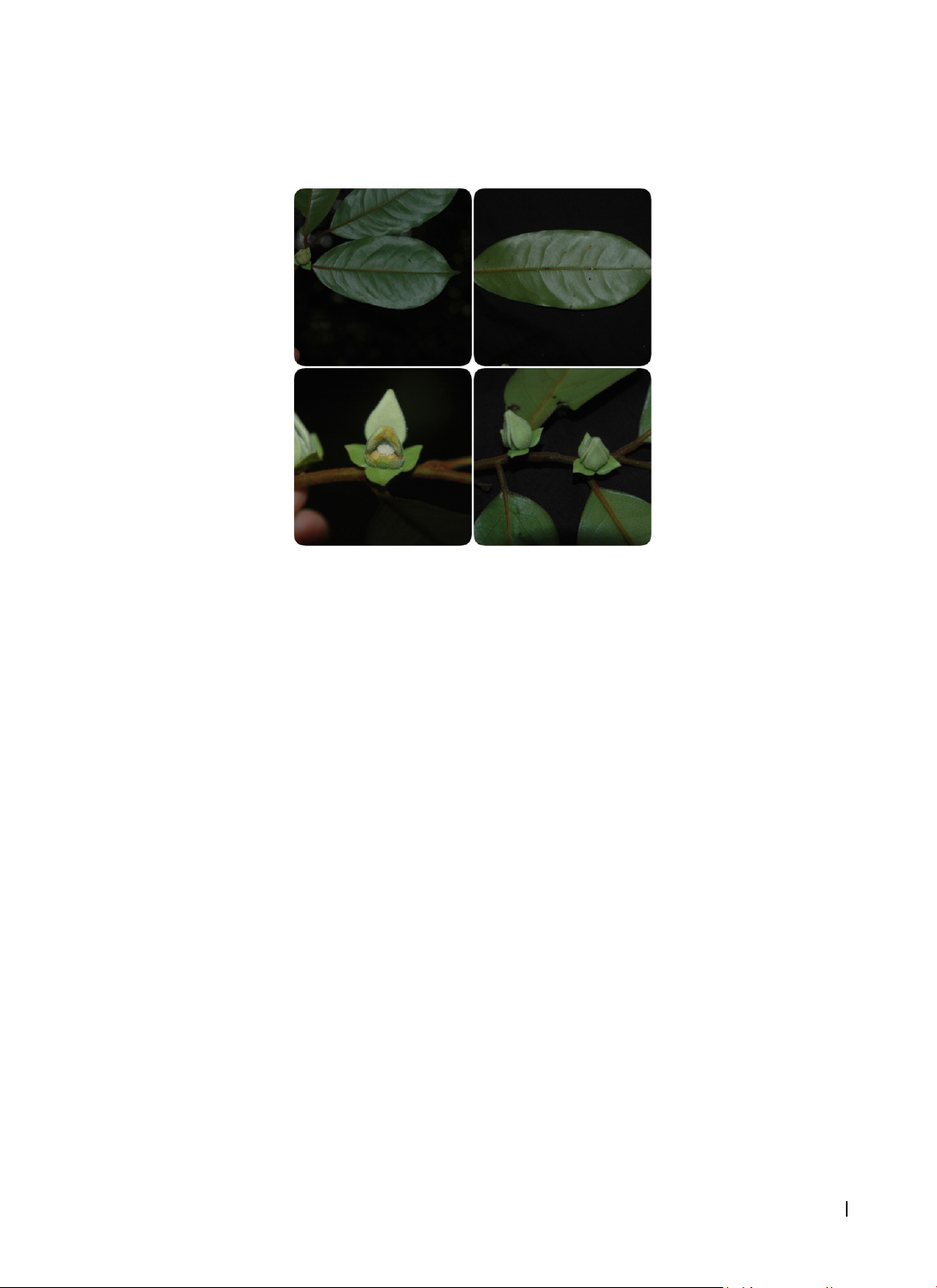
(EO) obtained from G. tamirensis leaves. Materials and methods: The leaves of G. tamirensis were collected
in Phong Dien, Thua Thien Hue, in August 2024. The chemical composition of the EO was identified using
GC-MS, and the anticancer potential of the EO was evaluated using the SRB assay against human cell lines
(HepG2, MCF7, A549). Results: The main compounds identified in G. tamirensis EO were oxygenated
sesquiterpenes. The EO showed significant anticancer activity against MCF, A549 and HepG2 cell lines with
IC50 of 8.79, 10.29 and 7.32 µg/mL, respectively. Conclusion: This study suggests that G. tamirensis leaf EO is
a promising candidate for use as an anticancer agent.
Keywords: Goniothalamus tamirensis, chemical composition, anticancer.
*Corresponding Author: Nguyen Khanh Thuy Linh. Email: nktlinh@huemed-univ.edu.vn
Received: 9/12/2024; Accepted: 10/3/2025; Published: 28/4/2025
DOI: 10.34071/jmp.2025.2.18
1. BACKGROUND
Cancer remains one of the leading causes of
mortality worldwide [1]. Although conventional
treatments like chemotherapy have advanced over
the years, they are still associated with significant
side effects. Moreover, the development of drug
resistance in cancer cells has diminished the efficacy
of many chemotherapeutic agents, highlighting the
urgent need for therapeutic alternatives. As a result,
there is a pressing demand for natural products to
be explored as potential cancer treatments. Several
bioactive compounds derived from natural sources,
including taxol, camptothecin, vincristine, and
vinblastine, have already demonstrated significant
therapeutic efficacy in oncology [2, 3]. These
examples emphasize the immense potential of
natural compounds as a rich and effective source of
medicinal agents for cancer treatment [4, 5].
Among the diverse array of natural compounds,
essential oils (EOs) have gained considerable
attention for their broad range of bioactivities,
which include antibacterial [6], antioxidant, anti-
inflammatory [7], and anticancer effects [8-10]. EO
is a mixture of many different chemical compounds.
Many studies have reported that EOs are considered
a potential anticancer agents. Besides their
effectiveness, EOs also have the ability to limit
side effects compared to conventioanl cancer
treatments such as chemotherapy [11]. Therefore,
research on the biological activity of Eos may open
up opportunities for the development of safe, plant-
based cancer therapies.
The genus Goniothalamus shows potential
as a source of medicinal compounds, although
researches have focused on only a few of its species.
Several studies have already demonstrated the
composition [12–16] and anticancer potential of
some Goniothalamus EOs on various cancer cell
lines [17]. However, many researches have focused
on other species within the genus, and relatively
little is known about Goniothalamus tamirensis,
a species native to Vietnam [18]. Given the lack of
comprehensive studies on the chemical profile and
anticancer activity of G. tamirensis EO, there is a
significant gap in our understanding of its potential
therapeutic applications. Research into the chemical
composition and biological activity of Goniothalamus
tamirensis EO is therefore crucial. Identifying and
characterizing the bioactive compounds present in
the EO could lead to the discovery of new anticancer
agents, which could complement or even provide an
alternative to conventional treatments. Additionally,
understanding the phytochemical composition of
G. tamirensis EO may contribute to the broader
body of knowledge on the therapeutic potential
of Goniothalamus species and their applicability in
cancer treatment.
Given the promising results from related species
and the limited research on G. tamirensis, this study
aims to fill this knowledge gap by investigating the
chemical composition and anticancer activity of G.
tamirensis EO.
HUE JOURNAL OF MEDICINE AND PHARMACY ISSN 3030-4318; eISSN: 3030-4326HUE JOURNAL OF MEDICINE AND PHARMACY ISSN 3030-4318; eISSN: 3030-4326
122 123
Hue Journal of Medicine and Pharmacy, Volume 15, No.2/2025 Hue Journal of Medicine and Pharmacy, Volume 15, No.2/2025
2. MATERIALS AND METHODS
2.1. Plant materials
The leaves of G. tamirensis were collected in
Phong Dien, Thua Thien Hue, in August 2024. A
voucher specimen has been deposited at the Faculty
of Pharmacy, Hue University of Medicine and
Pharmacy, Hue University, Vietnam.
Figure 1. G. tamirensis flower and leaf image
2.2. Methods
2.2.1. Distillation of the essential oil
The EO from G. tamirensis leaves was extracted
by hydro-distillation. After extraction, the EO was
dried over anhydrous sodium sulfate to remove any
residual water and then stored under refrigeration
until futher analysis. The obtained EO was used
to analyze its chemical composition and evaluate
its biological activities. The yield of the EO was
calculated using the formula: H = E/M × 100%,
where E is the volume of the extracted EO (mL) and
M is the initial rhizome biomass (g).
2.2.2. Gas Chromatography – Mass Spectrometry
(GC-MS) analysis
The chemical composition of the volatile
compounds was determined using gas
chromatography coupled with mass spectrometry
(GC-MS). The GC-MS analysis was performed on a
Shimadzu GCMS-QP2010 Plus system (Kyoto, Japan)
equipped with an Equity-5 capillary column (30 m
× 0.25 mm, 0.25 µm film thickness), and a mass
spectrometer (MSD QP2010 Plus). The EO (1 mg)
was diluted with n-hexane, and 1 µL was injected
for analysis. The oven temperature was initially set
to 60 °C for 2 min, followed by a temperature ramp
to 280 °C at a rate of 2 °C/min, with a total analysis
time of 110 min. Helium was used as the carrier at
a flow rate of 1.8 mL/min. The mass spectrometer
was operated with an interface temperature of
280 °C, the EI-mode was 70 eV and the mass
acquisition range was set from 40 to 500 m/z. A
splitless injection mode was employed. Compound
identification was performed by comparing the
obtained mass spectra with the Wiley 7 and National
Institute of Standards and Technology (NIST 11)
libraries. In addition, a standard solution of C8C38
alkanes was used to determine the retention index
of compounds and to compare them with literature
data [19]. The relative amounts of individual
components were calculated based on the GC peak
area without correction.
2.2.3. SRB assay for evaluating cytotoxic activity
The cytotoxic activity of the EO was evaluated
using the sulforhodamine B (SRB) assay, as outlined
by Monks et al., which quantifies the binding of
SRB dye to cellular proteins [20]. The cancer cell
lines MCF7 (breast cancer), A549 (human lung
adenocarcinoma), and HepG2 (hepatocarcinoma)
were cultured in Dulbecco’s modified Eagle’s
medium (DMEM) containing 2 mM L-glutamine,
1.5 g/L Na2CO3, and 10% fetal bovine serum, with
medium changes every 48 hours. The cells were
detached using 0.05% trypsin-EDTA solution,
subcultured at a 1:3 ratio every 3 to 5 days,
and incubated at 37 °C in a humidified 5% CO2
atmosphere for 48 hours. For the assay, 4 × 10⁴ cells
were seeded in each well of a 96-well microplate
with 180 μL of growth medium. After adding the
HUE JOURNAL OF MEDICINE AND PHARMACY ISSN 3030-4318; eISSN: 3030-4326HUE JOURNAL OF MEDICINE AND PHARMACY ISSN 3030-4318; eISSN: 3030-4326
124 125
Hue Journal of Medicine and Pharmacy, Volume 15, No.2/2025 Hue Journal of Medicine and Pharmacy, Volume 15, No.2/2025
test samples (10 μL), the plates were incubated
under the same conditions. Following 72 hours of
treatment with the EO, the medium was discarded,
and the cell monolayers were fixed with 20% (w/v)
cold trichloroacetic acid for 1 hour at 4 °C. SRB
solution was then added to each well, and the
plates were incubated at room temperature for
30 minutes. The wells were washed with 1% (v/v)
aqueous acetic acid to remove excess dye, and the
protein-bound dye was solubilized in 10 mM Tris
base solution. Absorbance was measured at 515
nm for both control and treated wells. Dimethyl
sulfoxide (DMSO) at 10% (DMSO concetration in
the well is 0.5%) served as the blank, and ellipticine
was used as the positive control. Cytotoxicity was
assessed at concentrations of 100, 20, 4, and 0.8
μg/mL, with IC50 value calculated using TableCurve
version 4.0. The inhibition rate of cancer cells
calculated by the following formula:
IR% = (100% - [(Abst – Abso)/ (Absc – Abso)]) × 100
Where, IR: inhibition rate of cell growth, Abst :
average optical density value at day 3; Abso : average
optical density value at time-zero; Absc : average optical
density value of the blank DMSO control sample.
All experiments were set up in triplicate. The IC50
value were presented as mean ± standard deviation.
Cytotoxicity categories based on the US National
Cancer Institute [21,22]: very toxic IC50 20 μg/mL,
moderate/toxic IC50 21-200 μg/mL, weak IC50 201-
500 μg/mL, and non toxic IC50 500 μg/mL.
3. RESULTS
3.1. Extraction and profilling
The yield of EO extraction from leaves of G.
tamirensis was 0.04% (v/w), calculated on a dry
weight basis. Table 1 displays the result obtained
from the chemical composition analysis of this EO
through GC-MS.
Table 1. Components of essential oil from leaves of Goniothalamus tamirensis
No RT CompoundsaType RIbRIcContent (%)
1 17,63 Terpinen-4-ol OM 1190 1174 1.29
2 18,67 iso-Dihydro carveol OM 1213 1212 8.92
3 21,32 neo-3-Thujanol acetate O1273 1273 5.73
4 21,65 ρ-Ethyl acetophenone O1280 1279 1.54
5 23,07 δ-Terpinyl acetate O1313 1316 9.18
6 23,29 neo-Verbanol acetate O1318 1319 5.74
7 23,68 cis-Piperitol acetate O1327 1332 1.34
8 24,64 α-Longipinene S1349 1350 1.70
9 32,13 10-epi-Cubebol OS 1530 1533 3.50
10 33,98 Allo-cedrol OS 1577 1589 4.61
11 35,02 1,10-di-epi-Cubenol OS 1604 1618 2.05
12 35,43 Ledol OS 1614 1602 1.38
13 35,67 2,(7Z)-Bisaboladien-4-ol OS 1621 1618 1.58
14 36,03 Hinesol OS 1631 1640 1.80
15 37,02 neo-Intermedeol OS 1657 1658 2.63
16 37,35 Intermedeol OS 1666 1665 6.08
17 37,73 Eudesma-4(15),7-dien-1β-ol OS 1676 1687 2.45
18 38,91 14-hydroxy-4,5-dihydro Caryophyllene OS 1708 1706 1.99
19 39,43 γ-Costol OS 1723 1745 2.88
20 40,81 (Z)-Lanceol OS 1762 1760 16.69
21 41,78 (E)-Isovalencenol OS 1789 1793 1.97
22 43,29 β-Vetivone OS 1833 1822 0.42
23 44,45 cis-Thujopsenic acid O1867 1863 3.72
24 46,17 Cembrene D 1919 1937 3.54
HUE JOURNAL OF MEDICINE AND PHARMACY ISSN 3030-4318; eISSN: 3030-4326HUE JOURNAL OF MEDICINE AND PHARMACY ISSN 3030-4318; eISSN: 3030-4326
124 125
Hue Journal of Medicine and Pharmacy, Volume 15, No.2/2025 Hue Journal of Medicine and Pharmacy, Volume 15, No.2/2025
Oxygenated monoterpene (OM) 10.21
Sesquiterpene hydrocarbon (S) 1.70
Oxygenated sesquiterpene (OS) 50.02
Dierpene (D) 3.54
Others (O) 27.25
Total 92.73
a Elution order on Equity-5 column
b Retention Indices on Equity-5 column
c Literature retention indices
- Not identified
In this study, the majority of the identified
compounds were oxygenated sesquiterpenes
(50.02%) and oxygenated monoterpenes (10.21%).
A total of 24 compounds were detected, with
(Z)-Lanceol representing the most abundant
component in G. tamirensis EO at 16.69%, followed
by δ-Terpinyl acetate (9.18%) and iso-dihydro
carveol (8.92%).
3.2. Anticancer activity
The anticancer activity of the EO of G. tamirensis was determined using the SRB method, and their results
are shown in Table 2.
Table 2. Cytotoxic activity of the leaves essential oil from G. tamirensis
C
(µg/mL)
G. tamirensis essential oil C
(µg/
mL)
Ellipticine
MCF7 A549 HepG2 MCF7 A549 HepG2
100 95.45 ± 2.05 90.92 ± 2.40 98.87 ± 1.64 10 90.83 ± 2.17 88.62 ± 3.24 92.79 ± 1.98
20 81.76 ± 1.28 88.19 ± 1.58 86.54 ± 1.32 2 78.14 ± 1.82 76.82 ± 1.20 79.68 ± 1.69
422.44 ± 1.61 10.80 ± 0.79 26.49 ± 1.28 0.4 52.66 ± 1.63 50.58 ± 1.23 53.06 ± 1.89
0.8 5.27 ± 0.14 1.75 ± 0.21 7.80 ± 0.61 0.08 22.32 ± 1.21 21.02 ± 1.12 22.26 ± 0.94
IC50 8.79 ± 0.44 10.29 ± 0.38 7.32 ± 0.24 IC50 0.34 ± 0.02 0.38 ± 0.03 0.33 ± 0.03
C: concentration
The IC50 values demonstrated that the EO from
the leaves exhibited significant cytotoxic effects
on the MCF7, A549, and HepG2 cell lines, with IC50
values of 8.79, 10.29, and 7.32 μg/mL, respectively.
4. DISCUSSION
This study was conducted to explore the
phytochemical composition and evaluate the
potential biological activities of the EO derived
from Goniothalamus tamirensis. Our findings
revealed that oxygenated sesquiterpenes were
the predominant components of G. tamirensis EO,
accounting for 50.02% of its composition. Among
these, the sesquiterpenoid (Z)-lanceol was identified
as the major constituent, comprising 16.69% of the
EO. Previous research has provided valuable insights
into the chemical composition of the EO from G.
tamirensis, with variations reported between oils
extracted from different parts of the plant. For
instance, a study by T.D. Thang identified α-pinene
(33.4%), viridiflorol (18.5%), and β-caryophyllene
(12.4%) as the dominant components in the EO from
the leaves of G. tamirensis. In contrast, the stem oil
was primarily composed of γ-gurjunene (11.2%),
β-caryophyllene (10.9%), and δ-cadinene (10.3%)
[18]. The chemical profile observed in our study
differed from these previous findings, which could
be attributed to several factors, such as variations
in the plants growing environment, differences in
the extraction methods employed, and advances in
the instrumental techniques used for analysis. These
factors can significantly influence the chemical
composition of EO, leading to discrepancies between
studies, even when investigating the same plant
species.
Extracts and isolated compounds from G.
tamirensis have demonstrated inhibitory effects
against various cancer cell lines. From the leaves of
G. tamirensis, 15 compounds were isolated. Among
them, (-)-5-acetoxygoniothalamine and (Z)-6-styryl-
5,6-dihydro-2-pyranone demonstrated inhibitory
activity against colon cancer cells (HCT116), with
IC₅₀ values of 8.6 µM and 22.1 µM, respectively.
Other species in this genus may also exhibit anti
cancer activity. For comparison, the positive control
HUE JOURNAL OF MEDICINE AND PHARMACY ISSN 3030-4318; eISSN: 3030-4326HUE JOURNAL OF MEDICINE AND PHARMACY ISSN 3030-4318; eISSN: 3030-4326
126 127
Hue Journal of Medicine and Pharmacy, Volume 15, No.2/2025 Hue Journal of Medicine and Pharmacy, Volume 15, No.2/2025
doxorubicin showed an IC₅₀ of 9.7 µM [23]. The
crude methanolic extract of G. elegans exhibited
pronounced cytotoxic activity against SW-480
(colorectal), AGS (gastric), and SK-LU-1 (lung) cancer
cell lines [24] with IC₅₀ values of 14.51; 19.57 and
24.69 µg/mL, respectively. Furthermore, two isolated
alkaloids-lysicamine and liriodenine-demonstrated
significant inhibitory effects against these cancer
cell lines, with the IC50 ranging from 9.84 to 31.72
µM, indicating their potential as lead compounds
for anticancer drug development [24]. However, the
anticancer potential of G. tamirensis EO has not yet
been investigated. This study represents the first
report on the cytotoxic activity of the EO from this
species. The results of this study suggest that the EO
from G. tamirensis possesses promising anticancer
properties, which may contribute to its ability to
inhibit the viability of cancer cells. The potent
cytotoxic effects observed in this study highlight
the potential of G. tamirensis EO as a candidate for
further investigation in cancer drug discovery. This
EO, with its bioactive compounds, may represent
a valuable natural source for developing new
therapies to treat cancer. The findings of this study
provide scientific evidence supporting the use of
G. tamirensis as a therapeutic agent in traditional
medicine, as well as its potential applications in
modern pharmaceutical research and development.
Although the results from this study are
encouraging, further research is needed to expand
our understanding of the anticancer potential of
G. tamirensis EO. To gain a more comprehensive
evaluation, it would be beneficial to test the oil
on a broader range of cancer cell lines, including
those derived from various tissue origins, to assess
its efficacy across different cancer types. To further
evaluate its therapeutic potential, it is recommended
that the antiproliferative activity of the EO be tested on
additional cancer cell lines from various tissue types.
Additionally, in vivo studies using animal models are
essential to confirm the observed anticancer effects
and to explore the potential therapeutic benefits
in a living organism. In-depth pharmacological
investigations, including dose-response studies and
toxicity profiling, are necessary steps to determine the
safety and efficacy of this EO as a potential anticancer
treatment. By expanding the scope of research, we
can better understand the full therapeutic potential
of G. tamirensis and its EO, paving the way for its
possible use as a natural adjunct or alternative to
conventional cancer therapies.
5. CONCLUSION
In conclusion, the chemical composition of
Goniothalamus tamirensis EO was analyzed using
GC-MS, revealing that oxygenated sesquiterpenes
(50.02%) and oxygenated monoterpenes (10.21%)
were the major constituents. The EO demonstrated
significant cytotoxic activity against the MCF7, A549
and HepG2 cancer cell lines with IC50 values of 8.79,
10.29, and 7.32 μg/mL, respectively. These findings
provide valuable insights into the potential of G.
tamirensis as a source of bioactive compounds with
anticancer properties.
Acknowledgements
The authors are grateful to University of Medicine
and Pharmacy, Hue University for financial support
to conduct this research (ID No. 15SV/24)
REFERENCES
1. Bray F, Laversanne M, Sung H, Ferlay J, Siegel
RL, Soerjomataram I, Jemal A. Global cancer statistics
2022: GLOBOCAN estimates of incidence and mortality
worldwide for 36 cancers in 185 countries. CA Cancer J
Clin [Internet]. 2024 [cited 2025 Apr 10];74(3):229–63.
Available from: https://onlinelibrary.wiley.com/doi/
abs/10.3322/caac.21834
2. Heinrich M, Bremner P. Ethnobotany and
ethnopharmacy-their role for anti-cancer drug
development. Curr Drug Targets. 2006 Mar;7(3):239–45.
3. Newman DJ, Cragg GM. Natural products as
sources of new drugs over the last 25 years. J Nat Prod.
2007 Mar;70(3):461–77.
4. Gezici S, Turkmen M, Karahan F. Exploring the
anti-cancer properties of essential oils from some
Lamiaceae species against human cancer cells with
multivariate analysis. South Afr J Bot [Internet]. 2024
Mar 1 [cited 2024 Nov 25];166:287–96. Available from:
https://www.sciencedirect.com/science/article/pii/
S0254629923006658
5. Ali Abdalla YO, Subramaniam B, Nyamathulla S,
Shamsuddin N, Arshad NM, Mun KS, Awang K, Nagoor
NH. Natural products for cancer therapy: A Review of their
mechanism of actions and toxicity in the past decade.
J Trop Med [Internet]. 2022 Mar 11 [cited 2024 Nov
25];2022:5794350. Available from: https://www.ncbi.nlm.
nih.gov/pmc/articles/PMC8933079/
6. Chouhan S, Sharma K, Guleria S. Antimicrobial
activity of some essential oils—Present status and future
perspectives. Medicines [Internet]. 2017 Aug 8 [cited 2024
Nov 30];4(3):58. Available from: https://www.ncbi.nlm.
nih.gov/pmc/articles/PMC5622393/
7. Miguel MG. Antioxidant and anti-Inflammatory
activities of essential Oils: A short review. Molecules
[Internet]. 2010 Dec 15 [cited 2024 Nov 30];15(12):9252–
87. Available from: https://www.ncbi.nlm.nih.gov/pmc/
articles/PMC6259136/