27
Journal of Medicine and Pharmacy, Volume 11, No.07/2021
Clinical and sub-clinical features in patients with systemic lupus
erythematosus
Nguyen Hoang Thanh Van*, Nguyen Tran Dieu Anh
Hue University of Medicine and Pharmacy, Hue University, Vietnam
Abstract
Objectives: To describe the clinical and sub-clinical features in patients with Systemic Lupus Erythematosus
(SLE) according to the criteria of the ACR/SLICC 2015, studying the relationship between clinical and sub-clinical
features. Methods: This was a descriptive cross - sectional study of 74 SLE patients admitted to Nephrology
Rheumatology Department of Hue Central Hospital and General Medicine - Endocrinology Department of
Hue University of Medicine and Pharmacy Hospital from March 2020 to March 2021. Results: Malar rash
70.3%, photosensitivity 66.2%, discoid rash 18.9%, non – scarring alopecia 75.7%, oral ulcers 21.6%, arthritis
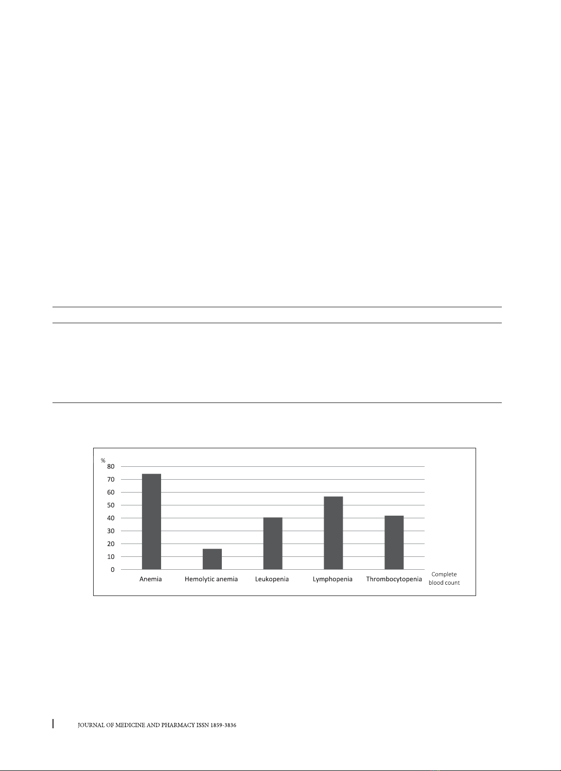
54.1%, serositis 2.7%, neurology and/or psychosis damage 5.4%, kidney involvement 75.7%, hematologic:
leukopenia 40.5%, lymphopenia 56.8%, thrombocytopenia 41.9%, hemolytic anemia 16.2%, positive ANA
74.3%, positive anti-dsDNA 64.9%. Positive ANA in non – scarring alopecia group was higher than the group
without (p < 0.05); positive anti-dsDNA in malar rash group was higher than the group without (p < 0.05); the
risk of kidney involvement was higher in the group with positive anti-dsDNA (OR = 3.1; p < 0.05), the rates
of anemia and thrombocytopenia in kidney involvement groups were higher than the group without (p <
0.05). Conclusions: In this study cohort, the clinical, subclinical features according to the criteria of the ACR/
SLICC 2015 that had the highest rate were non – scarring alopecia and kidney involvement, followed by malar
rash, photosensitivity. ANA positivity in the non-scarring alopecia group was higher. Anti-dsDNA positivity
in malar rash group was higher. The risk of potential kidney disorders was higher in the group with positive
anti-dsDNA. The rates of anemia and thrombocytopenia in the potential kidney disorders group were higher.
Key words: Systemic Lupus Erythematosus, ACR/SLICC 2015, ANA, anti ds DN.
1. BACKGROUND
Systemic Lupus Erythematosus (SLE) is a systemic
autoimmune disease with multisystem involvement
characterized by antinuclear antibodies and other
antigens. Organs that are often injured include
joints, skin, kidneys, hematologic abnormalities,
heart, lungs, nerves,... More than 90% of cases of SLE
occur in women, frequently starting at childbearing
age, between 20 and 40- year-olds [4].
Previously, the diagnosis of systemic lupus
erythematosus was based on the criteria of the
American College of Rheumatology 1997 (ACR
1997) [8]. This criteria was mainly based on clinical
organ damages. Therefore, it tended to diagnose
the disease at the late stage, when organ damages
were already shown. In 2012, the Systemic Lupus
International Collaborating Clinics (SLICC 2012)
published new criteria [7], classified into two
groups of clinical and biological manifestations,
emphasizing immunological criterion, which
allowed to diagnose systemic lupus erythematosus
even when there were only immunological changes
without clinical organ damages. In 2015, the ACR/
SLICC published criteria, based on the framework
of the 2012 SLICC criteria. Criteria were scored by
points, emphasizing the role of common symptoms.
Therefore, if a patient was admitted to the hospital
with many clinical symptoms pointing to systemic
lupus erythematosus without laboratory tests, the
diagnosis could be based on this criteria [6].
The early detection and diagnosis of systemic
lupus erythematosus based on clinical symptoms
will help early treatment for patients. Therefore,
we conduct the project: “Clinical and sub-
clinical features of patients with systemic lupus
erythematosus” with the following objectives:
1. To describe the clinical and sub-clinical features
in patients with systemic lupus erythematosus
according to ACR/SLICC 2015 criteria.
2. To study the relationship between clinical and
subclinical features in patients with systemic lupus
erythematosus
2. METHODS
2.1. Patients
It was a descriptive cross - sectional study
Corresponding author: Nguyen Hoang Thanh Van; email: nhtvan@huemed-univ.edu.vn
Received: 25/6/2021; Accepted: 28/10/2021; Published: 30/12/2021
DOI: 10.34071/jmp.2021.7.4
28
Journal of Medicine and Pharmacy, Volume 11, No.07/2021
including 74 SLE patients diagnosed in the
Nephrology Rheumatology Department of
Hue Central Hospital and General Medicine -
Endocrinology Department of Hue University of
Medicine and Pharmacy Hospital during the period
from March 2020 to March 2021.
All patients satisfied at least four of ACR/SLICC
2015 criteria for the classification of SLE [16].
2.2. Methods
Descriptive Cross - sectional study.
* Clinical variables
- Malar rash
- Photosensitivity
- Discoid rash
- Oral/nasal ulcers
- Arthritis
- Serositis
- Neurology and/or psychosis damage
- Non – scarring alopecia
- Kidney involvement
- Hemolytic anemia
*Sub-clinical variables:
- Complete blood count: anemia, hemolytic
anemia, leukopenia, lymphopenia, thrombocytopenia
- 24 hour urine protein
- ANA and anti-dsDNA
Methods of data processing: The collected data
were processed according to medical statistical
algorithms, using SPSS 26.0 software.
3. RESULTS
3.1. Clinical and sub-clinical features in patients with systemic lupus erythematosus
3.1.1. Clinical features
Table 1. Clinical features
Symptoms n % Symptoms n %
Malar rash 52 70.3 Non – scarring alopecia 56 75.7
Photosensitivity 49 66.2 Arthritis 2 2.7
Discoid rash 14 18.9
Oral/nasal ulcers 16 21.6 Kidney involvement 56 75.7
Arthritis 40 54.1 Neurology and/or psychosis damage 4 5.4
Comment: Kidney involvement and non – scarring alopecia accounted for 75.7%, malar rash accounted for
70.3%, photosensitivity accounted for 66.2%, arthritis accounted for 54.1%.
3.1.2. Peripheral blood cell disorders
Bar chart 1. Peripheral blood cell disorders
Comment: Anemia accounted for 74.3%, hemolytic anemia accounted for 16.2%, leukopenia accounted
for 40.5%, lymphopenia accounted for 56.8%, thrombocytopenia accounted for 41.9%.
29
Journal of Medicine and Pharmacy, Volume 11, No.07/2021
3.1.3. 24-hour proteinuria
Table 2. 24-hour proteinuria
24-hour proteinuria
(g/24h) n % ± SD
< 0.5 18 24.3 0.2 ± 0.13
0.5 – 3.5 27 36.5 7.12 ± 9.45
> 3.5 29 39.2
Comment: 56/74 patients who had proteinuria level > 0.5 g/24h accounted for 75.7% and the average of
24-hour proteinuria concentration in this group was 7.12 ± 9.45 g.
3.1.4. Distribution of ANA and anti-dsDNA
Table 3. Distribution of ANA and anti-dsDNA
Tests Positive Negative
n % ± SD n %
ANA (OD ratio) 55 7.3 2.92 ± 4.09 19 25.7
Anti-dsDNA (U/ml) 48 64.9 76.49 ± 104.75 26 35.1
Comment: Positive ANA accounted for 74.3% and the average of OD ratio was 2.92 ± 4.09; positive anti-
dsDNA accounted for 64.9% and the average concentration was 76.49 ± 104.75 U/ml
3.2. The relationship between clinical and subclinical features in patients with systemic lupus
erythematosus
3.2.1. The relationship between clinical features and immunological tests
Table 4. The relationship between clinical features and immunological tests
Symptoms
ANA
positivity p
Anti-dsDNA
positivity p
n % n %
Malar rash Yes 37 67.3 0.337 30 62.5 0.047
No 18 32.7 18 37.5
Photosensitivity Yes 35 63.6 0.425 28 58.3 0.051
No 20 36.4 20 41.7
Discoid rash Yes 11 20.0 0.686 12 25.0 0.070
No 44 80.0 36 75.0
Oral/nasal ulcers Yes 14 25.5 0.173 10 20.8 0.823
No 41 74.5 38 79.2
Non – scarring alopecia Yes 45 81.8 0.036 37 77.1 0.701
No 10 18.2 11 22.9
Arthritis Yes 32 58.2 0.225 25 52.1 0.644
No 23 41.8 23 47.9
Serositis
Yes 23.6
0.399
24.2
0.291
No 53 96.4 46 95.8
Neurology damage Yes 47.3
0.227
36.3 0.662
No 51 92.7 45 93.8
Comment:
+ Positive ANA in non scarring alopecia group was higher than the group without (81.8% 18.2%)
(p < 0.05).
30
Journal of Medicine and Pharmacy, Volume 11, No.07/2021
+ Positive anti-dsDNA in the malar rash group was higher than the group without (62.5% và 37.5%) (p
< 0.05).
3.2.2. The relationship between potential kidney disorders and immunological tests
Table 5. The relationship between potential kidney involvement and immunological tests
Antinuclear antibodies Potential kidney disorders OR p
n %
ANA positivity Yes 44 78.6 2.333
(0.744 – 7.314) 0.140
No 12 21.4
Anti-dsDNA positivity Yes 40 74.4 3.125
(1.045 – 9347) 0.037
No 16 28.6
Comment: The risk of potential kidney disorders was higher in the group with positive anti-dsDNA (OR =
3.1; p < 0.05).
3.2.3. The relationship between potential kidney disorders and peripheral blood cell disorders
Table 6. The relationship between potential kidney disorders and peripheral blood cell disorders
Kidney disorders Non-kidney disorders p
n % n %
Anemia 46 83.6 9 164 0.007
Leukopenia 24 80.0 620.0 0.474
Lymphopenia 34 81.0 8 19.0 0.225
Thrombocytopenia 19 613 12 38.7 0.014
Comment:
+ The rate of anemia in the group with kidney disorders was higher than the group without (83.6%
16.4%) (p < 0.05).
+ The rate of thrombocytopenia in the group with kidney disorders was higher than the group without
(61.3% và 38.7%) (p < 0.05).
4. DISCUSSIONS
Dermatological manifestations are one of the
most typical symptoms in SLE includes malar rash,
photosensitivity, discoid rash, oral/nasal ulcers,
non-scarring alopecia. In this study, we recorded the
rate of malar rash at 70.3%, higher than the study
of Ngo Thi Thuy Thanh (54.7%) [2]. Photosensitivity
accounted for 66.2%, similar to the study of Nguyen
Thi Kim Thanh (57.6%) [3]. Discoid rash accounted
for 18.9%, which is similar to the study of Nguyen
Thi Kim Thanh (19.5%) [3], but higher than the study
of Ngo Thi Thuy Thanh (3.8%) [2]. Oral/nasal ulcers
accounted for 21.6%, the oral mucosal ulcer is the
most popular one. As noted by author Abdulrahman
Maryam, who conducted a study at Nephrology
Rheumatology Department of Ain Sham University,
Greece in 2019 on 110 SLE patients reported the
rate of nasal/oral ulcers was 35.5% [5]. In our study,
75.7% of patients showed non scarring alopecia,
higher than some relative studies: Ngo Thi Thuy
Thanh (71.7%) [2], Mai Thu Huyen (43.8%) [1].
Musculoskeletal involvement: Arthralgia and
true synovitis are very common in SLE. In this study,
we recorded that 54.1% of patients had arthritis
symptoms, compared to some other studies: Ngo
Thi Thuy Thanh (57.0%) [2], Nguyen Thi Kim Thanh
(55.9%) [3]. This may be explained by the fact that
when the patients had joint pain, they had self-
treated with corticosteroids or analgesic drugs
at home, so when they came to the hospital, the
arthritis symptoms have been reduced significantly.
In 74 patients studied, we recorded 2 cases of
serositis, accounting for 2.7%, including 1 case of
pleurisy, 1 case of pericarditis. Serositis was shown
to be significantly lower in our series than that
reported in the study of Nguyen Thi Kim Thanh
(5.1% pleurisy, 5.1% pericarditis) [3].
Potential kidney disorders was diagnosed when
24- hour urine protein 3+ or > 0.5 g or hematuria or
lupus nephritis on histopathology [6]. We recorded
75.7% of cases met the criteria. This result is similar
to the study of Abdulrahman Maryam (72.7%) [5]
31
Journal of Medicine and Pharmacy, Volume 11, No.07/2021
and higher than the study of Nguyen Thi Kim Thanh
(57.6%) [3].
A wide array of neuropsychiatric manifestations
have been associated with SLE. However, only a few
of them are more specific for SLE and are helpful
for diagnosis. More importantly, these require the
exclusion of other known causes. Neurological
symptoms were not common in our study, accounting
for 5.4% (2 cases of cerebral infarction, 2 cases of
unexplained seizures), compared to the study of
Ngo Thi Thuy Thanh (15.1%) [2]. This difference
might be due to the small sample size and only a
cross-sectional study, which leads to limitations in
detecting other various types of neuropsychiatric
manifestations.
Anemia is a common hematological abnormality
in SLE, with many forms such as nonspecific anemia,
iron deficiency anemia, autoimmune hemolytic
anemia, chronic renal failure. In this study, we
recorded 74.3% of patients with anemia, while
hemolytic anemia was 16.2%. Compared to some
other studies: Mai Thu Huyen (anemia 70.8%,
hemolytic anemia 4.2%) [1], Ngo Thi Thuy Thanh
(anemia 83.0%, hemolytic anemia 7.5%) [2].
Leukopenia in patients with SLE may be
due to immune mechanisms, drugs (such as
cyclophosphamide or azathioprine), bone marrow
disorders,… In the classification criteria of ACR/
SLICC 2015 defined as WBC count < 4000/mm3 or
lymphocyte count < 1500/mm3 on 2 occasions or
WBC count < 4000/mm3 and along with lymphocyte
count < 1500/mm3 in one occasion [6]. Thus, the
rate of leukopenia was 40.5%, lymphopenia was
56.8% in our study. These figures are higher than
the study of Mai Thu Huyen (33.3% and 45.8%) [1],
Nguyen Thi Kim Thanh (13.6% and 16.9%) [3]
Thrombocytopenia accounted for 41.9%, which
was higher than other studies: Mai Thu Huyen
(18.8%) [1], Nguyen Thi Kim Thanh (13.6%) [3], this
could be explained by the difference in test kits used
and sample size.
ANA and anti-dsDNA are two very valuable
immunological tests in the diagnosis of SLE, in which
ANA has a sensitivity of 98-99%, considered the best
screening test and anti-dsDNA has a high specificity,
allowing the assessment of disease activity of SLE
[14]. According to table 3, the positivity rates of ANA
and anti-dsDNA were 74.3% and 64.9%, respectively.
This result was lower than other studies: Ngo Thi
Thuy Thanh (84.9% and 71.7%) [2], Nguyen Thi
Kim Thanh (98.3% and 72.9%) [3]. This could be
explained by the difference related to methods
used in antibody detection, thresholds for positive
determinations and the ethnic origin.
According to table 4, ANA positivity in non
scarring alopecia group was higher than the group
without (81.8% and 18.2%) (p < 0.05). Anti-dsDNA
positivity in malar rash group was higher than the
group without (62.5% 37.5%) (p < 0.05). A relative
study by V. Pradhan et al. showed the result was
the significant relationship between malar rash (p
= 0.046), oral/nasal ulcers (p = 0.0014), and anti-
dsDNA positivity [9]. This difference may be due to
the limitation of the study sample size, the variability
of sensitivity related to methods used in antibody
detection, and ethnic origin.
The risk of potential kidney disorders was higher
in the group with positive anti-dsDNA (OR = 3.1; p
< 0.05), compared to other studies: in the study of
Nguyen Thi Kim Thanh: the rate of potential kidney
disorders was higher in the group with positive ANA,
anti-dsDNA (p > 0.05) [3], the study of V. Pradhan
et al., the risk of potential kidney disorders was
higher in the group with ANA, anti-dsDNA positivity
(OR = 10, p = 0.0026) [9]. This could be explained
by the deposition of autoantibodies on renal tissue
in patients with lupus nephritis and anti-dsDNA is
primarily associated with the pathogenesis of lupus
nephritis.
The study also showed that the rates of
anemia, thrombocytopenia in groups with kidney
involvement were higher than the groups without
(83.6% and 16.4%), (61.3% and 38.7%) (p < 0.05).
These figures were similar to the study of Nguyen
Thi Kim Thanh: the rates of anemia, lymphopenia,
and thrombocytopenia in groups with kidney
involvement were higher than the groups without
(p < 0.05) [3].
5. CONCLUSIONS
Through the study of 74 patients diagnosed
with systemic lupus erythematosus according to
the 2015 ACR/SLICC classification criteria, we have
some conclusions:
- Non scarring alopecia 75.7%
- Potential kidney disorders 75.7%
- Malar rash 70.3%, photosensitivity 66.2%, discoid
rash 21.6%.
- Oral/nasal ulcers 21.6%
- Arthritis 54.1%
- Pleurisy and/or pericarditis 2.7%
- Neurology and/or psychosis damage 5.4%
-Hematologic abnormalities: leukopenia 40.5%,
lymphopenia 56.8%, thrombocytopenia 41.9%,