HUE JOURNAL OF MEDICINE AND PHARMACY ISSN 1859-3836 87
Hue Journal of Medicine and Pharmacy, Volume 13, No.6-2023
Correlating assessment between clinical features and morphologies
on CT scan of mandibular condyle fracture
Nguyen Van Minh1*, Hoang Vu Minh1, Vo Khac Trang1, Le Trung Thong1
(1) Faculty of Odonto Stomatology, Hue University of Medicine and Pharmacy, Hue University
Abstract
Background: Among mandibular fractures, condyle fractures are common injuries which directly affect the
occlusal function and aesthetics of the patients. Accurate diagnosis based on clinical and radiographic features
helps to choose the appropriate treatment. This study aims to evaluate clinical features, morphologies on CT
Scan of mandibular condyle fractures and analyze the relationship between these characteristics. Materials
and methods: A cross-sectional study on 30 patients with mandibular condyle fractures were conducted at
Department of ENT - Ophthalmology - Odonto Stomatology in the Hospital of Hue University of Medicine
and Pharmacy, from December 2021 to June 2023. Results: The male:female ratio was about 2:1, the main
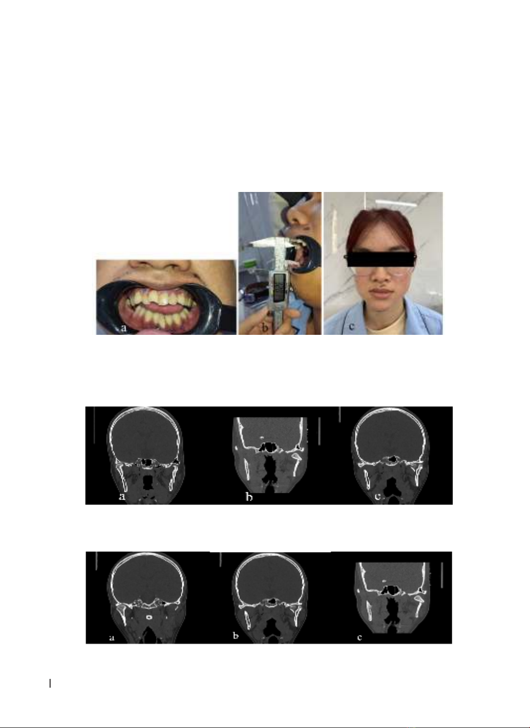
cause of fractures was traffic accidents (73.4%). The common clinical symptoms were malocclusion (96.7%)
with occlusal interferences on posterior teeth and limited mouth opening (83.3%). On CT Scan, condylar neck
fractures were the most common position (58.8%). There was a relationship between the side of deviated
mouth opening and the affected sides (p < 0.05). In cases of unilateral condyle fractures, there was a relation
between the side of premature contact on posterior teeth and the affected sides (p < 0.05). Conclusions:
Fractures of condylar neck was the most common fractures in the mandibular condyle, which resulted in
malocclusion, interferences on posterior teeth, limited and deviated mouth opening. There was a relationship
between deviated mouth opening and premature contacts on posterior teeth with the fractured side.
Keywords: Mandibular condyle fracture, clinical features, CT Scan.
Corresponding author: Nguyen Van Minh. Email: nvminh.rhm@huemed-univ.edu.vn
Recieved: 13/6/2023; Accepted: 20/8/2023; Published: 31/8/2023
DOI: 10.34071/jmp.2023.6.11
1. INTRODUCTION
The mandibular condyle is a component of the
mandible that contributes to the temporomandibular
joint. With its structure and position, the condyle
plays an important role in the masticatory function
and the growth of the mandible [1]. A fracture of the
mandibular condyle is not life-threatening, however,
it directly interferes the aesthetics and chewing of
the affected patient. If left untreated, it can lead to
complications such as joint dysfunction or stiffness,
impaired mandibular movement, and facial growth
disorders [2].
The incidence of condylar fractures varies among
studies, ranging from 17.5% to 52% of mandibular
fractures. In Vietnam, this rate is 14.03%, while in Hue,
it is reported to be 8.57% [3], [4]. The classification
systems of condylar fractures are relatively diverse.
Criteria for grouping fractures involves in fracture
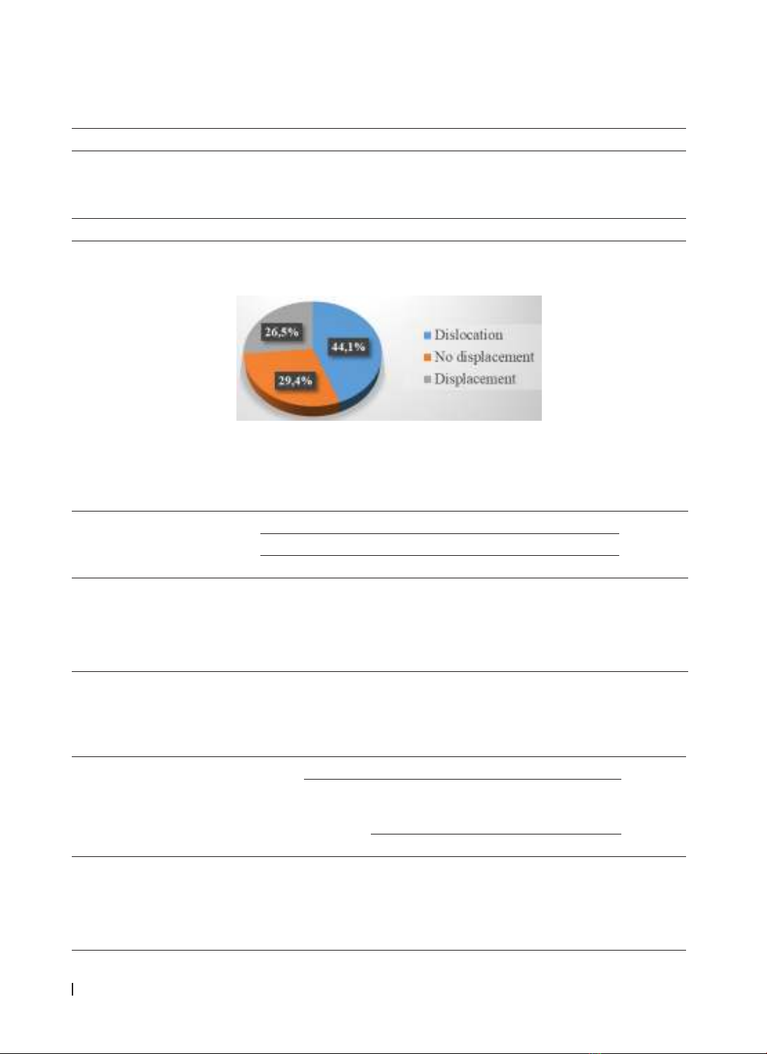
location, the relation between the condyle and the
glenoid fossa, affected side (unilateral or bilateral),
degree of displacement and whether other positions
of the mandible are fractured or not [5]. Clinical
symptoms commonly observed in patients with
condylar fractures of the mandible include tenderness
in the prearticular area, limited mouth opening,
deviated mouth opening, and malocclusion [6], [7],
[8]. A study of Duc Nguyen Quang (2022) reported
that limited mouth opening was present in all of the
patients, followed by malocclusion (74.7%). CT Scan
revealed that condylar neck fractures accounted for
65.2% of cases, and the percentage of combined
condylar fractures with other mandibular locations
was 77.9%. The ratio of unilateral to bilateral fractures
was 3.5:1 [9].
Although making an early diagnosis of
mandibular condylar fractures is not challenging,
it also requires a combination of clinical and
radiographic assessment to accurately determine
the location of the fractures. In order to enhance the
ability to diagnose early and precisely, we conducted
this study to assess clinical features, morphologies
on CT Scan, and analyze the relationship between
clinical characteristics and morphologies on CT Scan
of mandibular condyle fractures.
2. MATERIALS AND METHODS
2.1. Study design
We conducted a cross-sectional, descriptive study
on 30 patients diagnosed with mandibular condyle
fractures at the Department of ENT - Ophthalmology
- Odonto Stomatology, Hue University of Medicine
and Pharmacy Hospital from December 2021 to June