57
Journal of Medicine and Pharmacy, Volume 12, No.07/2022
Corresponding author: Vinh Khanh. Email: vkhanh@huemed-univ.edu.vn
Recieved: 12/10/2022; Accepted: 15/11/2022; Published: 30/12/2022
Research application of endoscopic ultrasound - fine needle aspiration
in diagnosis pancreas tumors
Vinh Khanh1*, Tran Van Huy1
Gastrointestinal Endoscopic Center, Hue University of Medicine and Pharmacy Hospital
Background: Pancreatic diseases is very multiform and complex, in which pancreatic tumors have often
poor prognosis, especially pancreatic cancer. Early detection and diagnosis of pancreatic tumors have great
significance in improving the quality of treatment and prognosis for patients. Endoscopic ultrasound has the
advantage of high-frequency ultrasound, an optimal approach to provide a possibility of EUS-FNA. This is
important evidence to confirm the diagnosis, guide to treatment and prognosis. This study was aimed at: (1)
To describe the characteristics of the pancreatic tumor by endoscopic ultrasound; (2) To evaluate the efficacy
and safety of endoscopic ultrasound fine needle aspiration in the diagnosis of pancreatic tumors. Subject
and methods: Cross-sectional study concludes 41 pancreatic tumor patients, which indicated endoscopic
ultrasound fine needle aspiration in Gastroenterology - Endoscopy Center, Hue University of Medicine and
Pharmacy Hospital from 2/2010 to 10/2022. Results: The size of the tumor was more than 2cm, tumors in
the pancreatic head accounted for 80.5% and solid tumors accounted for 80.5%. Besides, the main pancreatic
duct dilatation accounts for 39.0%, the biliary tract dilatation accounts for 46.3%, pancreatic tumor invades
adjacent organs accounts for 29.3%, vascular invasion accounts for 24.4%, with lymph nodes accounting
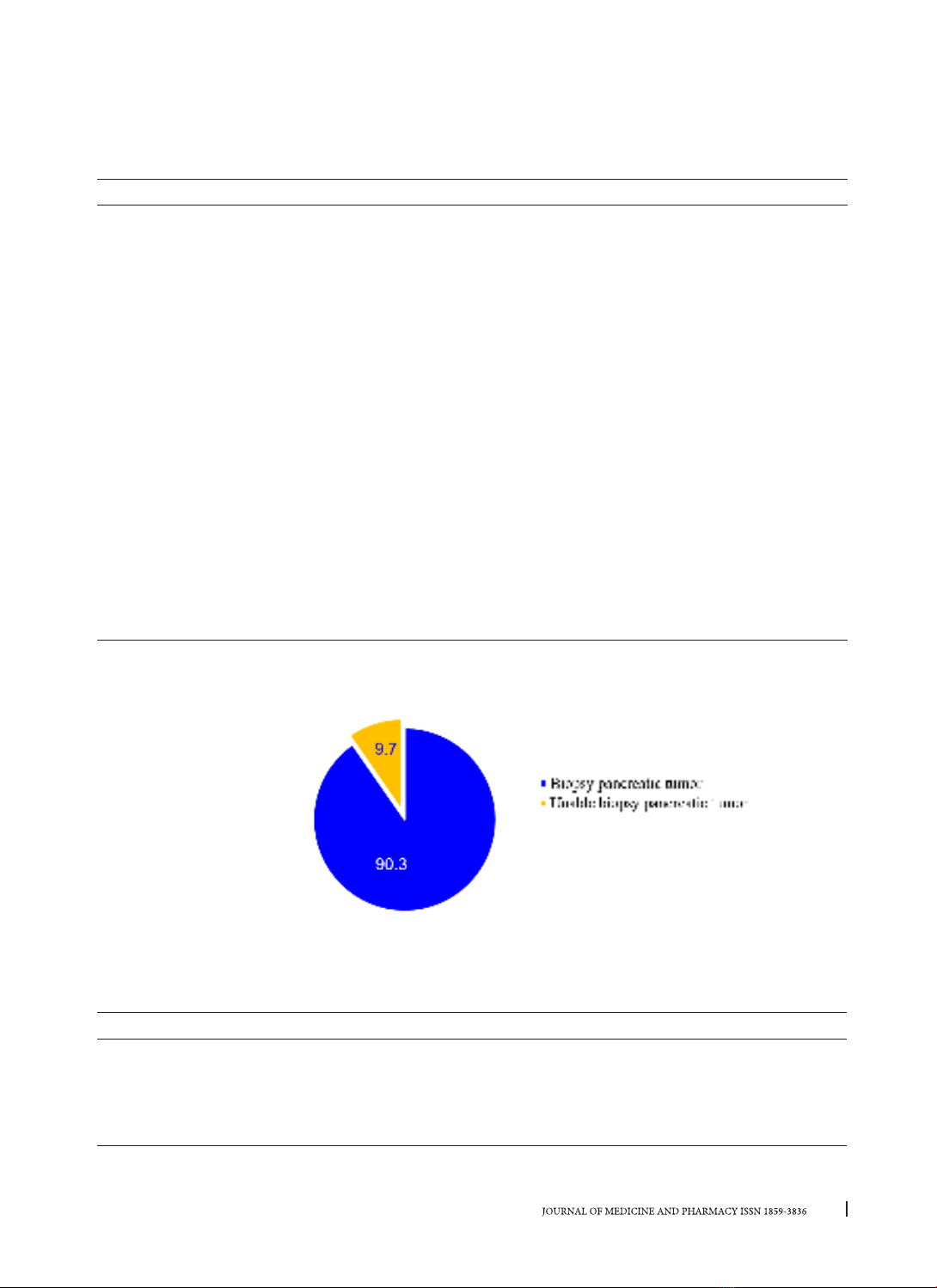
for 51.2%. Endoscopic ultrasound-guide fine needle aspiration pancreatic tumor was performed in 37/41
cases (90.3%). Pathological of pancreatic tumor: pancreatic cancer is highest about 59.5%, benign pancreatic
tumors accounted for 10.8%, mucinous cysts accounted for 5.4% and pancreatic tuberculosis accounted
for 2.7%. The complication rate of the procedure was 5.4%. Conclusion: Endoscopic ultrasound fine needle
aspiration pancreatic tumors showed relative safety and efficacy, the technical failure rate is very low.
Keywords: Pancreatic tumor, Endoscopic ultrasound fine needle aspiration.
1. INTRODUCTION
Pancreatic diseases are very diverse and complex,
in which pancreatic tumors have a very important
position, especially pancreatic cancer. Pancreatic
cancer is the seventh leading cause of cancer death
and one of the gastrointestinal cancers with the
worst prognosis [1]. Pancreatic cancer patients have
a 5-year survival rate lower than 10.0% even with
treatment [2]. Pancreatic tumor disease has often
asymptomatic in the early stages. Therefore, most
diseases are detected at a late stage due to treat
difficultly and have a poor prognosis [3]. Especially
for pancreatic cancer, if detected and treated early
(size 2cm), the survival rate over 5 years is quite high
(about 60.0%) [4]. Therefore, the pancreatic tumor
must detect and diagnose early which improves the
prognosis of the patients survival. Currently, there
are many methods to diagnose pancreatic tumors,
endoscopic ultrasound has the advantage of high-
frequency ultrasound, which has approached near
the tumor and can biopsy pancreatic tumors to
help diagnose the tumor. This is important evidence
to confirm the diagnosis, guide to treatment and
prognosis for patients. There are not many studies on
endoscopic ultrasound with fine needle aspiration to
diagnose pancreatic tumors in Vietnam, especially
in the central region. We made the study: Research
application of Endoscopic ultrasound- fine needle
aspiration in diagnosis pancreas tumors This study
was aimed at: (1) To describe the characteristics of
the pancreatic tumor by endoscopic ultrasound; (2)
To evaluate the efficacy and safety of endoscopic
ultrasound fine needle aspiration in the diagnosis of
pancreatic tumors.
2. SUBJECTS AND METHODS
2.1. Research subjects
Including 41 patients who treated at Hue
University of Medicine and Pharmacy Hospital from
2/2020 to 10/2022.
Criteria for choosing a disease
- There is a lesion in the pancreas or indirect
signs of pancreatic tumor on endoscopic ultrasound.
- All patients who perform endoscopic ultrasound
with fine needle aspiration
Exclusion criteria
- Patient does not agree to participate.
- Contraindications for upper gastrointestinal
DOI: 10.34071/jmp.2022.7.8
58
Journal of Medicine and Pharmacy, Volume 12, No.07/2022
endoscopy (heart failure, respiratory failure,
myocardial infarction…).
- Pyloric stenosis, duodenal stenosis.
- Coagulation disorders: Prothrombin ratio <50%,
INR >1.5.
- Platelet count: <50.000 G/L.
- Patients with contraindications to anesthesia.
2.2. Research Methods
Study design: Cross-sectional descriptive study
Data collected included patient demographics
(gender, age, and mass lesion location) and
procedure details (tumor characteristics and the
number of needle passes), pathological of tumor,
post-procedure complications were defined as
any symptoms requiring emergency department
evaluation, including bleeding, perforation,
pancreatitis and other severe complications.
The procedure of fine needle aspiration under
endoscopic ultrasound guidance was performed:
Step 1: Determine the lesion image at the
optimal location.
Step 2: Insert the needle through the biopsy
channel.
Step 3: Select the needle path into the lesion.
Step 4: Puncture the needle
Step 5: Moving the needle in the lesion
Step 6: Remove the needle from the endoscope [6].
Evaluation of results: Assessing the cytological
results according to Bellizzi’s standards [7].
+ No cells: only red blood cells, inflammatory
cells, and gastrointestinal tract cells.
+ Cell poverty: the number of cells is too small
to diagnose.
+ Benign cells: on the plate only benign cells.
+ Pancreatic cancer: enough cells are needed to
diagnose pancreatic cancer.
2.3. Statistical analysis
A multivariable binary logistic regression model
was created for variables that were statistically
relevant in the univariate analyses. p-values <0.05
were considered statistically significant. All statistical
analyses were conducted using SPSS Statistics.
3. RESULT
3.1. Patient characteristics
Table 1. Patient characteristics
Age, yr
Male Female Total
n%n%n%
≤ 40 1 5.0 3 14.2 4 9.8
41- 60 8 45.0 6 28.5 14 34.1
≥ 61 11 55.0 12 57.3 23 56.1
Total 20 100.0 21 100.0 41 100.0
Comment: The majority of patients were above 61 years old, which accounted for 55% of males and 57.3%
of females. The percentage of male patients was lower than females (48.7% versus 51.4%).
3.2. Characteristics of lesions by endoscopic ultrasound
Table 2. Characteristics of lesions by EUS
Characteristics n Rate %
Size (cm) ≥ 2 41 100.0
< 2 0 0,0
Location Head & neck 33 80.5
Body 6 14.6
Tail 2 4.9
Echoic Hypoechoic 40 97.6
Hyperechoic 1 2.4
Tumor border Regular 20 48.8
Irregular 21 51.2
Tumor structure Solid 33 80.5
Cystic 8 19.5
Total (%) 41 100.0
59
Journal of Medicine and Pharmacy, Volume 12, No.07/2022
Comment: The tumor detected by EUS was greater than 2 cm. Most of the lesions located in the pancreatic
head & neck region about 80.5%, hypoechoic about 97.6%, pancreatic tumor with irregular border accounting
for 51.2%, and solid structure accounting for 80.5%.
Table 3. Characteristics of beside of lesions by EUS
Characteristics n Rate %
Pancreatic parenchyma Atrophy 3 7.3
Normal 38 92.7
MDP dilation Yes 16 39.0
No 25 61.0
Pancreatic stone Yes 2 4.9
No 39 95.1
Tumor invades adjacent organs Yes 12 29.3
No 29 70.7
Tumor invades vessel Yes 10 24.4
No 31 76.6
Biliary tract dilation Yes 19 46.3
No 22 53.7
Gall bladder dilation Yes 10 24.4
No 31 75.6
Abdominal lymph nodes Yes 21 51.2
No 20 48.8
Total 41 100.0
Comment: the proportions of abdominal lymph nodes, main pancreatic duct dilatation, and biliary tract
dilatation were 51.2%, 39%, and 46,3% respectively
3.3. Efficacy and safety of EUS-FNA for diagnosing the pancreatic tumor
Figure 1. Tenichque of EUS - FNA for diagnosing pancreatic
Comment: In 41 patients were ordered to perform FNA, but only 37 patients were able to perform EUS
- FNA (90.3%).
Table 4. Characteristic of pancreatic tumor biopsy by endoscopic ultrasound
Characteristics n Rate %
Location Head & neck 29 78.4
Body 6 16.2
Tail 2 5.4
Total 37 100.0
60
Journal of Medicine and Pharmacy, Volume 12, No.07/2022
Tumor structure Solid 29 78.4
Cystic 8 21.6
Total 37 100.0
Comment: The most popular locations were the pancreatic head & neck (78.4%), followed by the pancreatic
body (16.2%). The pancreatic solid tumor accounted for 78.4%, the remainings were cystic tumors (21.6%).
Table 5. Characteristics and results of aspiration cytology
Characteristics n Rate %
Needle 19G 9 24.4
22G 28 75.6
Cytological No cells 2 5.4
Cell poverty 11 29.7
Benign cells 9 24.4
Pancreatic cancer 15 40.5
Pathological Yes 31 83.8
No 6 16.2
Total 37 100.0
Comment: Among 37 patients who are performing FNA, the 22G needle was used in 75.6% of cases, 24/37
cases have significant cytological results (64.9%) and 11/37 cases have poor cytological results (29.7%), 31/37
cases (83.8%) obtained adequate tissue samples for histopathology
Table 6. Pathological results
n Rate %
Pancreatic cancer 22 59.5
Benign pancreatic tumor 4 10.8
Mucinous cystic neoplasm 2 5.4
Pancreatic tuberculosis 1 2.7
Chronic pancreatitis 1 2.7
Unknown diagnosis 1 2.7
Small samples 6 16.2
Total 37 100.0
Comment: Pancreatic cancer was 22/37 (59.5%), 4 cases of benign pancreatic tumor (10,8%), 2 cases
of mucinous cyst neoplasm (5.4%), 1 case with pancreatic tuberculosis, chronic pancreatitis and unknown
diagnosis (2.7%) and 6 cases without clear pathology results due to small samples.
Table 7. Complication of EUS-FNA
Complication n (%)
Acute pancreatitis Yes 1 (2.7)
No 36 (97.3)
Bleeding Yes 1 (2.7)
No 37 (97.3)
Death Yes 0 (0.0)
No 37 (100.0)
Total 37 (100.0)
Comment: In most cases, EUS-FNA is performed without complications.
61
Journal of Medicine and Pharmacy, Volume 12, No.07/2022
4. DISCUSSION
4.1. Patient Characteristics
In our study, the majority of patients were above
61 years old, which accounted for 55% of males and
57.3% of females. The research from Okano and
Trinh Pham My Le also reported that the average
age of patients with pancreatic cancer was 65 and
63.5, respectively [8], [9].
The percentage of male patient was lower
than the female (48.7% versus 51.4%), included
pancreatic cancer and benign pancreatic tumors.
Several risk factors (such as alcohol consumption
and smoking), which are commonly seen in male
patients, have been proved to be related to
pancreatic disease, especially pancreatic cancer
[10].
4.2. Characteristics of lesions by endoscopic
ultrasound
In table 2, all of the tumors detected by EUS were
greater than 2 cm. The size of the lesions is a strong
predictor for malignancy, according to American
Joint Committee on Cancer 2017 [11]. In the report
of WHO, the size of pancreatic cancer ranges from
2.5 to 3.5 cm [12]. The advantages of EUS were
direct accessment, high-frequency probe, which
facilitate accessment small lesions. A recent meta–
analyses show that the sensitivity and specificity
were 92 - 100% and 89 - 100%, respectively, in
detecting malignant characteristics of pancreatic
tumors, especially small lesions [13]. Most of lesions
located in the pancreatic head region (80.5%), which
was a common site of pancreatic tumor and cancer, in
particularly. According to Meng et al, the proportion
of pancreatic tumor in the pancreatic head accounted
for 73.5% [14]. In addition, in our study, pancreatic
tumor with irregular border accounting for 51.2%
and solid structure accounting for 80.5% are the
features suggestive of malignancy on endoscopic
ultrasound.
In our study, the proportions of abdominal
lymph nodes, main pancreatic duct dilatation, and
biliary tract dilatation were 51.2%, 39%, and 46,3%
respectively. According to a study by Nguyen Truong
Son and Trinh Pham My Le on a group of pancreatic
cancer patients, the percentage of patients with
abdominal lymph nodes was 48.2% and 46.6%, with
the main pancreatic duct dilatation was 58.9% and
58.6%, with biliary dilatation was 55.4% and 37.9%,
respectively [8], [15].
4.3. Efficacy and safety of EUS-FNA for
diagnosing pancreatic tumor
In our study, 41 patients were ordered to
perform FNA pancreatic tumors, but only 37
patients were able to perform EUS - FNA. 4 patients
who could not go through the procedure were
those with a history of previous biliary-intestinal
anastomosis, so it was difficult to choose a favorable
site to access pancreatic tumors. Apart from general
contraindications of biopsy, the procedure also
depends on others factors. The procedure can be
performed step-by-step: the first step is to determine
the optimal location to approach the lesion, which
is the most important factor in deciding whether to
needle biopsy or not. The position is ideal if lesions
was visible at the largest size, the ultrasound probe
is closest to the lesion, especially there should be
nothing between the needle and the lesion (for
example: blood vessels, main pancreatic duct, and
biliary tract) [6].
In Table 4, 37/41 patients in our study had been
performed EUS-FNA. The most popular locations
that was accessible to the lesion was pancreatic head
(78.4%), followed by body of the pancreas (16.2%).
The pancreatic solid tumor accounted for 78.4%, the
remainings were cystic tumor (21.6%). Our results
are similar to that of Nguyen Truong Son et al.; 90.4%
of pancreatic tumors had been performed FNA in
the head and body of the pancreas, and 92.7% (n
= 51) cases were solid tumor [15]. With the ability
that almost directly approach the tumor, EUS-FNA
has great advantages in diagnosis and intervention
with small lesions under 2 cm [13].
Among 37 patients gone through the FNA, 22G
needle was used in 75.6% of cases. Technically, the
choice of needle depends on the location and the
nature of the lesions. Each type of needle did have
its pros and cons in approaching the lesions in the
pancreatic head and uncinate process, the ability
to puncture the tumor, the quality of the collecting
samples, and rate of adverse events. The 19G needle
obtains better tissue samples due to its large size,
but conversely, it is difficult to control and has more
complications when compared to the smaller 22G
needle, which is easy to control but has less tissue
samples. While the 25G needle is extremely safe but
is mainly used for obtaining cytology [16].
Our study uses two types of needles, 19G and
22G, in which the latter is used more. Both enable
us to not only obtain great tissue samples but also
ensure safety and control complications. The 19G
needle is preferable if the tumors are in easy-to-
access locations or previously usage 22G were
unable to obtain adequate sample. According to
Diogo T.H and CS (2020), the effectiveness of 19G