49
Journal of Medicine and Pharmacy, Volume 13, No.04/2023
Comparison of full-mouth and partial-mouth disinfection modalities
in nonsurgical periodontal treatment for periodontitis: a randomized
clinical trial in vietnam
Nguyen Thi Thuy Duong1,*, Tran Thi To Uyen1
(1) Faculty of Odonto-Stomatology, University of Medicine and Pharmacy, Hue University
Abstract
Background: Periodontitis is the most common form of periodontal disease, greatly affecting the aes-
thetics, function as well as patients quality of life. In the periodontal disease treatment strategy, nonsurgical
treatment is considered as the initial phase for anti-infection and soft-tissue management. Objective: This
study aims to compare the effect of two nonsurgical periodontal modalities: one-stage full-mouth and par-
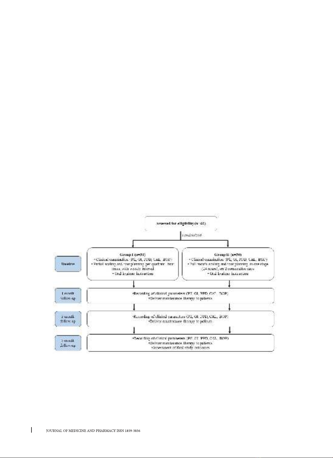
tial-mouth protocols for periodontitis. Materials and Methods: 60 patients with chronic periodontitis were
randomly allocated to 2 groups. Group I (n = 30) was treated according to partial-mouth therapy. Group II
(n = 30) was treated according to full-mouth disinfection therapy. Periodontal parameters were assessed at
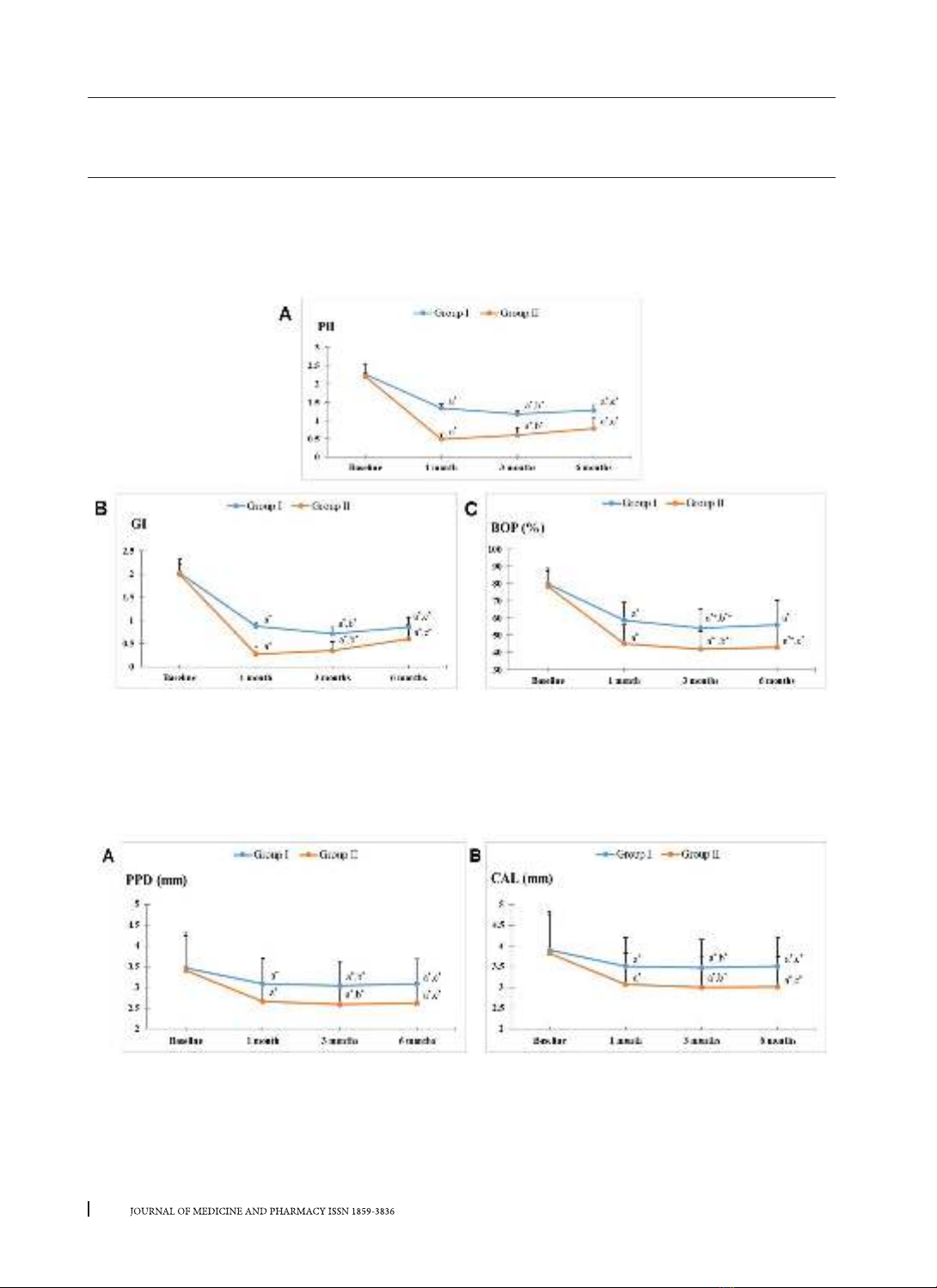
baseline and 1, 3, and 6 months, including plaque index, gingival index, periodontal probing depth, clinical
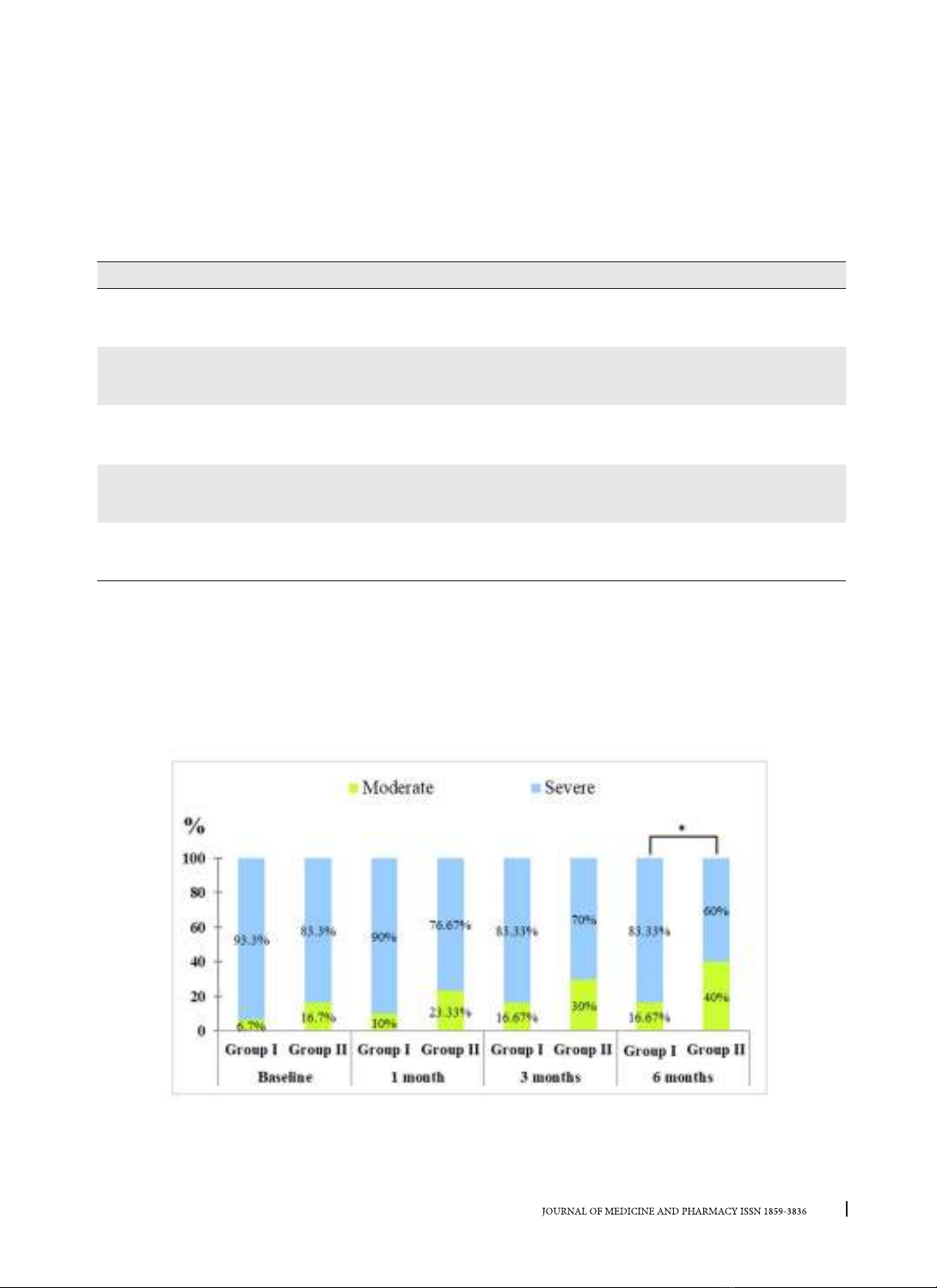
attachment loss, and bleeding on probing. Results: Both two treatment modalities resulted in significant
improvements in all clinical parameters over the entire duration of the study (p < 0.05). Full-mouth disinfec-
tion therapy showed significantly better improvements than the partial-mouth one during follow-up times
(p < 0.05). Conclusion: Nonsurgical periodontal treatment has positive effects on controlling periodontitis.
Full-mouth therapy shows clinical benefits over partial-mouth therapy in improving periodontal conditions.
Keywords: Periodontitis, nonsurgical periodontal therapy, full-mouth therapy, partial-mouth therapy.
Corresponding author: Nguyen Thi Thuy Duong, email: nttduong@huemed-univ.edu.vn
Recieved: 6/2/2023; Accepted: 5/5/2023; Published: 10/6/2023
1. INTRODUCTION
Periodontitis, the most frequent periodontal
disease in adults, is characterized by connective
tissue attachment loss and the resorption of coronal
alveolar bone due to dental plaque accumulation [1].
To treat and control periodontitis, nonsurgical therapy
has been considered as the priority treatment. The
objective of periodontal treatment is the reduction
and elimination of microbial load, and the removal
of dental plaque and calculus through scaling and
root planing (SRP) therapy [2]. According to partial-
mouth nonsurgical therapy, SRP is performed per
jaw quadrant at a 1- to 2-week interval [3]. This
protocol requires at least 4 appointments, thus time-
consuming for both patients and dentists. However,
most bacterial species exist not only in periodontal
pockets but also colonize several other oral niches
and the oropharyngeal area, such as the mucosa,
the tongue, the tonsils, and the saliva. They could
be transmitted from one of their niches to the
subgingival environment, leading to the reinfection
of treated periodontal pockets. Therefore, during
the time intervals of this therapy, the treated
pockets may be reinfected by the untreated pockets
[4]. To reduce this reinfection, Quirynen M. et
al. (1995) introduced the one-stage full-mouth
(OSFM) disinfection protocol that involves the use
of antiseptics (Chlorhexidine). The scaling and root
planing are conducted in two visits within 24 hours
with the use of Chlorhexidine solution and gel. The
OSFM disinfection showed a significantly higher
reduction of pocket depth and fewer pathogenic
organisms at one month recall, as compared to
partial therapy [5]. Regardless of Chlorhexidine
(CHX) use, the one-stage full-mouth scaling and root
planing showed more favorable reactions in patients
and clinical improvement in a long time follow-
up study [6]. Moreover, several studies supported
these clinical observations based on the reduction of
microbiology in the OSFM group [7],[8].
In Vietnam, previously published research were
conducted with partial-mouth SRP protocol in 1-2
visits or full-mouth modality without intensive
disinfection [9], [10]. Moreover, the term one-stage
full-mouth disinfection was not considered yet and
the effects of this modality on treating Vietnamese
periodontitis patients were not clarified. Therefore,
the present study aims to assess and compare two
nonsurgical treatment protocols: full-mouth therapy
and partial-mouth one.
50
Journal of Medicine and Pharmacy, Volume 13, No.04/2023
2. MATERIALS AND METHODS
Ethical approval
This study was conducted from June 2019 to
June 2021 in full accordance with the Helsinki
Declaration of 1975, as revised in 2000. The
protocol and the informed consent form were
reviewed and approved by the Institutional Ethics
Committee of Hue University of Medicine and
Pharmacy, Hue University, Hue, Vietnam (Number:
H2019/339). Sixty participants were recruited from
Hue University of Medicine and Pharmacy Hospital.
It was mandatory for consent participants have
to read and signed the consent form before being
included in the present study.
Study population
Participants were recruited from patients who
came for dental examination and were diagnosed
with chronic periodontitis.
The inclusion criteria were as follows:
- ≥ 18 years of age;
- Clinical signs of mild, moderate, and severe
periodontitis according to the 2014 update
Classification of Periodontal Disease by the American
Academy of Periodontology; [11]
- Having ≥ 20 teeth present on the dental arch;
- Commitment to participate in the study and
follow-up visits.
Exclusion criteria included:
- Teeth with combined endodontic and
periodontal lesions;
- Current smoking;
- Pregnancy and lactation;
- Having systemic diseases;
- Use of antibiotics or anti-inflammatory drugs
within 3 months of the start of the study;
- Periodontal therapy, including nonsurgical and
surgical, in the 6 months preceding the start of the
study.
After obtaining informed consent, the selected
subjects were randomly distributed into two
nonsurgical treatment groups:
- Group I (30 patients): subjects who received
the partial-mouth therapy.
- Group II (30 patients): subjects who received
the full-mouth therapy.
The patients were periodontally examined before
treatment (baseline) and after treatment (1-month,
3-month, and 6-month recall visits) (Figure 1).
Figure 1. Schematic diagram of the study design
Nonsurgical treatment
Group I (30 patients) received partial-mouth periodontal therapy. Group II (30 patients) received full-
mouth scaling and root planing. Both 2 groups received standard oral hygiene instructions at each visit.
+ Scaling and root planing: The sessions of SRP were performed by the same investigator using standard
periodontal curettes. The time needed per quadrant was approximately 1 hour.
- In Group I, the SRP process began in the upper right quadrant and continued clockwise, each quadrant
was treated at each appointment with an interval of 1 week.
51
Journal of Medicine and Pharmacy, Volume 13, No.04/2023
- In Group II, SRP was completed in two vis-
its within 24 hours, starting from the lower jaw.
During SRP, additional disinfection was sought by:
1) brushing the tongue dorsum (by the patient) for
60 seconds with a Chlorhexidine 1% gel; 2) rinsing
twice with Chlorhexidine 0.2% solution for 1 minute
(during the last 10 seconds patients had to gargle
in an attempt to reach the tonsils); 3) subgingival
irrigation of all the pockets 3 times within 10 min-
utes with a Chlorhexidine 1% gel. Additionally, the
subjects of this group were instructed to rinse twice
daily for 60 seconds with a 0.2% solution of Chlor-
hexidine for 2 months.
+ Oral hygiene instruction: Patients from both
groups received oral hygiene instruction at the first
visit for nonsurgical treatment and other recall ap-
pointments. All patients have been instructed on
toothbrushing (with the Bass technique), brushing
of the tongue dorsum (twice a day), and interdental
plaque control (by interdental brushes or dental floss).
Periodontal parameters assessment
At baseline, 1 month, 3 months, and 6 months
after treatment, the following clinical parameters
were recorded including probing pocket depth
(PPD) and clinical attachment loss (CAL) at six sites
per tooth. Plaque index (PlI), gingival index (GI), and
bleeding on probing (BOP) were recorded at four
sites per tooth. Clinical parameters were carried
out on a dental chair by a pre-calibrated examiner
who was blinded to the study with the appropriate
armamentarium.
Statistical Analysis: all data were collected in
an Excel database and analyzed by SPSS version
26.0. A p-value of <0.05 was considered statistically
significant.
Categorical variable like gender is expressed as
proportion and compared by use of a Chi-squared
test.
Quantitative variables were presented as mean
± standard deviation. The Student t-test (two-tailed,
independent) was used to find the significance of
study parameters on a continuous scale between
two groups (intergroup analysis). The Student
t-test (two-tailed, dependent) was used to find the
significance of study parameters on the continuous
scale within each group. The Mann-Whitney U-test
was used to find the significance of nonparametric
parameters probing depth between two groups and
the Wilcoxon signed-rank test was used to find the
significance of probing depth (nonparametric) in
paired conditions.
3. RESULTS
The study consisted of 22 men and 38 women.
The average age of subjects participating in the study
was 55.52 ± 12.95 years. No significant differences
were found between the 2 treatment groups (full-
mouth therapy and partial-mouth therapy) for all
participants (n=60) regarding the variables of sex,
age, and periodontitis severity (p>0.05).
The clinical parameters (PlI, GI, PPD, CAL, and
BOP) of both groups at baseline, 1 month, 3 months,
and 6 months are shown in Table 1, Figure 2, and
Figure 3.
Table 1. Comparison of clinical parameters at baseline, 1st month, 3rd month, and 6th month between
two groups
Clinical parameters Group Baseline
(Mean ± SD)
1 month
(Mean ± SD)
3 months
(Mean ± SD)
6 months
(Mean ± SD)
PlI
Group I 2.26 ± 0.27 1.34 ± 0.11 1.18 ± 0.10 1.28 ± 0.15
Group II 2.21 ± 0.35 0.49 ± 0.15 0.60 ± 0.21 0.78 ± 0.29
p*> 0.05 < 0.05 < 0.05 < 0.05
GI
Group I 2.02 ± 0.20 0.87 ± 0.09 0.71 ± 0.15 0.86 ± 0.21
Group II 1.99 ± 0.33 0.27 ± 0.15 0.35 ± 0.19 0.60 ± 0.30
p*> 0.05 < 0.05 < 0.05 < 0.05
PPD
(mm)
Group I 3.46 ± 0.78 3.08 ± 0.61 3.04 ± 0.59 3.08 ± 0.60
Group II 3.41 ± 0.93 2.66 ± 0.52 2.59 ± 0.47 2.61 ± 0.49
p*> 0.05 < 0.05 < 0.05 < 0.05
CAL
(mm)
Group I 3.89 ± 0.84 3.51 ± 0.69 3.47 ± 0.68 3.50 ± 0.70
Group II 3.82 ± 1.00 3.07 ± 0.74 3.00 ± 0.72 3.01 ± 0.72
p*> 0.05 < 0.05 < 0.05 < 0.05
52
Journal of Medicine and Pharmacy, Volume 13, No.04/2023
BOP
(%)
Group I 79.83 ± 7.44 58.63 ± 10.53 54.30 ± 11.03 56.03 ± 14.31
Group II 78.53 ± 10.45 45.03 ± 10.99 41.90 ± 10.30 42.97 ± 12.93
p*> 0.05 < 0.05 < 0.05 < 0.05
*p-value obtained with the Mann-Whitney U-test for PPD and the independent samples t-test for the other
variables for the intergroup comparison at the same time point.
PlI: plaque index; GI: gingival index; PPD: probing pocket depth; CAL: clinical attachment loss; BOP:
bleeding on probing; group I: partial treatment; group II: full-mouth disinfection.
Figure 2. Changes in plaque index (PlI), gingival index (GI), and bleeding on probing (BOP) in two treatment
groups after 1, 3, and 6 months. *: p<0.05 with paired sample t-test; a: compared to baseline; b: compared to
1st month; c: compared to 3rd month.
Figure 3. Changes in pocket depth (PPD) and clinical attachment loss (CAL) in 2 treatment groups after 1,
3, and 6 months. *: p < 0.05 with paired sample t-test; a: compared to baseline; b: compared to 1st month;
c: compared to 3rd month.
53
Journal of Medicine and Pharmacy, Volume 13, No.04/2023
At the first month follow-up, the results showed a significant reduction in all clinical parameters of both
groups (p<0.05). After 3 months, all recorded indexes of Group I continued to decrease (p<0.05). However, the
PlI and GI indexes of Group II illustrated a slight increase again, as compared to those in 1st month (p<0.05). For
the 6th-month assessment, most of the clinical parameters of both groups showed an increase in comparison
with the 3rd-month records (p<0.05). Only the BOP index of Group I at this time showed insignificant change
(p>0.05). Regarding the change of clinical parameters in two groups after treatment, all values at 3 assessed
point times show reduction compared to baseline (Table 2).
Table 2. Reduction of clinical parameters in two groups after treatment compared to baseline
Clinical parameters Group 1 2 3
PlI
Group I - 0.92 - 1.08 - 0.98
Group II - 1.72 - 1.61 - 1.43
GI Group I - 1.15 - 1.31 - 1.16
Group II - 1.72 - 1.64 - 1.39
PPD (mm)
Group I - 0.38 - 0.42 - 0.38
Group II - 0.75 - 0.82 - 0.80
CAL (mm)
Group I - 0.38 - 0.42 - 0.39
Group II - 0.74 - 0.81 - 0.80
BOP (%)
Group I - 21.20 - 25.53 - 23.80
Group II - 33.50 - 36.63 - 35.56
123: mean change of clinical parameters at 1st month, 3rd month, 6th month respectively, compared to baseline.
At baseline, there are no statistically considerable differences in periodontal parameters between the
two groups (p > 0.05). After treatment, the clinical indexes of Group I showed higher results as compared to
Group II at all recall visits (p < 0.05).
Following the AAP classification in 2014, the periodontitis severity of 60 participants showed a similar
distribution of moderate and severe status at baseline, 1 and 3-month follow-up (p>0.05) (Figure 4). However,
at the final assessment (6 months), the severe periodontitis percentage of Group II (60%) is lower than that
of Group I (83.3%) significantly (p<0.05).
Figure 4. Distribution of periodontitis severity in two groups at baseline, 1 month, 3 months, and 6 months
post-operation.* p < 0.05 with Chi-square test.