JOURNAL OF MILITARY PHARMACO-MEDICINE N04 - 2025
213
EVALUATION OF GASTRIC CONDUIT PERFUSION RESULTS
BASED ON REAL-TIME INDOCYANINE GREEN FLOW SIGNAL
IN THORACOSCOPIC SURGERY FOR ESOPHAGEAL CANCER
Nguyen Van Tiep1*, Le Thanh Son1, Nguyen Anh Tuan2
Abstract
Objectives: To evaluate gastric conduit perfusion (GCP) images based on
indocyanine green (ICG) flow signal timing in thoracoscopic surgery for
esophageal cancer (EsC). Methods: A cross-sectional descriptive study was
conducted on 70 patients who were applied ICG to evaluate GCP during
thoracoscopic surgery to treat EsC at 108 Military Central Hospital and Military
Hospital 103 from June 2022 to June 2024. Results: The mean age was 59.0 ± 7.9
(32 - 71) years old; 100% were male. The anastomotic leak rate was 7.1%, with a
mean gastric conduit (GC) width of 5.1 ± 0.2cm. Through ICG imaging, 17
patients with missing GC were detected. The average ischemic GC length was 2.7
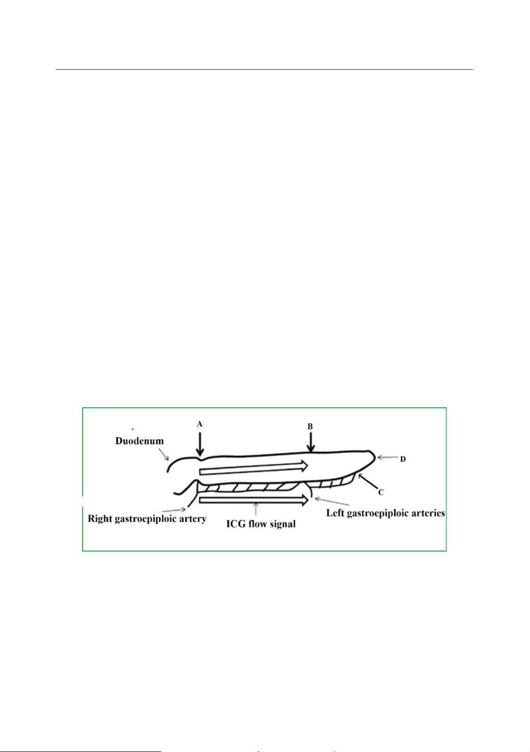
± 0.6cm. The time of appearance of the ICG signal in segments (B-C) and
segments (A-D) of the anastomotic leak group was longer than that of the group
without anastomotic leak (p < 0.05). Multivariable logistic regression analysis
found that the greater the rate of ischemic GC, the higher the anastomotic leak rate
(OR = 59.27; 95%CI= 1.25 - 2802.03; p = 0.04). Conclusion: Evaluation of GCP
images based on ICG flow signal timing is feasible, safe, and objective. ICG
current signal timing helps detect the location of the poorly perfused GC and select
the appropriate location to create the anastomosis with a low anastomotic leak rate.
Keywords: Esophageal cancer; Gastric conduit; Indocyanine green.
1Digestive Surgery Center, Gastrointestinal Surgery Department, Military Hospital 103,
Vietnam Military Medical University
2Department of Gastrointestinal Tract Surgery, 108 Military Central Hospital
*Corresponding author: Nguyen Van Tiep (chiductam@gmail.com)
Date received: 23/6/2024
Date accepted: 17/9/2024
http://doi.org/10.56535/jmpm.v50i4.872
JOURNAL OF MILITARY PHARMACO-MEDICINE N04 - 2025
214
INTRODUCTION
Anastomotic leak is a common
complication after esophagectomy for
EsC, with a rate of about 5 - 25% [1, 2].
Factors related to anastomotic leak
include patients’ nutritional status, the
tunnel where the GC is placed, the
location or technique of anastomosis,
and the tension of the anastomosis.
Among them, ischemia of the GC is an
important factor causing anastomotic
leak and anastomotic stenosis [3].
Around the world, the near-infrared
fluorescence method using ICG has
been applied as a valuable, objective
tool in surveying and evaluating the
perfusion status of the GC. To evaluate
GCP, the authors used one of the
following three methods: ICG signal
duration, flow rate, and ICG current
intensity in the GC. Many authors rely
on ICG flow signal duration as an
objective index to evaluate GCP. Noma
et al. reported that if the GC blood
supply is enhanced with ICG within 20
seconds, the GC is said to be well
perfused at that location, and anastomosis
performed in this area will ensure good
[4]. Kumagai et al. proposed the "90-
second rule" to confirm good perfusion
of the GC. All anastomoses performed
in the area of the ICG-enhanced GC
within 90 seconds of the ICG signal
appearing at the origin of the right
gastric omental artery [5]. EM de Groot
demonstrated that in patients with
anastomotic leakage, the mean time to
reach maximum ICG intensity at the tip
of the GC was 56 seconds (ranging
from 30 - 83 seconds) compared with
34 seconds (ranging from 12 - 66
seconds) in patients without anastomotic
leakage [6]. In Vietnam, thoracoscopic
esophagectomy is performed routinely
in many central hospitals, but there has
not been any work or research reported
on the use of ICG to evaluate GCP.
Therefore, we conducted this study to:
Evaluate the results of GC blood supply
based on ICG flow signal timing in
thoracoscopic surgery for EsC.
MATERIALS AND METHODS
1. Subjects
Including 70 EsC patients who
underwent thoracoscopic esophagectomy
and ICG imaging to evaluate the blood
supply of the replacement GC for EsC
from June 2022 to June 2024.
* Inclusion criteria: Diagnosis of
carcinoma in the thoracic esophagus
and undergoing thoracoscopic radical
esophagectomy for the preoperative
stage: cT1b-cT2, N0 or cT1b-cT3,
N+ after preoperative chemotherapy;
undergoing ICG imaging for evaluation
of the blood supply of the replacement
GC; ASA-PS 3.
JOURNAL OF MILITARY PHARMACO-MEDICINE N04 - 2025
215
* Exclusion criteria: Patients undergoing
thoracoscopic esophagectomy for non-
cancerous causes or those with tumor
invasion at the T4b level, according
to the American Joint Committee on
Cancer classification.
2. Methods
* Study design: A cross-sectional
descriptive study.
* Surgical procedure using ICG to
assess GCP:
Thoracoscopic radical esophagectomy
and 2-region lymph node dissection
were applied to all patients.
Thoracic step: Thoracoscopic surgery.
Abdominal surgery: Laparoscopic or
open surgery was possible in cases with
a history of previous abdominal surgery.
* Technique to create a large GC:
The reconstructed GC was approximately
5cm wide, retaining the right and left
gastroepiploic arteries, some of the first
branches of the right gastric artery, and
the arcade between the right and left
gastroepiploic arteries.
The GC was created using an open
stapler with open surgery, assisted
laparoscopic surgery, and an endoscopic
stapler with laparoscopic surgery.
* Fluoroscopy uses ICG to evaluate GCP:
Measure the dimensions of the GC with a tape measure:
Figure 1. Image of marking positions to measure GC dimensions.
Source: According to Kazuo Koyanagi in 2016 [7].
Point (A) is the pylorus;
Point (B) is the location of the
connection between the right and left
gastroepiploic arteries;
Point (C) is the last point of the
pulsatile GC to the final tip of the
stomach;
Point (D) is the final point of the GC.
JOURNAL OF MILITARY PHARMACO-MEDICINE N04 - 2025
216
The camera had a near-infrared light
source placed in front of the GC at a 3 -
5cm distance to ensure clear recorded
images.
The injection technique in the study
was applied according to author
Rao-Jun Luo (2020) [5] as follows:
Injection dose: 2.5mg ICG injection/
1 time.
Injection site: Can be injected into a
central vein (right external jugular vein)
or peripheral vein (right cephalic vein).
Injection time: Rapid bolus injection
of ICG in about 3 - 5 seconds.
Number of injections: 1st ICG injection:
After completing the shaping of the GC.
The purpose is to evaluate the GCP
status when the shaping is completed.
Second ICG injection: Recheck GCP
after the GC is inserted through the
posterior mediastinal tunnel to the neck.
The time to appear ICG was
calculated when the blue signal begins
to appear at point a (origin of the right
gastroepiploic artery).
Based on the time of ICG appearance
calculated from point a to the distal end
of the GC in the arterial phase at the 1st
injection.
The GC segment was considered to
be well perfused when the time to
appear ICG is < 60 seconds from point
A. The GC segment was considered
poorly perfused when the time to appear
ICG is 60 seconds from point A.
* Data analysis: All statistical
analyses were performed using SPSS
software (version 26.0, 64-bit from
IBM Corporation, NY, USA).
3. Ethics
This study was conducted in accordance
with the declaration of Helsinki, approved
by the Ethics Council of Military
Hospital 103, on December 9th, 2022,
approval number 193/CNChT - HĐĐĐ.
Participants fully agreed and voluntarily
participated in the study. Written
informed consents were obtained with
full signatures. Military Hospital 103
granted permission for the use and
publication of the research data. The
authors declare to have no conflicts of
interest in the study.
RESULTS
70 patients participated in the study
from June 2022 to June 2024. The average
age was 59.0 ± 7.9 (32 - 71) years old;
100% were male. ASA = 2, accounted
for 64.3%. ASA = 3, accounted for
35.7%. Patients received preoperative
chemotherapy and radiotherapy,
accounting for 77.1%. The anastomotic
leak rate was 7.1% (5 patients)
* Characteristics of GC esophageal
replacement used in research:
GC width: 5.1 ± 0.2cm; GC length
(AD): 31.4 ± 1.3cm; length of GC
segment (AB): 18.9 ± 1.3cm; length of
GC segment (AC): 29.1 ± 1.2cm; length
of GC segment (BC): 10.2 ± 1.0cm.
JOURNAL OF MILITARY PHARMACO-MEDICINE N04 - 2025
217
Table 1. Real-time ICG flow signals in the GC (arterial phase).
Real-time ICG flow signals
in the GC (arterial phase)
N0 of
patients
Min
Max
Mean ± SD/
Median (Q1-Q3)
First injection
Segment (A-B) (sec)
70
3
14
6.2 ± 2.0
Segment (A-C) (sec)
70
9
32
18.4 ± 5.1
Segment (A-D) (sec)
70
14
120
24.0 (21.0 - 59.3)
70
5
24
12.2 ± 4.2
70
2
105
6.0 (4.0 - 40.3)
Second injection
70
12
32
18.9 ± 4.6
70
17
120
25.0 (22.0 - 48.0)
70
1
105
7.0 (5.0 - 31.3)
When injecting ICG for the first time, the GC immediately after shaping, the
time to appear ICG in the entire GC (A-D) was 24.0 (21.0 - 59.3) seconds. The
appearance time of ICG segments (A-C) and (B-C) were 18.4 ± 5.1 seconds and
12.2 ± 4.2 seconds, respectively. When the GC was placed in the posterior
mediastinal tunnel, the ICG appearance time of the entire GC (A-D) and segment
(A-C) was 25.0 (22.0 - 48.0) seconds and 18.9 ± 4.6 seconds, respectively.
Table 2. Comparison of determining the GC with poor blood supply between
visual observation and ICG fluorescence imaging.
Perfusion of the GC
ICG imaging
Total
n (%)
No
Yes
Visual observation
(The first observation)
No
53
8
61 (87.1)
Yes
0
9
9 (12.9)
Visual observation
(The second observation)
No
53
8
61 (87.1)
Yes
0
9
9 (12.9)
Total
53
17
70 (100)
Management of the ischemic portion of the GC observed by ICG imaging
Excision of the ischemic portion of GC
17 (24.3)
Suture burying the ischemic part of GC
0
The average length of the GC that was removed (cm): 2.7 ± 0.6 (2 - 4)