
147
JOURNAL OF MEDICAL RESEARCH
JMR 184 E15 (11) - 2024
Corresponding author: Do Thu Thao
Hanoi Medical University Hospital
Email: dothuthao.nm@gmail.com
Received: 20/09/2024
Accepted: 13/10/2024
I. INTRODUCTION
INTRATHYROIDAL PARATHYROID CYST PRESENTING WITH
RECURRENT KIDNEY STONES: A CASE REPORT
Do Thu Thao1,2,, Nguyen Thi Thanh Huong1,2
1Hanoi Medical University Hospital
2Hanoi Medical University
Parathyroid cysts are rare and can be the cause of persistent hypercalcemia leading to kidney stones.
The aim of this article is to present a case presenting to the hospital with recurrent kidney stones and
incidentally found hypercalcemia. From there, we discovered a cystic lesion in the thyroid gland. Combining
ultrasound and cyst fluid aspiration to measure the concentration of parathyroid hormone in the fluid
helped us confirm that the cystic lesion belonged to the parathyroid gland. The patient underwent surgery
to remove the cyst and the blood calcium and parathyroid hormone levels returned to normal after surgery.
Keywords: Parathyroid cyst, parathyroid hormone, surgery.
Parathyroid cysts are rare lesions, which
represent less than 0.5% of parathyroid glands
pathologies and account for only 1 - 5% of neck
masses.1-3 They are classified as functioning
and nonfunctioning cysts.4,5 Cysts are common
in women and are usually asymptomatic.
Parathyroid cysts are usually detected by
ultrasound imaging, but they are easily confused
with thyroid cysts. Cyst fluid aspiration and
detection of parathyroid hormone in the cyst fluid
help diagnose parathyroid cysts.4 Parathyroid
cyst removal surgery is the optimal method. We
report a case of a patient with recurrent kidney
stones due to persistent hypercalcemia. We
found a cyst located in the lower third of the left
thyroid lobe, which was difficult to differentiate
from a thyroid or parathyroid lesion. Cyst fluid
aspiration revealed a very high concentration of
parathyroid hormone in the cyst fluid, confirming
a parathyroid cystic lesion. This is also different
from the commonly reported diagnostic
approach of a solid parathyroid tumor.
II. CASE REPORTS
A 65-year-old woman came to the hospital
because of dull back pain for 2 months.
She had a history of 3 times kidney stone
surgery. About 2 months, the patient had dull
back pain without fever, painful urination,
or urinary frequency. Her vital signs were
normal. Abdominal examination showed mild
tenderness in the flanks bilaterally,with no
abdominal wall reaction. Examination of the
cardiovascular, pulmonary, neurological, and
peripheral systems was normal.
The patient was admitted in the hospital
and examination revealed that the right kidney
stones and ureteral stones on both sides caused
dilation of the ureteral calyces. Biochemical
examination at the clinic showed elevated serum
calcium (3.1 mmol/l). Parathyroid hormone also
increased to 79.73 pmol/l.
A neck ultrasound did not detect parathyroid
adenomas in four common locations. The left
lobe thyroid gland in the lower third had a
partially cystic nodule, consisting of a solid and
a fluid, 4.5x3.0cm in size, developing mainly in
148
JOURNAL OF MEDICAL RESEARCH
JMR 184 E15 (11) - 2024
the lower and mediastinum. The patient’s chest
radiograph was normal.
A neck computed tomography (CT) scan
showed that the position behind the left lobe
of the thyroid gland had a heterogeneous
density; after injection, the enhancement was
uneven measuring 4.8x3.9x4.0cm. The border
of the left lobe of the thyroid gland and the
left wall of the esophagus was not clear. The
tumor was well confined to the larynx, the left
carotid space, and the great vessels in the
superior mediastinum. It displaced the larynx
and trachea and the head of the esophagus to
the right. The mass had partially extended into
the superior mediastinum and left vocal cord
paralysis was detected.
We suspect that this mass, located in the
lower third of the left lobe of the thyroid gland,
may be a parathyroid tumor. The fluid mass
occupied > 50% of the tumor volume, so we
performed fine-needle aspiration to collect
the fluid and measured the PTH. An elevated
PTH result (> 530 pmol/l) confirmed that it was
a parathyroid cyst. The results of parathyroid
Tc99m scintigraphy were commensurate with
the increased radioactivity localized in the left
lobe of the thyroid gland.
The tumor was surgically removed. WE
observed that the tumor had many fibrous
tissues attached around and to the left recurrent
laryngeal nerve. The immediate histopathology
also confirmed that the tumor was a parathyroid
tumor. Preoperative parathyroid hormone was
89.39 pmol/l; PTH measured at 20 minutes
after tumor removal decreased to 18.39 pmol/l.
PTH at 5 days after surgery returned to normal
at 4.1 pmol/l. Serum calcium returned to
normal.The final pathology was a parathyroid
adenoma. The patient was present on the 5th
postoperative day.
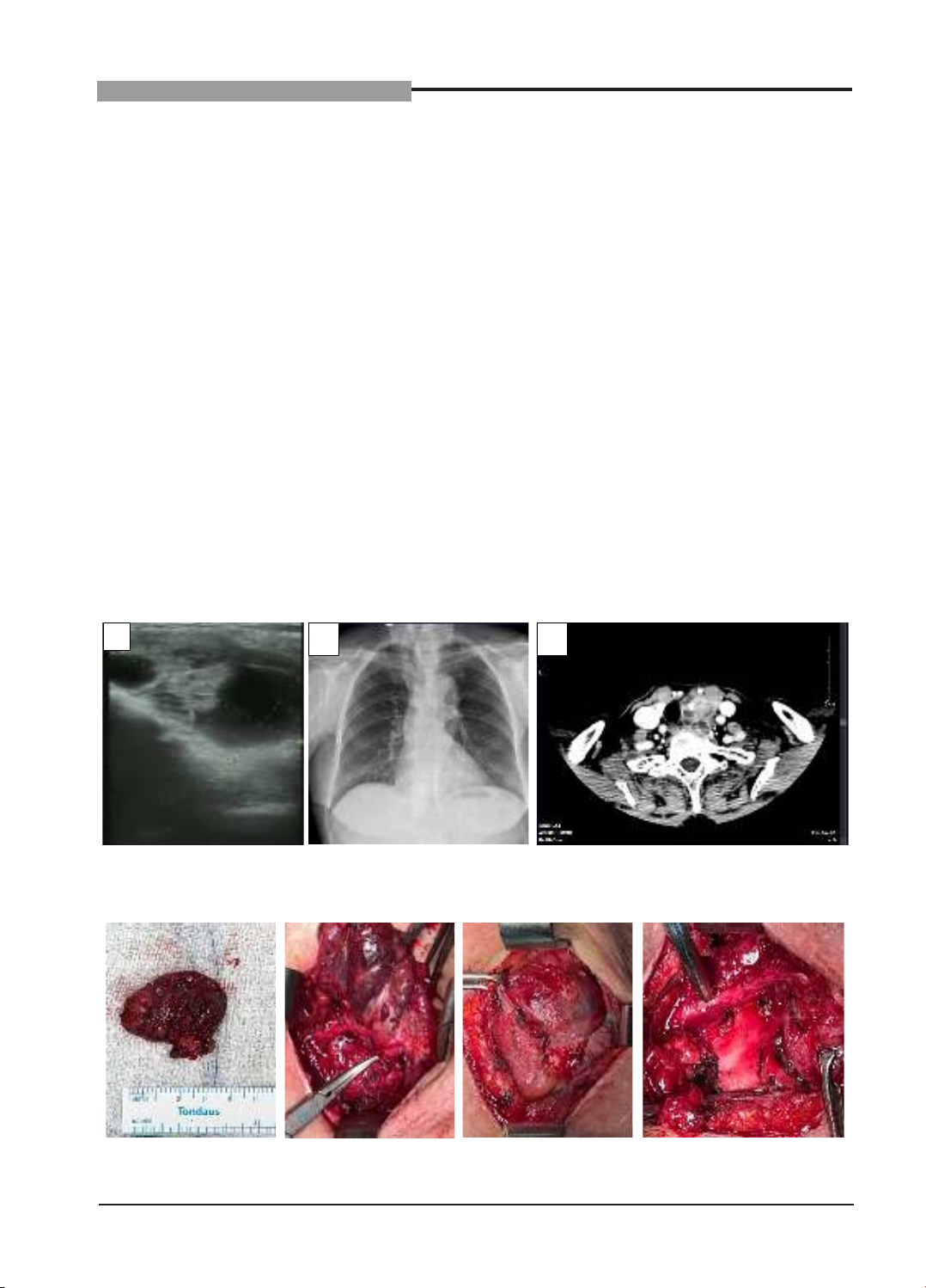
A
B
C
Figue 1. (A) Ultrasound image of the neck region. (B) Chest radiograph. (C) CT scan of the
neck: an 4.8x3.9x4.0cm mass was found in the left lobe of the thyroid gland
Figue 2. Open surgery to remove the tumor, the tumor has fibrous tissue attached to the
surrounding area, while removing the tumor while preserving the left recurrent laryngeal nerve

149
JOURNAL OF MEDICAL RESEARCH
JMR 184 E15 (11) - 2024
III. DISCUSSION
Parathyroid cysts are rare lesions. They
were first described by Sandstrom in 1880.6
DeQuervain published the first case of a
mediastinal PC in 1925, whereas the first
functioning parathyroid cysts was described by
Greene in 1952.7,8 They are subdivided into two
kinds: non-functioning and functioning.
Most of tumors have no symptom and are
discovered incidentally. Most patients have
clinical signs when the tumor enlarges, causing
compression. The larger cysts can cause
compressive symptoms and mediastinal ones
can cause recurrent laryngeal nerve palsy.
Compressive symptoms include: dysphagia,
odynophagia, dyspnea, hoarseness, choking
sensation, caused by displacement of
the adjacent structures. Parathyroid cysts
may present with hyperparathyroidism,
hypercalcemia and hypophosphatemia,
nephrolithiasis, constipation, bone changes,
osteomalacia, or there are only incidental
laboratory findings.9-11
The study of Papavramidis TS showed
that parathyroid cysts can be found from the
angle of the mandible until the mediastinum.12
The most common site is the left thyroid lobe
(31.6%), the second most common site is the
superior mediastinum (19.3%). Therefore, it
is necessary to differentiate parathyroid cysts
from thyroid cysts, thymic cysts and parathyroid
cancer.
Diagnostic methods for parathyroid cysts
are neck ultrasound, plain radiograph, CT
scan, Tc-scintigraphy. Neck ultrasound is
important in assessing the cystic nature of
the mass and its size, assisting in aspiration
of cyst fluid. Radiograph are often used when
the tumor is located in the mediastinum or the
lower neck. CT scan of the neck readily detects
cystic structures, especially when the cyst
extends into the mediastinum, and can help
differentiate it from solid and vascular lesions.
The sensitivity of 99mTc sestamibi scans for
functioning parathyroid cysts is lower (29%)
than for non-cystic parathyroid adenomas
(68% - 95%).5,13 Compared with solid lesions,
aspirate fluid from parathyroid cysts is usually
colorless, clear, and has few or no cells. Fluid
aspiration from parathyroid cysts and detection
of parathyroid hormone are important tool to
confirm the diagnosis.4
Our patient presented for recurrent kidney
stones and was found to have hypercalcemia.
Primary hyperparathyroidism is the most
common cause of hypercalcemia.14 To
investigate the cause of hyperparathyroidism,
we performed an ultrasound of the neck but
did not detect parathyroid tumors in the four
common locations but we found a cystic lesion
in the lower third of the left lobe. The patient had
cyst fluid aspirated and parathyroid hormone
was found in the cyst fluid, indicating that the
cyst was a parathyroid lesion[
Surgery to excise Active parathyroid cysts
is recommended. However, postoperative
complications may include: hypocalcemia,
hypercalcemia, hemorrhage, and laryngeal
nerve paralysis. It should be noted that surgery
must avoid rupture of the cyst because of the
risk of recurrence. Because these cysts have
very thin walls, it is difficult to excise them
as a whole and without rupture. In general,
parathyroid cyst has a good prognosis, with low
recurrence and metastasis.4
IV. CONCLUSION
Parathyroid cysts are rare, asymptomatic,
and may lead to primary hyperparathyroidism.
The location of parathyroid cysts can be
mistaken for thyroid lesions. Neck ultrasound
and cyst aspiration to measure parathyroid
hormone levels are useful tools in the diagnosis

150
JOURNAL OF MEDICAL RESEARCH
JMR 184 E15 (11) - 2024
of parathyroid cysts. Functional parathyroid
cysts are surgically resectable, but surgical
rupture of the cyst should be avoided to reduce
the risk of recurrence.
REFERENCES
1. Cappelli C, Rotondi M, Pirola I, et
al. Prevalence of parathyroid cysts by neck
ultrasound scan in unselected patients. J
Endocrinol Invest. 2009;32:357-9.
2. Arduc A, Tutuncu YA, Dogan BA, et
al. Parathyroid cysts. Am Surg. 2015;81:e163-5
3. Ciuni R, Ciuni S, Monaco G, et
al. Parathyroid cyst: A case report. Ann Ital
Chir. 2010;81:49-52
4. Papavramidis TS, Chorti A, Pliakos
I, et al. Parathyroid cysts: a review of
359 patients reported in the international
literature. Medicine (Baltimore). 2018;97(28):
e11399.
5. Xu P, Xia X, Li M, et al. Parathyroid cysts:
experience of a rare phenomenon at a single
institution. BMC Surg. 2018;18:9.
6. Sandström I. On a new gland in man
and several mammals. Upsula Lak Foren
Forh. 1879;15:441.
7. deQuervain F. Chirugishe demonstrationen
(Epithel-Kuperchen-Cysti). Schweiz Med
Wochenschr. 1925;55:1169.
8. Greene EK, Greene JM, Busch
EC. Unusual manifestations after removal of
parathyroid cyst. JAMA. 1952;150:853-5.
9. Linos DA, Schoretsanitis G, Carvounis
E. Parathyroid cysts of the neck and mediastinum:
case report. Acta Chir Scand. 1989;155:211-6.
10. Noble JF, Borg JF. Hyperparathyroidism
complicated by hyperthyroidism. Report of a
case. Arch Intern Med. 1936;58:846-59.
11. McKay GD, Ng TH, Morgan GJ,
et al. Giant functioning parathyroid cyst
presenting as a retrosternal goitre. ANZ J
Surg. 2007;77:297-304.
12. Papavramidis TS, Chorti A, Pliakos
I, Panidis S, Michalopoulos A. Parathyroid
cysts: A review of 359 patients reported in the
international literature. Medicine (Baltimore).
2018;97(28):e11399.
13. Hirano H, Miyamoto Y, Tsubota N, et
al. Two patients with mediastinal parathyroid
cysts. Nihon Kyobu Shikkan Gakkai
Zasshi. 1997;35:82-8.
14. Luu Thi Thao, Nguyen Quang Bay,
Dang Thi Hoa. Hypercalcemia associated with
hyperparathyroidism: A case study. Journal
of Medical Research. 2022;152(4):230-236.
https://doi.org/10.52852/tcncyh.v152i4.666