HUE JOURNAL OF MEDICINE AND PHARMACY ISSN 3030-4318; eISSN: 3030-4326
150
Hue Journal of Medicine and Pharmacy, Volume 14, No.6/2024
Evaluation of occlusal contact patterns obtained by red-colored sheets
in adult sleep bruxers
Nguyen Gia Kieu Ngan1*, Le Thi Khanh Huyen1, Hoang Anh Dao1,
Nguyen Thi Nhat Vy1, Truong Thi Anh Nhue1, Nguyen Ngoc Tam Dan1
(1) Faculty of Odonto-Stomatology, University of Medicine and Pharmacy, Hue University
Abstract
Background: A color-stained sheet was recommended to evaluate various occlusal contact patterns during
sleep. Objectives: The study aimed to assess the occlusal contact patterns and to survey the status of TMD
symptoms related to occlusion patterns in sleep bruxers. Materials and methods: 30 patients who visited
Hue University of Medicine and Pharmacy Hospital were diagnosed using criteria suggested by American
Association of Sleep Medicine and the EMG Logger. Then, they were fitted with a Bruxchecker® to examine
the occlusal contact patterns. The Diagnostic Criteria for Temporomandibular Disorders (DC/TMD) was
utilized to detect temporomandibular disorders. Results: The average bruxism index in the male group was
higher than in the female group, 10.42 ± 4.47 and 9.38 ± 2.32 respectively (p>0.05). The ICPM (incisor-canine-
premolar-molar) + MG (mediotrusive guiding) pattern occupied the largest proportion (93,3%). There were
no IC, IC + MG, or ICP patterns. Nearly all of the quadrants (98,3%) showed an MG pattern. The percentage
of sleep bruxers with clicking, arthralgia, masseter myalgia, and temporalis myalgia were 50%, 33.3%, 80%,
and 33.3% respectively. Conclusions: The ICPM and MG (when evaluating laterotrusive and mediotrusive
contact respectively) were common occlusal contact patterns in adult sleep bruxers. The proportion of TMD
symptoms in adult sleep bruxers was relatively high.
Keywords: sleep bruxers, Bruxchecker®, occlusal contact patterns, temporomandibular disorders.
Corresponding Author: Nguyen Gia Kieu Ngan. Email: ngkngan@huemed-univ.edu.vn
Received: 8/7/2024; Accepted: 14/11/2024; Published: 25/12/2024
DOI: 10.34071/jmp.2024.6.21
1. INTRODUCTION
The definition of bruxism has changed
significantly over the years. In 2018, an International
Consensus Conference proposed two definitions
for sleep and awake bruxism. Sleep bruxism (SB) is
defined as the activity of the masticatory muscles
during sleep characterized by rhythmic (phasic) or
non-rhythmic (tonic) contraction of these muscles
[1]. SB might be diagnosed by many different
methods. Polysomnography (PSG) is still the gold
standard among definitive diagnostic modalities [2].
However, PSG has many limitations in clinical practice
(high cost, changing sleep environment during the
testing procedure, and so on), therefore, various
alternative tools are proposed. The device that is
considered highly accurate is the electromyography
of masticatory muscles (masseter or temporalis
muscle), followed by devices that record tooth
contacts or bite force in the mouth [3], [4]. Recently, a
new tool using screening questionnaires and clinical
examination (Standardised tool for the Assessment
of Bruxism - STAB) has been introduced and is under
a validating process [5].
A systematic review found that sleep disturbances
had the strongest association, whereas few occlusal
characteristics had a moderate association with
adolescent sleep bruxism [6]. However, some
studies found a relationship between sleep bruxism,
TMD signs and symptoms, and occlusal factors [7-
9]. Another review when referring to the causes of
bruxism, suggests that specific occlusal interferences
might trigger bruxism, despite emphasizing that
bruxism is a multifactorial and central-nervous-
driven process [10]. The occlusal factors that are
paid attention to the most include occlusal contact
patterns and mediotrusive (MT) or nonworking-
side occlusal contacts. Occlusal contact patterns are
the status of occlusal contact during sleep bruxism,
which is usually revealed by evaluating an intraoral
color-stained sheet. The 9th Edition of the Glossary
of Prosthodontic Terms defines MT contacts as
“contact on the teeth on the side opposite to the
direction of laterotrusion of the mandible” [11].
Bruxism has caused excessive force on the
muscles, joints, and dentition, which is believed to
be associated with many potential consequences.
The possible damage includes tooth wear (e.g.
mechanical wear of enamel and dentin); loosening
or fractures of the tooth (crown or root); fractures
or failures of dental restorations and implants;
and temporomandibular disorders (e.g. pain and
dysfunction of the masticatory muscles and/ or

HUE JOURNAL OF MEDICINE AND PHARMACY ISSN 3030-4318; eISSN: 3030-4326 151
Hue Journal of Medicine and Pharmacy, Volume 14, No.6/2024
temporomandibular joint) [10]. Certain occlusal
patterns that might be common in sleep bruxers
or SB in patients with particular occlusal patterns
might also cause symptoms of temporomandibular
disorders (TMD) [3], [7], [8]. The above issues have
been mentioned in some previous studies [7], [9].
However, in these studies, the criteria for diagnosing
participants with sleep bruxism have not been
clearly stated. Therefore, we conducted this study
to clarify the occlusal patterns in sleep bruxism
patients diagnosed by an electromyographic device,
and to survey the status of TMD symptoms related
to occlusion patterns in sleep bruxers.
2. MATERIALS AND METHODS
Diagnosis sleep bruxers: This study was
conducted at the Dental Clinic in Hue University of
Medicine and Pharmacy Hospital. Participants over
18 year olds who visited the hospital with a suspicion
of sleep bruxism were subjected to a thorough clinical
examination to diagnose this behavior. Firstly, ASSM
(American Association of Sleep Medicine) criteria
were utilized for screening sleep bruxism. Then a
surface electromyograph (EMG) device (EMG Logger,
GC Corporation, Japan) was used to diagnose sleep
bruxism. Patients who were evaluated as having
sleep bruxism with both AASM criteria and EMG
device were included in this study.
Bruxchecker® preparation: The Bruxchecker®
(BC) used in this study is a 0.1-mm-thick polyvinyl
chloride sheet. It was coated with red food colorant
and its color-stripped portion indicates the occlusal
contact patterns during sleep bruxism. The maxillary
arch of the participant was taken impression by
alginate and the stone cast was poured. BC was
customized for each subject by heating it at 2300 C
for 15 seconds in a thermoforming machine named
Ministar® (Scheu-Dental, Iserlohn, Germany), then
compressing it over the upper stone cast. It was
trimmed before being fitted into the maxillary arch
of the patient [3], [12].
Experimental procedure: On the first night of
the experiment, patients were instructed to wear
an unactivated EMG Logger (EL) device on the
masseter region and a maxillary transparent splint
with a thickness of 0.1 mm. This first night set-up
allowed the patient to adapt to EL and BC to remove
the bias of the equipment-induced irritations during
sleep. In the two following consecutive nights, EL
was activated to collect muscle activity data and BC
was fitted intraorally. After three nights, EL and BC
were returned to the dentist for data analysis. The
study was approved by the Ethics Committee in
Biomedical Research of the University of Medicine
and Pharmacy, Hue University.
Data collection:
Patients were detected as sleep bruxers by AASM
if they met two following requirements [2]: (1) The
Questionnaire consists of 6 questions: “1. Has anyone
heard you grinding your teeth at night? 2. Is your jaw
ever fatigued or sore on awakening in the morning?
3. Are your teeth or gums ever sore on awakening
in the morning? 4. Do you ever experience temporal
headaches on awakening in the morning? 5. Are you
ever aware of grinding your teeth during the day? 6.
Are you ever aware of clenching your teeth during
the day?”. Patient must be indicated having bruxism
sound at night (positive response for the first
question) and must have at least one yes-answer
for the rest five questions. (2) Clinical examination:
having bruxofacet (compulsory) and one or more
than one of these symptoms: joint clicking sound,
joint pain, muscle pain, muscle tenderness, limited
mouth movements.
Patients who satisfied AASM criteria would be
confirmed as having sleep bruxism by EMG Logger.
We instructed patients to wear EL prior to going
to bed. Patients did basic movements, including
a clenching last 3 seconds, 3 times firmly biting at
the maximal intercuspal position, and ended with
a 3-second clenching. Sleep bruxism was evaluated
based on the average number of bruxism per hour
(bruxism index) over two nights (at which the EF was
activated for collecting data). If the bruxism index
is from 5.5 to 7.6, the patient has moderate sleep
bruxism and severe bruxism when it is over 7.6 [13].
Evaluation of tooth contact pattern: Each
maxillary quadrant of patients was evaluated (60
sides in 30 patients). Occlusal contact patterns
in sleep bruxers obtained by Bruxchecker® were
classified based on a combination of laterotrusive-
side tooth contact and mediotrusive-side tooth
contact [7]. Laterotrusive grinding patterns include
IC (incisor-canine), ICP (incisor-canine-premolar),
and ICPM (incisor-canine-premolar-molar): if only
the IC area was worn off, it was categorized as an IC
pattern; if tooth contact appeared in the P area, it
was called an ICP pattern; if tooth contact occurred
in the M area (despite the ICP area was stripped off
or not), it was classified as ICPM pattern. On the
mediotrusive side, the MG (mediotrusive grind)
pattern was observed in the internal inclined plane
to the lingual cusp tip or the ridge of the lingual cusp
of premolars or molars. Therefore, occlusal contact
patterns were classified into 6 types, including IC, IC
+ MG, ICP, ICP + MG, ICPM, and ICPM + MG. Occlusal
HUE JOURNAL OF MEDICINE AND PHARMACY ISSN 3030-4318; eISSN: 3030-4326
152
Hue Journal of Medicine and Pharmacy, Volume 14, No.6/2024
contact patterns on both sides (right and left) were
also evaluated for
TMD symptoms were assessed by one
calibrated dentist based on Diagnostic Criteria
for Temporomandibular Disorders (DC/TMD)
suggested by Schiffman in 2014, including TMJ
clicking sound, arthralgia, and myalgia (masseter or
temporalis) [15].
Data were processed using SPSS 22.0 software
(IBM, SPSS Inc., Chicago, IL, USA), with a p-value of
0.05 for statistical significance.
3. RESULTS
3.1. Distribution of sleep bruxers
Thirty patients participated in this study, including 11 males and 19 females. There was no difference in
the distribution of severity of bruxism in the gender group. Bruxism index in males and females were 10.42 ±
4.47 and 9.38 ± 2.32 respectively, with no statistically significant difference (p > 0.05).
Table 1. Distribution of bruxism index in participants according to gender.
Number of bruxism
episodes per hour
Participants
Gender - number (%) Bruxism index
Male Female Male Female
5.5 - 7.6
(Moderate) 5 (45.5) 5 (26.3) 6.72 ± 0.37 6.32 ± 0.58
> 7.6
(Severe) 6 (54.5) 14 (73.7) 13.51 ± 3.84 10.47 ± 1.58
Total 11 (100) 19 (100) 10.42 ± 4.47 9.38 ± 2.32
p - value 0.425 * 0.403**
* Fisher exact Test; **: Independent – Samples T test
3.2. Occlusal contact patterns
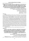
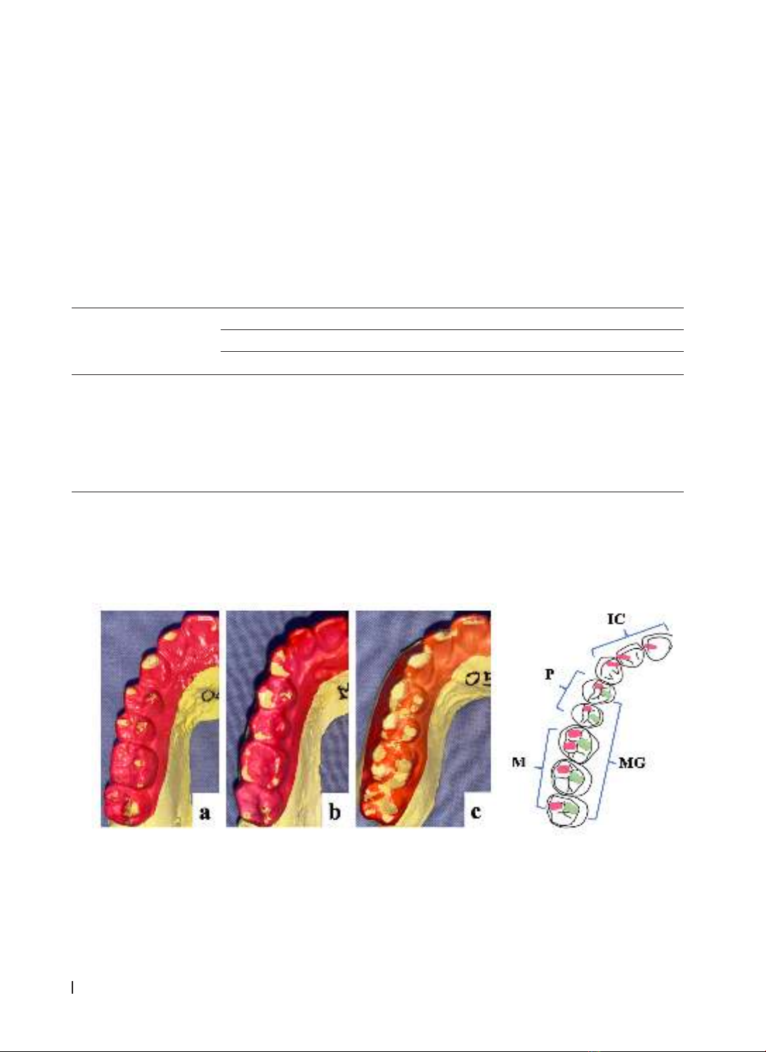
Figure 1 demonstrated occlusal contact patterns assessed by Bruxchecker®. In the category of occlusal
patterns, the ICPM + MG pattern occupied the largest proportion (93,3%). There were no IC, IC + MG, or
ICP patterns observed in this experiment. Nearly all of the quadrants (98,3%) showed an MG pattern in
mediotrusive griding (Table 2).
A B
Figure 1. Occlusal contact patterns inspected by Bruxchecker®
A. Observed area to evaluate cclusal contact patterns on right side: IC (incisor-canine), P (premolar), M
(molar) and MG (Mediotrusive Guide)
B. Occlusal contact patterns on Bruxchecker®: a. ICP+MG, b. ICPM, c. ICPM + MG
HUE JOURNAL OF MEDICINE AND PHARMACY ISSN 3030-4318; eISSN: 3030-4326 153
Hue Journal of Medicine and Pharmacy, Volume 14, No.6/2024
Table 2. Occlusal contact patterns during sleep bruxism assesed via Bruxchecker®
Occlusal contact pattern Number of side
(n = 60)
Frequency
(%)
IC 0 -
IC + MG 0 -
ICP 0 -
ICP + MG 35.0
ICPM 1 1.7
ICPM + MG 56 93.3
MG 59 98.3
IC: incisor – canine, ICP: incisor – canine – premolar;
ICPM: incisor – canine – premolar- molar, MG: mediotrusive guide
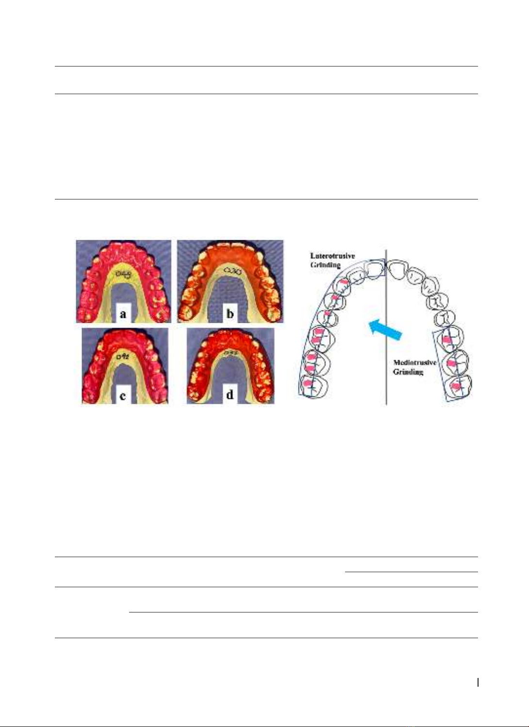
A B
Figure 2. Both sides tooth contact patterns
A. Observe area when evaluating grinding pattern on the right side
B. Laterotrusive contact pattern: a, c. ICP+ICPM; b, d. ICPM+ICPM;
and Mediotrusive contact pattern: c. Unilateral MG; a, b, d. MG+MG
3.3. State of both sides tooth contact pattern and temporomandibular disorders’ symtoms
In Table 3, the tooth contact pattern of both sides was evaluated. The majority of bruxism patients (90%)
had ICPM patterns on both sides (ICPM + ICPM). The rest three bruxers (10%) had ICP + ICPM pattern. In terms
of mediotrusive guiding, an extremely high percentage of patients showed an MG + MG pattern (96.7%).
Only one bruxers had unilateral MG. A relatively high proportion of patients experienced TMD symptoms,
such as clicking, arthralgia, or myalgia.
Table 3. State of laterotrusive and mediotrusive tooth contact
and temporomandibular disorders symptoms
Both sides
contact pattern
Number
(%) Clicking Arthralgia Myalgia
Masseter Temporalis
Laterotrusive
contact pattern
ICP + ICPM 3
(10) 2 2 31
ICPM +
ICPM
27
(90) 13 8 21 9

HUE JOURNAL OF MEDICINE AND PHARMACY ISSN 3030-4318; eISSN: 3030-4326
154
Hue Journal of Medicine and Pharmacy, Volume 14, No.6/2024
Mediotrusive
contact pattern
MG + MG 29 (96.7) 14 9 23 9
Unilateral
MG
1
(3.3) 1 1 1 1
No MG 0 - - - -
ICP: incisor - canine - premolar; ICPM: incisor - canine - premolar - molar, MG: mediotrusive guide.
4. DISCUSSION
In this study, 30 sleep bruxers were diagnosed
using ASSM criteria and the EMG Logger, were fitted
with a Bruxchecker to evaluate the occlusal contact
patterns. We also examined the clinical symptoms
of TMD to define whether there is a relationship
between occlusal contact patterns and TMD
symptoms.
There were more women bruxers than men, and
the average grinding index in the male group was
higher than in the female group (Table 1). However,
there was no difference in the distribution of severity
or index of bruxism in the gender group. Saczuk K.
(2019) showed no difference between genders in
terms of SB status [16]. The reason for the higher
proportion of women observed in the study might be
when teeth grinding occurs, women are more likely to
be worried and want to seek medical care than men.
Electromyography devices are increasingly used
in the diagnosis of SB [4], [16]. In this research, we
used an EMG Logger device. When comparing the
results measured by the EMG Logger with the gold
standard PSG, with a cut-off point of 5.5 episodes/
hour (equivalent to 2 episodes/hour when using
PSG), the sensitivity and specificity of the device is
100 % [13]. In this study, we chose the cut-off point
of 5.5 episodes/hour to determine a subject with
SB. Therefore, the diagnostic results of SB using
EMG Logger in the study are highly reliable. We use
Bruxchecker® to record occlusal contact patterns.
It was first introduced by Onodera and colleagues
and could be useful for screening occlusal contact
patterns during sleep bruxism [3]. In our study,
first-night data were excluded to remove the bias. A
previous study excluded 2 first nights and just used
the third-night data for analysis [17]. However, our
patient reported adapting the device and 0.1mm
thick splint quickly right in the first night. None of
the participants complained of the irritation caused
by EF or BC in their sleep. Measuring 2 consecutive
nights, EF and BC at the same time made it reliable
for the collected results. Onodera also suggested
wearing BC for two consecutive nights to evaluate
grinding patterns during sleep bruxism [3].
Occlusal contact patterns
We evaluated occlusal contact patterns on 60
maxillary quadrants of 30 patients. ICPM + MG was
the most prevalent pattern (93,3%). Interestingly,
there were no IC, IC + MG, and ICP patterns
observed in this experiment (Table 2). According
to Park B. (2008), the highest proportion of ICPM +
MG is 59%, followed by ICP + MG at 33% [7]. In the
study of Nguyen VTQ (2014), the rate of ICPM + MG
was 52.5%, followed by ICP + MG at 15%, then ICPM
and IC + MG at 12.5% [12]. Another study showed
that in sleep bruxers, the ICPM and ICPM + MG
grinding types are significantly more common than
the IC and ICP types [18]. This means that grinding
motions involving the molars will result in greater
muscle activities. According to Park B. (2008), the
average maximum separation distance of excursion
and incursion and the average maximum condylar
lateral deviation during protrusion/retrusion and
open/close movements in the ICPM and ICPM+MG
types were larger than those of the IC and ICP types
[7]. Bruxism, a common cause of microtrauma, often
leads to the lengthening of the capsular ligaments,
the thinning of the articular disc, and loss of muscle
coordination. The condylar lateral movement is
wider and the displacement of the condyle is more
prolonged. Therefore, bruxism might result in the
displacement of the articular disc [7]. Tago C. (2017)
mentioned that the ICPM and MG contacts observed
in Bruxchecker might be higher than those observed
during intraoral examination. Because SB involves
severe powerful contractions of the masticatory
muscles, these tooth contacts are difficult to see
by examining patients while awake, even using
articulating papers [14]. Nearly all of the quadrants
(98,3%) showed an MG pattern in mediotrusive
griding contact (Table 2). The proportion of MG
in studies of Nguyen VTQ (2014), Onodera K
(2006), Tago C. (2017) were 80%, 84%, and 95.9%
respectively [3], [12], [14]. In our study, participants
were moderate and severe bruxers (evaluated by
EMG Logger), therefore, the proportion of MG could
be probably higher than others (in which diagnostic
criteria were not mentioned nor clarified).