129
Journal of Medicine and Pharmacy, Volume 12, No.07/2022
Map of perforators of the posterior tibial artery and peroneal artery using
handheld doppler ultrasound evaluating clinical outcome of perforator
flaps which cover soft tissue defects of the lower leg and foot
Le Hong Phuc1*, Le Nghi Thanh Nhan1
(1) University of Medicine and Pharmacy, Hue University
Abstract
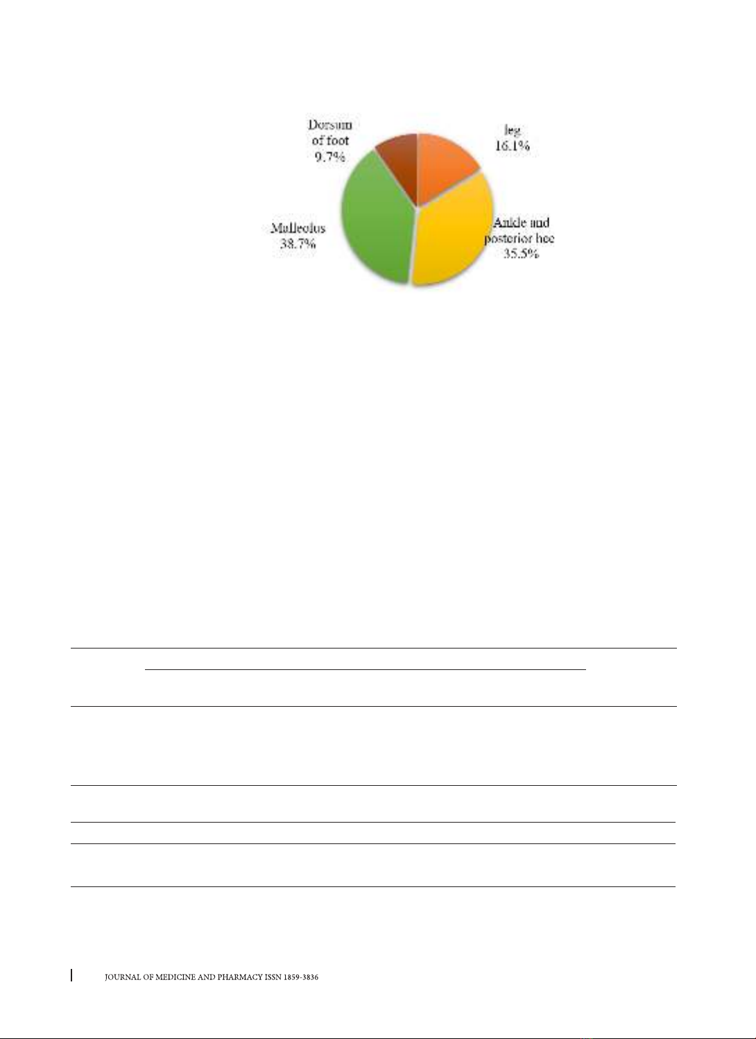
Background: Soft tissue defects of the lower leg and foot are complicated injuries with numerous causes
including trauma, ulcers, and Gout. Widespread treatment of these defects has been effectively applied
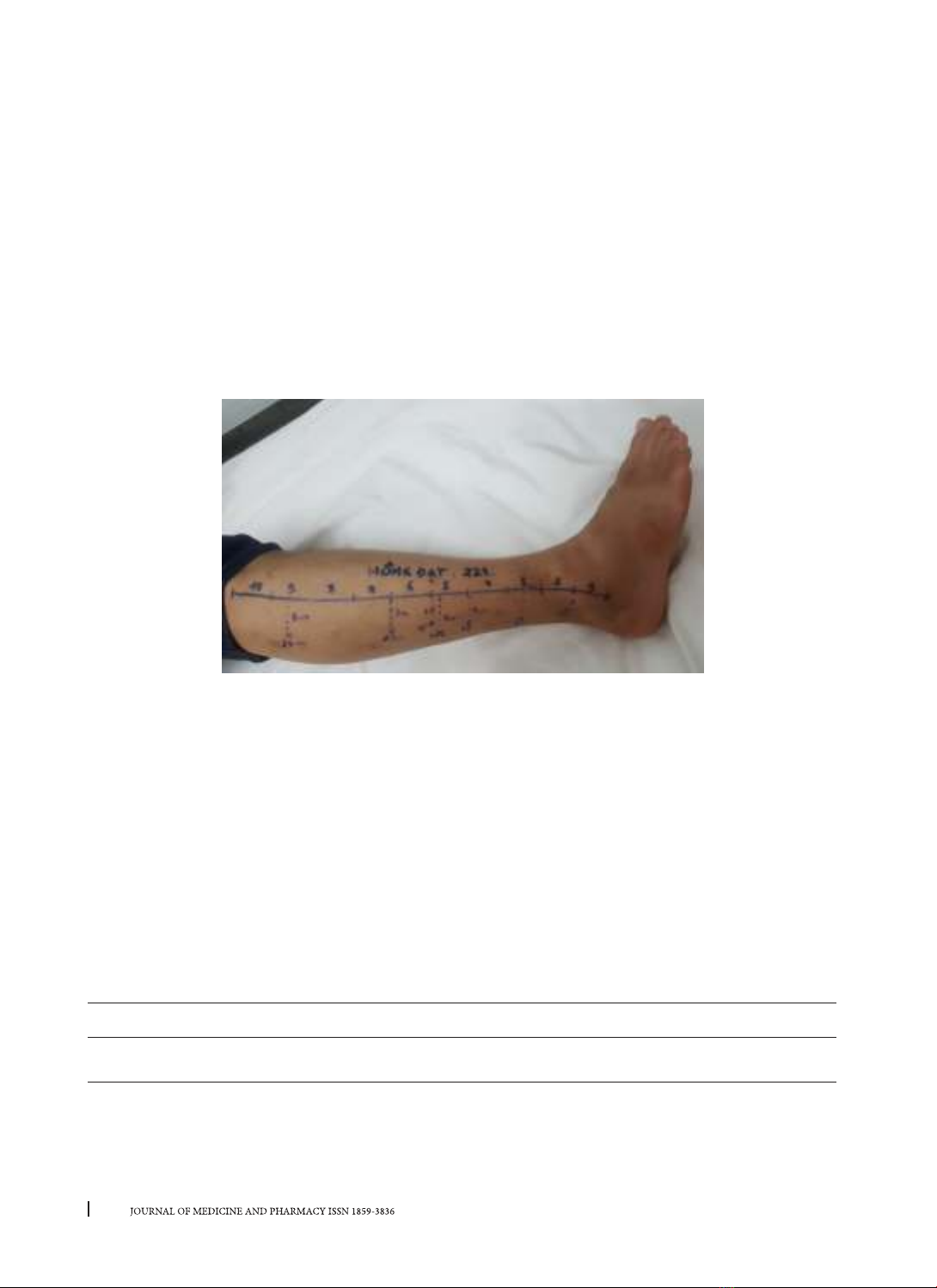
with perforator flaps of the peroneal artery and posterior tibial artery. Objectives: 1. To construct a map
of the perforators of the peroneal artery and posterior tibial artery using a handheld Doppler ultrasound.
2. To evaluate the clinical outcome of perforator flaps to cover soft tissue defects in the lower leg and foot.
Materials and method: Cross-sectional study of 34 volunteers with no previous history of vascular diseases
and the prospective study of 31 patients with soft tissue defects treated with peroneal artery perforator flap
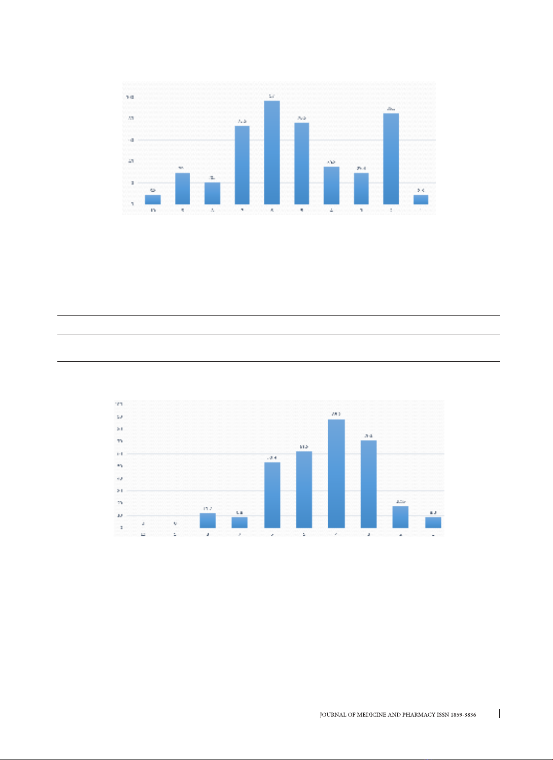
and posterior tibial artery perforator flap. Results: There are, on average, 4.7 peroneal perforating arteries.
From the lateral malleolus to the fibula’s head, the percentage of perforating arteries in the 2/10 and 6/10
segments is 85.3% and 97%, respectively. In the 2-3/10 segment, 94.1% have perforator arteries. There are,
on average, 3.3 posterior tibial artery perforators. From the medial malleolus to the medial tibial plateau, it
was found that 100% of volunteers had perforating arteries in the 3-4/10 segments and 61.7% in the 5/10
segment. Evaluation of postoperative results in 31 patients: 77.49% showed the right flap. The most common
complications were edge necrosis (12.9%), partial necrosis (6.44%), and infection (3.23%). The donor sites
showed good survival in 96.4% of patients, while partial necrosis resulted in 3.6%. A follow-up examination
revealed that 90.32% of flaps had a good result, 9.68% had an average result, and no poor results were
shown. 100% of donor sites had good results. Conclusion: An average of 4.7 perforators of the peroneal
artery is detected by handheld Doppler ultrasound. Also, there are 1-2 relatively constant perforators in
segments 2/10 and 5-6/10 from the lateral malleolus. On average, there are 3.3 perforators of the posterior
tibial artery, primarily in the 3-4/10 and 5/10 segments proximally from the medial malleolus. 90.32% of the
flap had good results.
Keywords: perforator flap, soft tissue defect, lower leg, foot, reconstruction.
Corresponding author: Le Hong Phuc, email: lhphuc@huemed-univ.edu.vn
Recieved: 12/10/2022; Accepted: 1/12/2022; Published: 30/12/2022
1. INTRODUCTION
Soft tissue defects of the lower limb are common
injuries, frequently associated with bone injuries,
osteomyelitis, or bone necrosis, often leading to
limb amputation. The risk of flap failure in the lower
limb appears to be greater than in other locations
due to the lack of elastic material and limited
perfusion in this region.
Reconstruction of defects in the lower leg
with either peroneal artery perforator (PAP) flap
or posterior tibial artery perforator (PTAP) flap
is a flexible and efficient therapy. A handheld
Doppler to identify the perforator arteries and
design the flap is a simple and accurate solution,
up to 91.9%, according to Blondeel research [1]. A
map of perforators being developed by a Doppler
ultrasound is helpful. Furthermore, complications
and treatment outcomes of PAP flaps and PTAP flaps
in the lower limb have not been extensively studied.
Therefore, we conducted the research:
“Constructing a map of the perforators of the
posterior tibial artery and peroneal artery using
handheld Doppler ultrasound and evaluating the
clinical outcome of perforator flaps to cover soft
tissue defects in the lower leg and foot with two
objectives:
1. Construct of the perforators of the peroneal
and posterior tibial arteries using a handheld
Doppler.
2. Evaluating the treatment outcome of PAP and
PTAP flaps.
2. MATERIALS AND METHOD
2.1. Materials: 34 volunteers (18-50 years old)
with no previously confirmed vascular disease and
31 patients with soft tissue defects in lower limbs
treated with PAP and PTAP flaps were selected. All
of the patients were treated in the Orthopedics -
DOI: 10.34071/jmp.2022.7.18
130
Journal of Medicine and Pharmacy, Volume 12, No.07/2022
Thoracic Surgery Department, Hue University of
Medicine and Pharmacy Hospital, from 7/2019 to
4/2011.
2.2. Method:
* Objective 1:
A cross-sectional study was developed to inves-
tigate the number and location of perforators using
handheld Doppler. Steps on how to perform this are
discussed below:
Step 1: Draw the baseline from the fibula’s head
to the lateral malleolus, and divide into ten equal
segments. The segments are marked distally to
proximally.
Step 2: Identify the peroneal perforating arteries
with handheld Doppler (Hadeco Bidop Es100v3 Bi-
Directional Doppler)
Step 3: Find the correlation between the Location
of detected perforating arteries and the previously
divided segments.
Step 4: Draw the baseline from the medial tibial
plateau to the medial malleolus, and divide into ten
equal segments. The segments are marked distally
to proximally.
Step 5: Identify the peroneal perforating arteries
with a handheld Doppler.
Step 6: Find the correlation between the Location
of detected perforating arteries and the previously
divided segments.
Picture 1. Location of the peroneal artery perforators by handheld Doppler.
* Objective 2: A prospective study was
conducted on 31 patients. The treatment result and
postoperative complications were monitored. The
results were evaluated after 1-3 months based on
Touam C and Vo Tien Huys research, which classifies
flaps as good, average, and poor. [2] [3]
* Flap: - Good: Good flap survive, primary heal.
- Average: Malnutrious flap, bluster or partial
necrosis of flap edge (< 1/3 site of flap), skin graft
required or not, hematoma or infection,
- Bad: Flap necrosis from 1/3 to total flap,
required another reconstructive treatment
* Donor site: - Good: Good primary heal, not
infected, grafted skin survive
- Complications: Infection, fistula formation,
necrosis of skin graft.
3. RESULTS
3.1. The map of perforating arteries detected by handheld Doppler:
3.1.1. The number of peroneal perforating arteries
Table 1. Number of peroneal artery perforators:
Number of perforator arteries 3 4 5 6 7 8
Number of calves (patients) 7
(20.6%)
9
(26.5%)
10
(29.4%)
5
(14.7%)
2
(5.9%)
1
(2.9%)
3-5 perforators per leg were found in the majority of volunteers (26/34 cases). On average, we found 4.7
± 1.3 perforator arteries per leg.
131
Journal of Medicine and Pharmacy, Volume 12, No.07/2022
3.1.2. Location of peroneal artery perforators:
Chart 1. Proportion of the peroneal artery perforators by location.
The perforating arteries mainly concentrated on segments 2 and 6 from the lateral malleolus, 85.3%, and
97.0%, respectively, followed by segment 5 with 76.5%. The average distance between the lateral malleolus
and the fibula’s head was 33.4±1.81 cm. A higher density of perforating arteries was found between 3.3 and
6.7 cm (segment 2), 13.4 and 16.7 cm (segment 5), 16.7 and 20.1 cm (segment 6).
3.1.3. The number of posterior tibial artery perforators:
Table 2. Number of posterior tibial artery perforators:
Number of perforator arteries (n=34) 23 4 5 6
Number of calves (patients) 8
(23.5%)
14
(41.2%)
7
(20.6%)
4
(11.75)
1
(2.9%)
2-4 perforator arteries were found per leg in the majority of volunteers (29/34 cases). On average, each
leg had 3.3 ± 1.06 perforator arteries.
3.1.4. Location of posterior tibial artery perforators:
Chart 2. The proportion of the posterior tibial artery perforators by location.
Perforators of the posterior tibial artery mainly concentrate on segments 4,3 and 5, from the medial
malleolus, with 88.2%, 70.6%, and 61.8%, respectively. If segments 3 and 4 were considered as a whole,
perforating arteries can be found in 100% of volunteers.
The average distance between the medial malleolus to the medial plateau of the tibia was 33.9±1.78 cm.
A high density of perforators was found between 6.8 and 10.2 cm (segment 3), 10.2 and 13.6 cm (segment
4), 13.6 and 16.9 cm (segment 5).
3.2. Preoperative characteristics:
3.2.1. Etiology of the Defect:
The two most frequently reported causes of defects in the lower extremity were tophaceous ulcers (35.5%)
and trauma (32.3%), which were mostly due to traffic accidents. Several other causes included infected ulcers
(22.6%), burn injuries (6.5%), and carcinoma (one case, 3.2%).
132
Journal of Medicine and Pharmacy, Volume 12, No.07/2022
3.2.2. Location of the Defect:
Chart 3. Location of soft tissue defects (n=31)
Most of the defects were found in the heel and malleolus (35.5% and 38.7%). The leg and dorsal foot had
a lower rate of soft tissue defects.
3.2.3. The base of the defects:
Underlying tissue was exposed in 48.4% of cases. The most common issues were tendon (19.4%) and
bone (16.1%).
3.3. Evaluation of treatment results:
3.3.1. Types of flaps and methods of transfer:
The PAP flaps were commonly used (67.8%), while posterior tibial artery perforators flaps were in 32.2%.
The most common method was V-Y advancement (58.1%). The rotation flap in 9 cases (29.0%), and the rest
were transposition flaps (12.9%).
3.3.2. Closure of the donor site
The primary closure was performed in 80.6%. A skin graft was in 6 cases (19.4%).
3.3.3. Flap and donor site status in the postoperative period:
3.3.3.1. Status of flap:
22.6% of patients had complications (n=7). The most frequent complications were edge necrosis in 12.9%
(n=4), partial necrosis in 6.44% (n=2) and infection in 3.23% (n=1). The four flaps with distal necrosis were
further corrected with either a skin graft or dissection, which all led to successful healing. The 2 cases with
partial necrosis were treated with a full-thickness skin graft.
Table 3. Flaps in postoperative period (n =31)
Flap
Flaps in postoperative period Total
(n=31)
Good survival Edge necrosis Infected Partial
necrosis
Complete
necrosis
PAP 16 4 10 0 21
PTA P 8 0 0 20 10
Total 24
(77.41%)
4
(12.90%)
1
(3.23%)
2
(6.44%)
0
(0%)
31
(100.0%)
3.3.3.2. Donor site result:
Table 4. Donor site survival
Donor site Number of case (n=31)
Survival 30 (96.77%)
Partial necrosis 1 (3.23%)
In most of the cases, the donor site showed a good result in the postoperative period. Only one patient
(3.23%) with partial necrosis was further treated with a skin graft.
133
Journal of Medicine and Pharmacy, Volume 12, No.07/2022
3.3.4. Flap status after 1-3 month of monitoring:
Table 5. 1-3 months monitoring
Evaluation Number (n=31)
Flap Good 28 (90.32%)
Average 3 (9.68%)
Poor 0
Donor site Good 31 (100%)
Bad 0
The rate of complete flap survival was 90.32% (n=28). There were 2 cases with partial necrosis and 1 case
with edge necrosis, which was then classified as average result (9.68%). There was no case of complete
necrosis.
The donor site showed promising results, with 100% of skin graft survival.
4. DISCUSSION
The handheld Doppler had excellent reliability
in many researches. In Khan and G.Millers study,
handheld Doppler could identify the perforator ar-
tery with a sensibility of 87%, and Blondeel had an
80.6% right positive rate and a positive predictive
value of 91.9% in discovering perforators [4][5]
An average of 4.7±1.3 peroneal artery perforators
was found in each lower limb. This is similar to the
result found by Lykoudis (4.54 perforators in 52
cadavers), and Yooseok Ha (4.8 perforators) [6], [7]
Perforator vessels are consistently found in
segment 2 (3.3 - 6.7 cm); segment 5 (13.4 - 16.7 cm)
and 6 (16.7 - 20.1 cm) from the lateral malleolus.
According to Fu-Chan Wei, there are 1-2 constant
perforators on the posterior side of the fibula at the
intersection of the middle third and the lower third
of the leg [8].
The constant perforators in segments 5 and 6
discovered in our study were also found in the study
of Metthew L.Iorio [9], and Nguyen Quang Duc [10]
(the study of Vietnamese cadavers)
Table 6. Comparison of study on the peroneal artery perforators.
Study 10 9 8 7 6 5 4 3 2 1
Current study 8.8 29.4 20.5 73.5 97.0 76.5 35.3 29.4 85.3 8.8
Nguyen Quang Duc 12.5 53.1 56.3 87.5 68.75 96.8 71.9 18.75
Matthew L. Iorio 3.6 8.2 36.7 68.9 62.5 79.1 73.5 55.6 21.2 5.6
3.3 ± 1.06 posterior tibial artery perforators
were found. This was not similar to the conclusion
of Drimouras [11] (5 perforating arteries). The
reason for this may be due to the lower sensitivity
of handheld Doppler compared with the study of
cadavers.
In our study, segments 3-4 were quite similar
when compared with the study by Liu and Koshima.
Segment 5 was also similar to another study by
Boriani and Schaverien. Even though these studies
used cadavers, some similarity was found in the
results of both methods.
Table 9. Comparison of study on the posterior tibial artery perforators:
Study (year of study) 10 9 8 7 6 5 4 3 2 1
Current study x x x
Boriani(2010) [12] x x x x x
Schaverien (2008) [13] x x x x x
Liu (1990) [14] x x x
Koshima (1992) [15] x x x
Our study showed that the two most common
causes were ulcers and trauma, which is comparable
to Vo Tien Huy [2] (the most common cause was
trauma, 84.4%) and Nguyen Anh Tuan [16] (ulcers
occupied the highest percentage of 41.2%).
Complications of the flap in the postoperative
period were 22.57%, which was lower than several
other studies: Bekara (25.2%) and Innocenti (42.4%).