HUE JOURNAL OF MEDICINE AND PHARMACY ISSN 3030-4318; eISSN: 3030-4326HUE JOURNAL OF MEDICINE AND PHARMACY ISSN 3030-4318; eISSN: 3030-4326
170 171
Hue Journal of Medicine and Pharmacy, Volume 15, No.2/2025 Hue Journal of Medicine and Pharmacy, Volume 15, No.2/2025
Phenolic Compounds and Carotenoids from the Leaves of Gymnosporia
chevalieri Tard.
Doan Thi Ai Nghia1,4, Hoang Thi Nhu Hanh2, Le Tuan Anh3, Le Thi Hong Van4, Ho Viet Duc1*
(1) Faculty of Pharmacy, University of Medicine and Pharmacy, Hue University
(2) Faculty of Engineering & Food Technology, University of Agriculture and Forestry, Hue University
(3) Mien Trung Institute for Scientific Research, Vietnam National Museum of Nature, VAST
(4) Faculty of Pharmacy, University of Medicine and Pharmacy at Ho Chi Minh City
Abstract
Background: The Gymnosporia genus holds substantial potential for bioactive compound discovery, yet
its phytochemical composition remains underexplored relative to its botanical diversity. In Vietnam, aside
from preliminary studies on G. stylosa, systematic research on Gymnosporia species is notably lacking.
This study provides the understanding of the chemical constituents of G. chevalieri, establishing a basis for
future research on its potential applications and bioactive properties. Materials and methods: The leaves
of G. chevalieri were macerated in methanol to produce a crude extract, which was further fractionated via
liquid-liquid partitioning using n-hexane and ethyl acetate as solvents. Compounds were then isolated and
purified using various chromatographic techniques. Structural elucidation was achieved through 1D- and
2D-NMR and HR-ESI-MS analysis, supported by comparison with previously reported spectroscopic data.
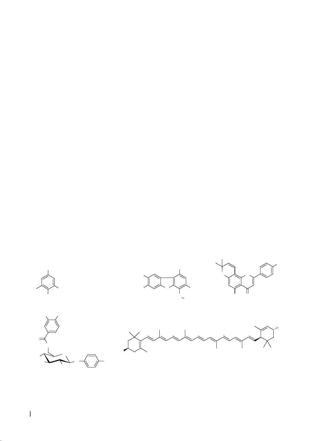
Results & Conclusion: Seven phenolic compounds and two carotenoids were isolated from G. chevalieri
leaves. These were identified as syringaldehyde (1), 3,7-dihydroxy-6-methoxy-2,7-dimethyldibenzofuran (3),
atalantoflavone (4), lutein (6), lutein 3′-methyl ether (7), and two mixtures: 4-hydroxybenzaldehyde (2a) with
4-hydroxy-3-methoxybenzaldehyde (2b), and breynioside B (5a) with 6′-O-vanilloylarbutin (5b). Notably,
compounds 2-7 were isolated from the Gymnosporia genus for the first time.
Keywords: Gymnosporia chevalieri, phenolic compounds, carotenoid.
*Corresponding author: Ho Viet Duc. Email: hvietduc@hueuni.edu.vn
Received: 15/11/2024; Accepted: 15/4/2025; Published: 28/4/2025
DOI: 10.34071/jmp.2025.2.24
1. INTRODUCTION
The genus Gymnosporia (Celastraceae family)
comprises approximately 116 species worldwide
[1]. In Vietnam, the genus Gymnosporia includes 8
recorded species, among which G. chevalieri is an
endemic species located in the Binh Tri Thien area
[2]. In the search for potential bioactive compounds
from the genus Gymnosporia, phenolics were found
alongside other active groups [3, 4]. This article
reports, for the first time, the extraction, isolation,
and structural identification of seven phenolic
compounds and two carotenoids from the leaves of
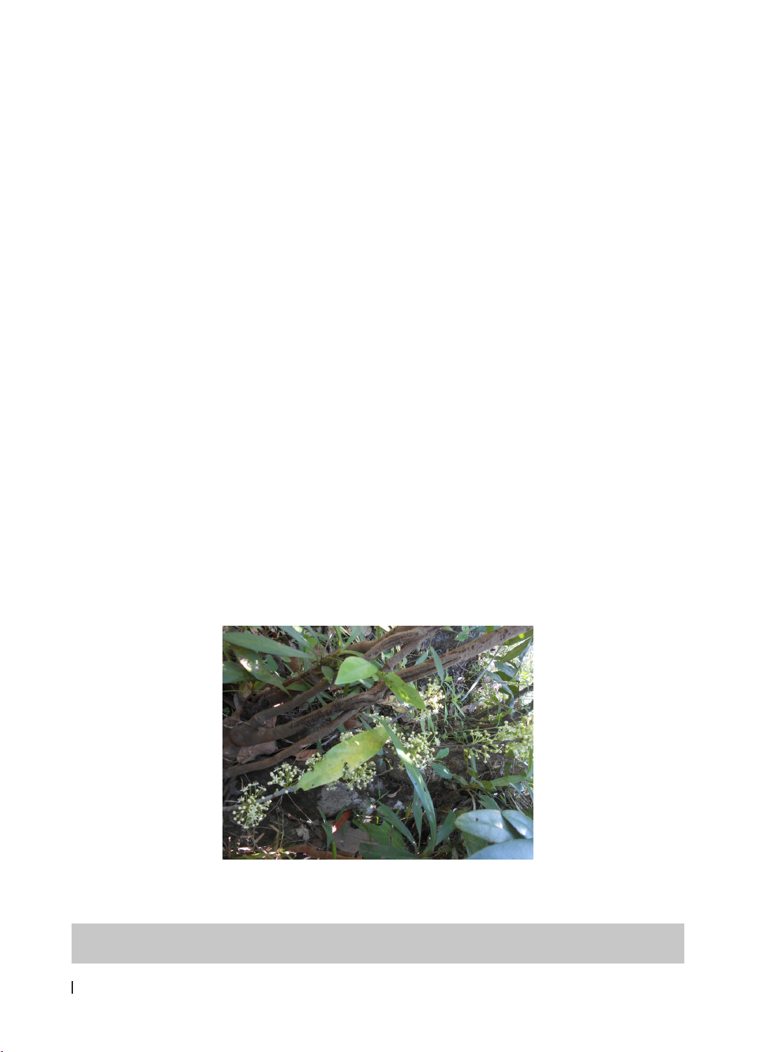
G. chevalieri (Figure 1).
Figure 1. The leaves and flowers of Gymnosporia chevalieri Tard.
Phenolics and carotenoids are two major
groups of plant compounds celebrated for their
significant health benefits, especially for their roles
in antioxidant defense, reducing inflammation, and
HUE JOURNAL OF MEDICINE AND PHARMACY ISSN 3030-4318; eISSN: 3030-4326HUE JOURNAL OF MEDICINE AND PHARMACY ISSN 3030-4318; eISSN: 3030-4326
170 171
Hue Journal of Medicine and Pharmacy, Volume 15, No.2/2025 Hue Journal of Medicine and Pharmacy, Volume 15, No.2/2025
protecting cells [5, 6]. The presence of phenolic
compounds and carotenoids in G. chevalieri is not
only significant as a phytochemical database but also
as a basis for orienting studies on the antioxidant
and anti-inflammatory activities of this species.
2. MATERIALS AND METHODS
Materials
The species Gymnosporia chevalieri was collected
in October 2023 from Đakrong District, Quang Tri
Province. Dr. Anh Tuan Le from the Mien Trung
Institute for Scientific Research (Vietnam National
Museum of Nature, VAST) identified the species. A
voucher specimen (GC-01) has been stored at the
Faculty of Pharmacy, Hue University of Medicine and
Pharmacy, Vietnam.
Methods
The leaves of G. chevalieri were cleaned, air-dried,
and ground into powder. The powdered material was
extracted by maceration with MeOH (10 L x 3 times),
and the solvent was removed under reduced pressure
to yield a crude extract. The total extract undergoes
sequential liquid-liquid distribution with solvents
of increasing polarity, after which the solvents are
evaporated to yield the corresponding fractions.
The isolation process involves a combination
of thin-layer chromatography (TLC) and column
chromatography (CC). TLC was conducted on pre-
coated DC-Alufolien 60 F254 and RP18 F254 plates
(Merck, Germany). Compounds were detected
by UV light at wavelengths of 254 and 365 nm or
by spraying the plates with a 10% H₂SO₄ solution
and heating them until color developed. Column
chromatography was performed using different
stationary phases, including normal silica gel (40–63
µm, Merck, Germany), reverse-phase RP-18 (30–50
µm, Fuji Silysia Chemical, Japan), sephadex LH-20,
and MCI gel (Sigma-Aldrich, USA).
The chemical structures were elucidated using
1D-, 2D-NMR, HR-ESI-MS spectra, in conjunction
with comparison to reference spectral data. NMR
spectra were recorded on a Bruker Avance Neo
600 Spectrometer (Bruker, Massachusetts, USA).
Tetramethylsilane was employed as the internal
standard. High-resolution electrospray ionization
mass spectrometry (HR-ESI-MS) data were acquired
on SCIEX X500 QTOF spectrometers (SCIEX,
Massachusetts, USA).
3. RESULTS
3.1. Extraction and isolation
A total crude extract (GCM, 930 g) was achieved
by extraction of dry leaves powder (4 kg) of G.
chevalieri. After performing liquid-liquid distribution
extraction with n-hexane and EtOAc (each, 4 L x 3
times). Three fractions were abtained including
n-hexane (GCH; 201.8 g), EtOAc (GCE; 97.9 g), and
aqueous extract (GCW; 525 g).
The GCH extract was subjected to silica gel column
using a gradient elution of n-hexane/acetone (100:0,
80:1, 40:1, 20:1, 10:1, 5:1, 2:1, 1:1, and 0:100, v/v)
to afford six fractions, GCH1-GCH6. Fraction GCH5
(30.3 g) was separated by silica gel column, eluting
with a gradient of n-hexane/acetone (10:1, 5:1, 2:1,
1:1, v/v), yielding four fractions: GCH5.1–GCH5.4.
Fraction GCH5.3 (7.67 g) was separated using a RP-
18 column, eluted with acetone/water (5:1, v/v) to
obtain six fractions: GCH5.3.1–GCH5.3.6.
Fraction GCH5.3.1 (770.8 mg) was carried out
over the sephadex eluted with CH2Cl2/MeOH (1:1,
v/v) yielded four fractions: GCH5.3.1.1–GCH5.3.1.4.
Fraction GCH5.3.1.3 (47.8 mg) was further separated
using a RP-18 column, eluted successively with MeOH/
water (3:2, v/v) and acetone/water (2:1, v/v), yielding
five fractions: GCH5.3.1.3.1–GCH5.3.1.3.5. Fraction
GCH5.3.1.3.1 (9.4 mg) was separated using a sephadex
column, eluted with CH₂Cl₂/MeOH (1:1, v/v), yielding
two fractions: GCH5.3.1.3.1.1–GCH5.3.1.3.1.2.
Fraction GCH5.3.1.3.1.1 (4.8 mg) was purified using
a sephadex column, eluted with MeOH 100% to give
compound 1 (3 mg). Fraction GCH5.3.1.3.1.2 (4.7 mg)
was purified using a silica gel column, eluted with
n-hexane/acetone (5:1, v/v), yielding compound 2 (3
mg). Fraction GCH5.3.1.3.4 (4.8 mg) was crystallized,
resulting in compound 4 (1.3 mg).
Fraction GCH5.3.1.4 (9.8 mg) underwent
purification using a RP-18 column with acetone/
water (2:1, v/v), followed by further purification
on a sephadex column using MeOH 100%, yielding
compound 3 (3 mg).
Fraction GCH5.3.4 (630 mg) was separated using
a silica gel column, eluted with CH2Cl2/acetone
(30:1, v/v) to give three fractions: GCH5.3.4.1–
GCH5.3.4.3. Fraction GCH5.3.4.3 (230 mg) was
further separated using a silica gel column, eluted
with an n-hexane/CH₂Cl₂/acetone (1:1:0.1, v/v),
yielding three fractions: GCH5.3.4.3.1–GCH5.3.4.3.3.
Fraction GCH5.3.4.3.1 (76 mg) was carried out over
a RP-18 column by eluted with acetone/water
(5:1, v/v) to afford four fractions: GCH5.3.4.3.1.1–
GCH5.3.4.3.1.4. Compound 6 (11.7 mg) was obtained
from sub-fraction GCH5.3.4.3.1.4 (21 mg).
Fraction GCH5.1 (3.6 g) was subjected to silica
gel column with a gradient of n-hexane/EtOAc (5:1,
HUE JOURNAL OF MEDICINE AND PHARMACY ISSN 3030-4318; eISSN: 3030-4326HUE JOURNAL OF MEDICINE AND PHARMACY ISSN 3030-4318; eISSN: 3030-4326
172 173
Hue Journal of Medicine and Pharmacy, Volume 15, No.2/2025 Hue Journal of Medicine and Pharmacy, Volume 15, No.2/2025
3:1, v/v), giving eight fractions: GCH5.1.1–GCH5.1.8.
Fraction GCH5.1.6 (650 mg) was separated by
MCI gel, eluting with a gradient of MeOH/water
(100:0, 8:2, and 6:4, v/v), yielding four fractions:
GCH5.1.6.1–GCH5.1.6.4. Fraction GCH5.1.6.4 (116.3
mg) was separated using a sephadex column with
CH₂Cl₂/MeOH (1:1, v/v), followed by a RP-18 column
with acetone/MeOH/water (1:1:0.1, v/v), yielding
two fractions: GCH5.1.6.4.1 and GCH5.1.6.4.2.
Fraction GCH5.1.6.4.2 (83.9 mg) was purified using
a silica gel column, eluted with an n-hexane/acetone
(6:1, v/v), yielding compound 7 (8.4 mg).
The EtOAc extract (GCE; 97.9 g) was loaded on
a silica gel column eluted with a stepwise solvent
gradient of CH2Cl2/MeOH (20:1, 10:1, 5:1, 2:1, 1:2,
0:100, v/v) to obtain six fractions: GCE1–GCE6.
Fraction GCE3 (38 g) was separated on a RP-18 column
by eluted with MeOH/water (1:2, v/v) to yield four
sub-fractions: GCE3.1–GCE3.4. Fraction GCE3.1 (6.33
g) was separated on the silica gel column and eluted
sequentially with CH2Cl2/EtOAc/MeOH (3:1:0.4, v/v)
and CH2Cl2/EtOAc/MeOH (3:1:0.6, v/v) to obtain six
fractions: GCE3.1.1–GCE3.1.6. Fraction GCE3.1.3
(62.0 mg) was carried out over the sephadex eluted
with MeOH 100% to afford two fractions: GCE3.1.3.1–
GCE3.1.3.2. Fraction GCE3.1.3.1 (12.0 mg) was purified
sequentially using a RP-18 column with MeOH/water
(2:3, v/v), followed by a sephadex column with MeOH
100%, yielding compound 5 (4.9 mg).
3.2. Structural determination of isolates
Compound 1 was isolated as a white powder.
The 1H-NMR spectrum showed a characteristic
resonance signal for a formyl group at δH 9.82 (s),
two aromatic protons of a 1,3,4,5-tetrasubstituted
benzene ring at δH 7.16 (2H, s), and two methoxy
groups at δH 3.98 (6H, s). The presence of the formyl
and methoxy groups was further confirmed by the
13C-NMR spectrum, which showed characteristic
signals at δC 190.7 and 56.5, respectively. Signals
at δC 106.8 (C), 128.5 (2CH), 147.4 (2C), and 141.3
(C) were consistent with a substituted benzene
ring. Comparison with reference data [7] identified
compound 1 as syringaldehyde, also known as
4-hydroxy-3,5-dimethoxybenzaldehyde (Figure 2).
The 1H-NMR spectrum of 2 showed resonance
signals for two formyl groups at δH 9.87, 9.83 (each,
1H, s); four aromatic protons of a 1,4-disubstituted
benzene ring at δH 7.81, 6.96 (each, 2H, d, J = 8.6
Hz); three aromatic protons of a 1,3,4-trisubstituted
benzene ring at δH 7.42 (1H, d, J = 1.9 Hz), 7.05 (1H,
d, J = 8.5 Hz), and 7.43 (1H, dd, J = 8.5, 1.9 Hz); and
one methoxy group at δH 3.97 (3H, s). The 13C-NMR
spectrum displayed 15 carbon signals, including two
carbonyl carbons (δC 191.0, 190.8); three oxygenated
sp2 carbons (δC 161.3, 151.7, 147.2); nine sp2
carbons [δC 132.4 (2CH), 130.1 (C), 129.9 (C), 127.6
(CH), 115.9 (2CH), 114.4 (CH), 108.8 (CH)]; and one
methoxy carbon (δC 56.2) (Table 1). Comparison with
reference data [8], [9] led to the identification of
compound 2 as a mixture of 4-hydroxybenzaldehyde
(2a) and 4-hydroxy-3-methoxybenzaldehyde (2b)
(also known as vanillin).
O
OH
O
OH O
2
4
5
7
8a 1'
4'
3''
1''
4a
4''
5''
46
9
5a
4a
O
H
3
C
HO
CH
3
OH
OCH
3
1
9a
9b
2
3
7
8
10
11
12
1 R
1
= R
2
= OCH
3
2a R
1
= R
2
= H
2b R
1
= OCH
3
R
2
= H
OH
R
1
R
2
CHO
1
2
3
4
5
6
7
O
HO
HO
OH
O
O
OH
O
OH
R
5a R = H
5b R = OCH
3
14
1'
3'
5'
1''
4''
7''
HO
OR
1
35
7
9
11
13
15
16 17
18
19 20
1'
3'5'
7'
9'
11'
13'
15'
16'17'
18'
19'
20'
6 R = H
7 R = CH
3
34
Figure 2. Structures of phenolic compounds (1-5) and carotenoids (6, 7) isolated
from Gymnosporia chevalieri
HUE JOURNAL OF MEDICINE AND PHARMACY ISSN 3030-4318; eISSN: 3030-4326HUE JOURNAL OF MEDICINE AND PHARMACY ISSN 3030-4318; eISSN: 3030-4326
172 173
Hue Journal of Medicine and Pharmacy, Volume 15, No.2/2025 Hue Journal of Medicine and Pharmacy, Volume 15, No.2/2025
Compound 3 was isolated as a white powder.
The 1H-NMR spectrum displayed characteristic
resonance signals of three aromatic protons at δH
7.61, 7.00, and 6.71 (each, 1H, s); one methoxy
group at δH 4.19 (3H, s); and two methyl groups at δH
2.64 and 2.38 (each, 3H, s). The 13C-NMR and HSQC
spectra indicated that molecule 3 consists of 15
carbons, categorized as three methyls (δC 61.2, 19.4,
16.2), three methines (δC 122.6, 111.8, 98.3), and
nine quaternary carbons. Signals at δC 155.8, 152.3,
146.9, and 145.9 were assigned to oxygenated sp2
carbons. The δC/δH 61.2/4.19 signal confirmed
the presence of a methoxy group attached to the
aromatic ring (Table 2). These data suggest that
compound 3 is a dibenzofuran derivative.
The HMBC correlations between H-1 (δH 7.61)/H-
4 (δH 7.61) and C-2 (δC 119.2)/C-3 (δC 152.3)/C-4a
(δC 155.8)/ C-9b (δC 117.6) confirm the presence of
a 1,2,4,5-tetrasubstituted benzene ring (Figure 3).
The remaining singlet at δH 6.71 was attributed to
a 1,2,3,4,5-pentasubstituted benzene ring. Notably,
strong HMBC correlations between H3-10 (δH 2.38)
and C-1 (δC 122.6)/C-2/C-3; between H3-11 (δH 2.64)
and C-8 (δC 111.8)/C-9 (δC 122.6)/C-9a (δC 117.6); and
between H3-12 (δH 4.19) and C-6 (δC 129.8) located
two methyl groups and the methoxy group at C-2,
C-8, and C-6, respectively, on the dibenzofuran
skeleton. These analyses identified the structure
of compound 3 as 3,7-dihydroxy-6-methoxy-2,7-
dimethyldibenzofuran [10].
Table 1. NMR data of 1, 2 and reference compounds in CDCl3 [δ (ppm), J (Hz)]
C 1 2a 2b
δC
#δC
aδH
b
δC
*δC
aδH
b
δC
$δC
aδH
b
1 106.9 106.8 128.7 129.9 130.1 130.1
2128.6 128.5 7.16s 132.6 132.4 7.81 d
(8.6)
108.9 108.8 7.42 d
(1.9)
3 147.5 147.4 116.1 115.9 6.96 d
(8.6)
147.3 147.2
4 141.1 141.3 161.1 161.3 151.8 151.7
5 147.5 147.4 116.1 115.9 6.96 d
(8.6)
114.5 114.4 7.05 d
(8.5)
6 128.6 128.5 7.16s 132.6 132.4 7.81 d
(8.6)
127.7 127.6 7.43 dd
(8.5, 1.9)
7 190.6 190.7 9.82s 191.1 191.0 9.83 s 191.0 190.8 9.87 s
OCH3 56.5 3.98s 56.3 56.2 3.97 s
OH 5.30 s 6.19 s
#, *, $δC values of syringaldehyde, 4-hydroxybenzaldehyde, vanillin, respectively [7], [8], [9], a150 MHz, b600
MHz
Compound 4 was isolated as a yellow powder.
The 1H-NMR spectrum showed a characteristic
resonance signal for a hydroxyl proton at δH 12.85
(s); four aromatic protons of a 1,4-disubstituted
benzene ring at δH 7.79, 6.97 (each 2H, d, J = 8.4 Hz);
four olefinic protons at δH 6.78, 5.61 (each 1H, d, J
= 10.0 Hz), 6.56, 6.28 (each 1H, s). Additionally, two
equivalent methyl groups were observed in the high-
field region at δH 1.49 (6H, s) (Table 2).
The 13C-NMR spectrum showed resonances
mainly in the downfield region (δC > 100) except
for an oxygenated sp3 carbon at δC 78.0 and methyl
carbons at δC 28.2. A carbonyl group appeared at
δC 182.6, and signals between δC 150-165 indicated
oxygenated sp2 carbons. Furthermore, HSQC data
indicated eight sp2 CH groups and two CH3 groups.
This evidence suggests that compound 4 is a
prenylated flavone.
Key HMBC correlations were observed between
H-3 (δH 6.56) and C-2 (δC 163.5)/C-4 (δC 182.6)/C-
4a (δC 105.2)/ C-1′ (δC 124.0); between H-2’,6’ (δH
7.79) and C-2/C-4’ (δC159.1); and between 5-OH
(δH 12.85) and C-4a/C-5 (δC 161.8)/C-6 (δC 100.3),
confirming the flavone skeleton of compound 4.
Additionally, correlations between the two methyl
groups (δH 1.49) and C-2” (δC 127.5)/C-3” (δC 78.0);
between H-1” (δH 6.78) and C-7 (δC 159.7)/C-3”; and
between H-2” (δH 5.61) and C-8 (δC 101.3) enabled
the construction of a 2,2-dimethyl-2H-pyran ring.
The attachment of this ring to the flavone moiety
at C-7/C-8 was confirmed by the correlations H-6
(δH 6.28) with C-7/C-8, H-1” with C-7/C-8a, and
HUE JOURNAL OF MEDICINE AND PHARMACY ISSN 3030-4318; eISSN: 3030-4326HUE JOURNAL OF MEDICINE AND PHARMACY ISSN 3030-4318; eISSN: 3030-4326
174 175
Hue Journal of Medicine and Pharmacy, Volume 15, No.2/2025 Hue Journal of Medicine and Pharmacy, Volume 15, No.2/2025
H-2” with C-8. From these analyses, compound 4 is
proposed to be 2-(4-hydroxyphenyl)-5-hydroxy-8,8-
dimethyl-4H,8H-pyrano[2,3-f]chromen-4-one, also
known as atalantoflavone [11].
O
H
3
C
HO
CH
3
OH
OCH
3
O
OH
O
OH O
HO
OCH
3
1
35
7
9
11
13
15
16 17
18
19 20
1'
3'
5'
7'
9'
11'
13'
15'
16'17'
18'
19'
20'
34
7
Figure 3. Selected HMBC (arrow), COSY (bold line) correlations of 3, 4 and 7
The 1H-NMR spectrum of 5 showed characteristic
resonance signals for three 1,4-disubstituted
benzene rings with 12 protons at δH 7.92, 6.95, 6.94,
6.88, 6.62, and 6.60 (each 2H, d, J = 8.8 Hz), as well
as one 1,3,4-trisubstituted ring with three protons
at δH 7.61 (1H, dd, J = 8.2, 2.0 Hz), 7.58 (1H, d, J =
2.0 Hz), and 6.90 (1H, d, J = 8.2 Hz). Resonances
corresponding to two β-anomeric protons for sugar
units were observed at δH 4.76 and 4.75 (each 1H,
d, J = 7.5 Hz), and a signal at δH 3.89 (3H, s) was
assigned to a methoxy group (Table 3).
The 13C-NMR and HSQC spectra showed two
carboxyl carbons (δC 167.95, 167.91), fifteen sp2
methine carbons [δC 132.9 (2C), 119.6 (2C), 119.6 (2C),
116.6 (4C), 116.2 (2C), 125.3, 116.0, 113.8], ten sp3
methine carbons [δC 103.7 (2C), 75.0 (2C), 75.6 (2C),
78.0 (2C), 72.1 (2C)], two sp3 oxymethylene carbons
(δC 65.2, 65.1), and one methoxy carbon (δC 56.5).
Overall, the 1H- and 13C-NMR signals appeared as
pairs with an approximate ratio of 1.5:1, indicating
that compound 5 is a mixture of two phenolic
glycosides. The presence of two carboxyl carbons
(δC 168.0, 167.9) along with the downfield shift of
oxymethylene carbons suggested esterification
at the C-6’ of the sugar moiety. Comparison
with reference data allowed the identification of
compound 5 as a mixture of breynioside B (5a) [12]
and 6′-O-vanilloylarbutin (5b) [13].
Table 2. NMR data of 3, 4 (in CDCl3) and reference compounds [δ (ppm), J (Hz)]
C3C4
δC
#δC
aδH
b
δC
*δC
aδH
b
1 123.4 122.6 7.61 s 2164.9 163.5 -
2121.6 119.2 -3 104.4 104.4 6.56 s
3 155.4 152.3 -4 183.3 182.6 -
4 98.5 98.3 7.00 s 5 162.9 161.8 -
4a 157.3 155.8 -6 100.7 100.3 6.28 s
5a 149.6 146.9 -7 160 159.7 -
6 132.2 129.8 -8 102.2 101.3 -
7 148.4 145.9 -8a 152.6 152.1 -
8 114 111.8 6.71 s 4a 106.1 105.2 -