
RESEARC H Open Access
Membrane proteomic analysis of pancreatic
cancer cells
Xiaojun Liu
1
, Min Zhang
1
, Vay Liang W Go
2
, Shen Hu
1,3*
Abstract
Background: Pancreatic cancer is one of the most aggressive human tumors due to its high potential of local
invasion and metastasis. The aim of this study was to characterize the membrane proteomes of pancreatic ductal
adenocarcinoma (PDAC) cells of primary and metastatic origins, and to identify potential target proteins related to
metastasis of pancreatic cancer.
Methods: Membrane/membrane-associated proteins were isolated from AsPC-1 and BxPC-3 cells and identified
with a proteomic approach based on SDS-PAGE, in-gel tryptic digestion and liquid chromatography with tandem
mass spectrometry (LC-MS/MS). X! Tandem was used for database searching against the SwissProt human protein
database.
Results: We identified 221 & 208 proteins from AsPC-1 and BxPC-3 cells, respectively, most of which are
membrane or membrane-associated proteins. A hundred and nine proteins were found in both cell lines while the
others were present in either AsPC-1 or BxPC-3 cells. Differentially expressed proteins between two cell lines
include modulators of cell adhesion, cell motility or tumor invasion as well as metabolic enzymes involved in
glycolysis, tricarboxylic acid cycle, or nucleotide/lipid metabolism.
Conclusion: Membrane proteomes of AsPC-1 (metastatic) and BxPC-3 (primary) cells are remarkably different. The
differentially expressed membrane proteins may serve as potential targets for diagnostic and therapeutic
interventions.
Introduction
Pancreatic cancer is one of the most aggressive human
malignancies. Despite the advances in therapeutic strate-
gies including surgical techniques as well as local and
systemic adjuvant therapies, the overall survival in
patients with pancreatic cancer remains dismal and has
not improved substantially over the past 30 years. Med-
ian survival from diagnosis is typically around 3 to
6 months, and the 5-year survival rate is less than 5%.
As a result, in 2003, pancreatic cancer surpassed pros-
tate cancer as the 4
th
leading cause of cancer-related
death in the US [1]. The main reason for the failure of
current conventional therapy to cure pancreatic cancer
and the major cause for cancer-related mortality in gen-
eral, is the ability of malignant cells to detach from the
primary tumor site and to develop metastasis in
different regions of the same organ and in distant
organs [2,3]. Pancreatic cancer usually causes no symp-
toms early on, leading to locally advanced or metastatic
disease at time of diagnosis [4]. In this regard, it is
important to identify the functional proteins that regu-
late/promote metastasis in pancreatic cancer. This
would facilitate the development of strategies for thera-
peutic interventions and improved management of
cancer patients.
The purpose of this study is to compare the membrane
proteins expressed in pancreatic cancer cells of primary
and metastatic origins using a proteomics approach. Mem-
brane proteomics can be defined as analysis and character-
ization of entire complement of membrane proteins
present in a cell under a specific biological condition [5,6].
In fact, membrane proteins account for more than two-
thirds of currently known drug targets. Defining
membrane proteomes is therefore important for finding
potential drug targets. Membrane proteomics can also
serve as a promising approach to human cancer biomarker
* Correspondence: shenhu@ucla.edu
1
UCLA School of Dentistry & Dental Research Institute, Los Angeles, CA,
90095, USA
Full list of author information is available at the end of the article
Liu et al.Journal of Biomedical Science 2010, 17:74
http://www.jbiomedsci.com/content/17/1/74
© 2010 Liu et al; licensee BioMed Central Ltd. This is an Open Access article distributed under the terms of the Creative Commons
Attribution License (http://creativecommons.org/licenses/by/2.0), which permits unrestricted use, distribution, and reproduction in
any medium, provided the original work is properly cited.

discovery because membrane proteins are known to have
implication in cell proliferation, cell adhesion, cell motility
and tumor cell invasion [7-9].
Materials and methods
Cell culture
AsPC-1 and BxPC-3 cell lines were obtained from
American Tissue Culture Collection (ATCC, Rockville,
MD). These cell lines were initially generated from
patients with pancreatic ductal adenocarcinoma (PDAC)
[10-12]. The cells were maintained at 5% CO
2
-95% air,
37°C, and with RPMI 1640 (ATCC) containing 10% FBS,
100 μg/ml penicillin G and 100 mg/ml streptomycin.
When the confluence reached 80-90%, the cells were
harvested and washed with PBS for three times.
Sample preparation
Membrane proteins from AsPC-1 and BxPC-3 cells were
isolated with the ProteoExtract Native Membrane Pro-
tein Extraction Kit (EMD Chemicals, Gibbstown, NJ). In
brief, the cell pellet was washed three times with the
Washing Buffer, and then incubated with ice-cold
Extract Buffer |at 4°C for 10 min under gentle agitation.
After the pellet was centrifuged at 16,000 g for 15 min
(4°C), the supernatant was discarded and 1 mL ice-cold
Extract Buffer|| was added to the pellet. This membrane
protein extraction step was allowed for 30 min at 4°C
under gentle agitation. Then the supernatant was
collected after centrifugation at 16,000 g for 15 min 4°C.
SDS-PAGE and proteolytic cleavage
Total membrane protein concentration was measured
with the 2-D Quant Kit (GE Healthcare, Piscataway, NJ).
In total, 20 μg of membrane proteins from each cell line
were loaded into a 4-12% NuPAGE Bis-Tris gel (Invitro-
gen, Carlsbad, CA) for SDS-PAGE separation. The gel
was stained with the Simply Blue staining solution (Invi-
trogen) to visualize the proteins. Each gel was then cut
into 15 sections evenly and proteolytic cleavage of pro-
teins in each section was performed with enzyme-grade
trypsin (Promega, Madison, WI) as previously described.
Tandem MS and database searching
Liquid chromatography (LC) with tandem MS (LC/MS/
MS) of peptides was performed using a NanoLC system
(Eksigent Technologies, Dublin, CA) and a LTQ mass
spectrometer (Thermo Fisher, Waltham, MA). Aliquots
(5 μL) of the peptide digest derived from each gel slice
were injected using an autosampler at a flow rate of 3.5
μL/min. The peptides were concentrated and desalted
on a C
18
IntegraFrit Nano-Precolumn (New Objective,
Woburn, MA) for 10 min, then eluted and resolved
using a C
18
reversed-phase capillary column (New
Objective). LC separation was performed at 400 nL/min
with the following mobile phases: A, 5% acetonitrile/
0.1%formic acid (v/v); B, 95% acetonitrile/0.1% formic
acid (v/v). The chosen LC gradient was: from 5% to 15%
B in 1 min, from 15% to 100% B in 40 min, and then
maintained at 100%B for 15 min.
Database searches were performed using the X! Tandem
search engine against the SwissProt protein sequence data-
base. The search criteria were set with a mass accuracy of
0.4 Da and semi-style cleavage by trypsin. Proteins with
two unique peptides are considered as positively identified.
Western blot analysis
AsPC-1 and BxPC-3 cells were lysed with a lysis buffer
containing 8 M urea, 2 M Thiourea and 4% CHAPS.
Cell lysates with a total protein amount of 40 μgwere
separated with 8-12% NuPAGE gels at 100 V for about
2 hours and then transferred to polyvinylidene difluoride
membrane using an iBlot system (Invitrogen, Carlsbad,
CA, USA). After saturating with 2% slim milk, the blots
were sequentially incubated with primary antibody
(1:100 dilution) and horseradish peroxidase-conjugated
antimouseIgGsecondaryantibody(1:1000dilution,
Applied Biological Materials Inc, Richmond, Canada).
Anti-annexin A1 was obtained from Abcam (Cambridge,
MA, USA) whereas anti-phosphoglycerate kinase 1 was
obtained from Santa Cruz Biotechnology (Santa Cruz,
CA, USA). Finally, the bands were visualized by
enhanced chemiluminescence detection (Applied Biolo-
gical Materials).
Results
The purpose of this study was to demonstrate a mem-
brane proteomic analysis of PDAC cells and to identify
differentially expressed membrane proteins between pri-
mary and metastatic PDAC cells, which may have a
potential role in metastasis of pancreatic cancer. Two
PDAC cell lines, AsPC-1 and BxPC-3, were used in this
study. AsPC-1 is a cell line of metastatic origin from a
62 year-old female Caucasian whereas BxPC-3 is a cell
line of primary PDAC from a 61 year-old female Cauca-
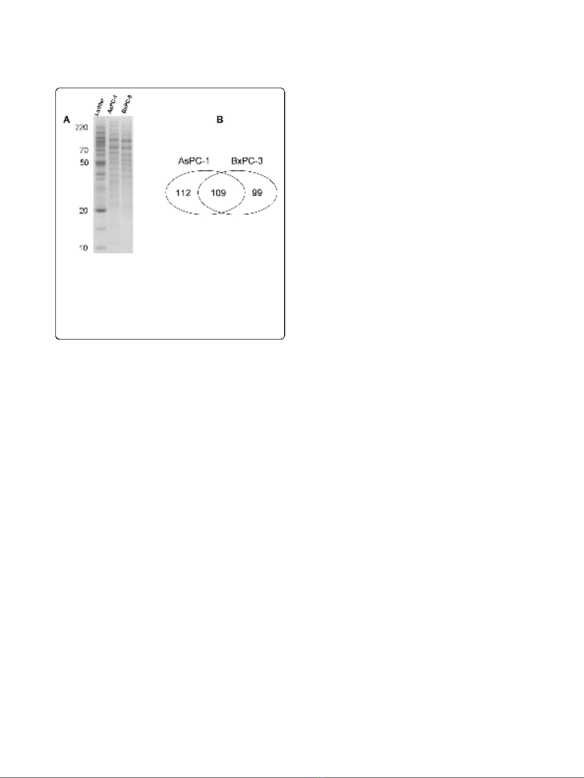
sian [10-12]. Membrane proteins of AsPC-1 and BxPC-3
cells were isolated and then resolved with SDS-PAGE
(Figure 1A). Proteins in each gel slices were proteolyti-
cally cleaved and the resulting peptides were analyzed
with LC-MS/MS. In total, we identified 221 and 208
membrane or membrane-associated proteins from
AsPC-1 and BxPC-3 cells, respectively, based on at least
2 unique peptides. A hundred and nine proteins were
present in both cell lines but others were only found in
AsPC-1 or in BxPC-3 cells (Figure 1B). All the identified
proteins and matched peptides from the two cell lines
are summarized in Additional file 1, Tables S1 and S2.
Proteins with single matched peptide were not tabulated
although previous publications reported identification of
Liu et al.Journal of Biomedical Science 2010, 17:74
http://www.jbiomedsci.com/content/17/1/74
Page 2 of 13
membrane proteins based on single unique peptide
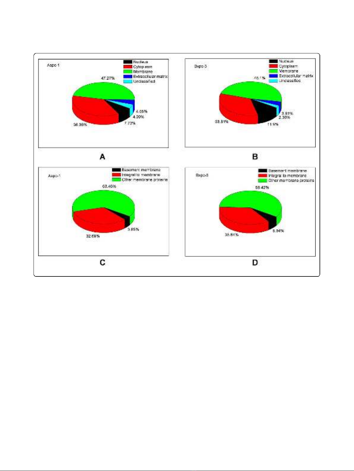
[13,14]. The identified proteins were then sorted accord-
ing to the Gene Ontology Annotation database
(Figure 2). A hundred and four proteins were assigned
as membrane proteins in AsPC-1 cells whereas 101 pro-
teins were assigned as membrane proteins in BxPC-3
cells. Table 1 lists the “integral to membrane”proteins
found in AsPC-1 and BxPC-3 cells. Besides the mem-
brane proteins, the proteomic analysis also identified
many membrane-associated proteins, e.g., extracellular
matrix (ECM) proteins. To confirm the proteomic find-
ing, we verified the differential levels of Annexin A1 and
PGK1 between AsPC-1 and BxPC-3 cells using Western
blotting (Figure 3). Annexin A1 was found to be over-
expressed in BxPC-3 cells whereas phosphoglycerate
kinase 1 was over-expressed in AsPC-1 cells, which
agrees to the results obtained by the proteomic
approach.
Discussion
Metastasis is a highly organ-specific process, which
requires multiple steps and interactions between tumor
cells and the host. These include detachment of tumor
cells from the primary tumor, intravasation into lymph
and blood vessels, survival in the circulation, extravasa-
tion into target organs, and subsequent proliferation and
induction of angiogenesis. Many proteins are critically
involved in this process, such as cell-cell adhesion mole-
cules (CAMs), members of the cadherins and, integrins,
metalloproteinases (MMPs) and the urokinase plasmino-
gen activator/urokinase plasminogen activator receptor
(uPA/uPAR) system. As modulators of metastatic
growth, these molecules can affect the local ECM,
stimulate cell migration, and promote cell proliferation
and tumor cell survivals [15]. Furthermore, hypoxia can
drive genomic instability and lead to a more aggressive
tumor phenotype [16,17], which may partially explain
the highly metastatic nature of PDAC [18]. Last but not
least, angiogenesis plays a critical role in invasion and
metastasis in terms of tumor cell dissemination. Based
on these new insights in mechanism of tumor invasion
and metastasis, novel therapies are currently investigated
for therapy of patients with pancreatic cancer [19-21].
Nevertheless, proteomic analysis of primary and meta-
static PDAC is required to reveal additional functional
proteins that regulate or promote tumor metastasis, as
detailed in previous studies [22-24]. These signature
molecules are predictors of metastatic risk and also pro-
vide a basis for the development of anti-metastatic
therapy.
Our proteomic analysis has revealed a large number of
differentially expressed membrane/surface proteins
between metastatic and primary PDAC cells, and the
validity of such a proteomic approach has been verified
by Western blot analysis. In fact, the differential expres-
sion of membrane proteins between AsPC-1 and BxPC-
3 can be observed from the SDS-PAGE patterns of
membrane proteins from the two cell lines (Figure 1).
The proteins showing differential levels include cadher-
ins, catenin, integrins, galectins, annexins, collagens and
many others, which are known to have roles in tumor
cell adhesion or motility. Cadherins are a class of type-1
transmembrane proteins that depend on calcium ions to
function.Theyplayimportantrolesincelladhesion,
ensuring that cells are bound together within tissues.
Catenins, which are proteins found in complexes with
cadherins, also mediate cell adhesion. Our study identi-
fied cadherins (protocadherin-16 and protocadherin
alpha-12) and alpha-2 catenin in primary tumor cells
(BxPC-3) but not in metastatic tumor cells (AsPC-1),
suggesting a defect in cell-to-cell adhesion in metastatic
AcPC-1 cells.
Integrins are members of a glycoprotein family that
form heterodimeric receptors for ECM molecules. These
proteins are involved in an adhesive function, and they
provide traction for movement in cell motility [25]. In
total, there are 18 a-subunits and 8 b-subunits, which
arepairedtoform24differentintegrinsthroughnon-
covalent bonding. Among these proteins, integrin-b
1
,a
2
,
a
5
,anda
6
represent major adhesion molecules for the
adhesion of pancreatic cancer cells to ECM proteins
[26]. In our study, integrin-b
1
and integrin-b
4
was found
in both tumor cell lines while integrin a
2
and a
5
only
identified in BxPC-3 cells. Collagens are major ECM
proteins. Cell surface-expressed portion of collagens
Figure 1 Analysis and identification of membrane proteins in
AsPC-1 and BxPC-3 cells using a proteomics approach based on
SDS-PAGE, in-gel digestion and LC-MS/MS. (A) Membrane
proteins were isolated, separated with SDS-PAGE and detected with
Simply Blue stain. The gel bands were then excised and digested
with trypsin, and the resulting peptides were extracted for LC-MS/MS
analysis. (B) 221 and 208 proteins were identified from AsPC-1 and
BxPC-3 cells, respectively, with 109 proteins present in both cell lines.
Liu et al.Journal of Biomedical Science 2010, 17:74
http://www.jbiomedsci.com/content/17/1/74
Page 3 of 13
may serve as ligands for integrins, mediating cell-to-cell
adhesion. Twelve members of collagen family were
found in the BxPC-3 cells whereas only four members
found in AsPC-1 cells.
Conversely, galectin-3 and galectin-4 were found in
AsPC-1 but not in BxPC-3 cells. Galectins are carbohy-
drate-binding proteins and have an extremely high affinity
for galactosides on cell surface and extracellular glycopro-
teins. Galectins, especially galectin-3, are modulators of
cancer cell adhesion and invasiveness. Galectin-3 usually
exists in cytoplasm, but can be secreted and bound on the
cellsurfacebyavarietyofglycoconjugate ligands. Once
localized to the cell surface, galectin-3 is capable of oligo-
merization, and the resultant cross-linking of surface
glycoproteins into multimolecular complexes on the
endothelial cell surface is reported to mediate the adhesion
of tumor cells to the vascular endothelium [27]. Lyso-
some-associated membrane glycoprotein 1 (LAMP1) is a
receptor for galectin-3, and was found on the cell surface
of highly metastatic tumor cells [28]. Our study revealed
LAMP1 in AsPC-1 cells but not in BxPC-3 cells. The cell
surface-expressed portion of LAMP1 maybe serve as a
ligand for galectin 3, mediating cell-cell adhesion and
indirectly tumor spread. FKBP12-rapamycin complex-
associated protein (a.k.a., mTOR) was also identified in
AsPC-1 cells but not in BxPC-3 cells. mTOR is a down-
stream serine/threonine protein kinase of the phosphatidy-
linositol 3-kinase/Akt pathway that regulates cell
proliferation, cell motility, cell survival, protein synthesis,
and transcription. Rapamycin, a specific inhibitor of
mTOR, suppresses lymphangiogenesis and lymphatic
metastasis in PDAC cells [29].
The described proteomic approach is reproducible for
analysis of membrane proteins in cultured pancreatic
cancer cells. We observed consistent SDS-PAGE gel pat-
terns for membrane proteins isolated from cultured
AsPC-1 or BxPC-3 cells. To examine the reproducibility
of LC-MS/MS for identification of membrane proteins,
we repeated LC-MS/MS analysis of the peptides yielded
from 3 gel bands. Compared to single LC-MS/MS,
which identified 45 proteins in total, the duplicate LC-
MS/MS analyses identified 47 proteins (~4% increase).
Figure 2 Sorting of the identified proteins according to their subcellular localization.
Liu et al.Journal of Biomedical Science 2010, 17:74
http://www.jbiomedsci.com/content/17/1/74
Page 4 of 13

This suggested that the observed difference in mem-
brane protein profiles between the two PDAC cell lines
is meaningful. Our adopted approach is valid to identify
large membrane proteins, which are usually difficult to
analyze with 2-D gel electrophoresis (2-DE) method. In
AsPC-1 cells, 35% of the identified proteins have a
molecular weight above 70 kDa, whereas 43% of the
proteins are larger than 70 kDa in BxPC-3 cells. In addi-
tion to the proteins either present in AsPC-1 or in
BxPC-3 cells, many other proteins were found in both
cell types with a differential number of peptides
matched. This may reflect the differential level of a
Table 1 Integral to membrane proteins identified in AsPC-1 & BxPC-3 cells
AsPC-1 BxPC-3
Accession # Protein name Accession # Protein name
1A25_HUMAN HLA class I histocompatibility antigen, A-25 alpha chain 4F2_HUMAN 4F2 cell-surface antigen heavy chain
4F2_HUMAN 4F2 cell-surface antigen heavy chain ACSL3_HUMAN Long-chain-fatty-acid–CoA ligase 3
AAAT_HUMAN Neutral amino acid transporter B(0) ACSL4_HUMAN Long-chain-fatty-acid–CoA ligase 4
ACSL5_HUMAN Long-chain-fatty-acid–CoA ligase 5 ADT2_HUMAN ADP/ATP translocase 2
ADT2_HUMAN ADP/ATP translocase 2 ALK_HUMAN ALK tyrosine kinase receptor precursor
ANPRC_HUMAN Atrial natriuretic peptide clearance receptor APMAP_HUMAN Adipocyte plasma membrane-associated protein
AOFB_HUMAN Amine oxidase [flavin-containing] B AT1A1_HUMAN Sodium/potassium-transporting ATPase subunit alpha-1
APMAP_HUMAN Adipocyte plasma membrane-associated protein CALX_HUMAN Calnexin
AT1A1_HUMAN Sodium/potassium-transporting ATPase subunit alpha-1
precursor
CEAM1_HUMAN Carcinoembryonic antigen-related cell adhesion
molecule 1
ATP7B_HUMAN Copper-transporting ATPase 2 CEAM6_HUMAN Carcinoembryonic antigen-related cell adhesion
molecule 6
CALX_HUMAN Calnexin CKAP4_HUMAN Cytoskeleton-associated protein 4
CEAM1_HUMAN Carcinoembryonic antigen-related cell adhesion
molecule 1
CLCN1_HUMAN Chloride channel protein
CEAM6_HUMAN Carcinoembryonic antigen-related cell adhesion
molecule 6
CMC2_HUMAN Calcium-binding mitochondrial carrier protein Aralar2
CMC2_HUMAN Calcium-binding mitochondrial carrier protein Aralar2 CODA1_HUMAN Collagen alpha-1(XIII) chain
CY1_HUMAN Cytochrome c1, heme protein CSMD2_HUMAN CUB and sushi domain-containing protein 2
EGFR_HUMAN Epidermal growth factor receptor precursor EAA1_HUMAN Excitatory amino acid transporter 1
FLNB_HUMAN Filamin-B GP124_HUMAN Probable G-protein coupled receptor 124
FLRT1_HUMAN Leucine-rich repeat transmembrane protein FLRT1 GRP78_HUMAN 78 kDa glucose-regulated protein
FZD8_HUMAN Frizzled-8 precursor HNRPM_HUMAN Heterogeneous nuclear ribonucleoprotein M
GRP78_HUMAN 78 kDa glucose-regulated protein ITAV_HUMAN Integrin alpha-V
IL4RA_HUMAN Interleukin-4 receptor alpha chain KCNQ3_HUMAN Potassium voltage-gated channel subfamily KQT
member 3
IMMT_HUMAN Mitochondrial inner membrane protein L2HDH_HUMAN L-2-hydroxyglutarate dehydrogenase
KCNK3_HUMAN Potassium channel subfamily K member 3 M2OM_HUMAN Mitochondrial 2-oxoglutarate/malate carrier protein
KTN1_HUMAN Kinectin MUC16_HUMAN Mucin-16
LAMP1_HUMAN Lysosome-associated membrane glycoprotein 1 MYOF_HUMAN Myoferlin
LRC59_HUMAN Leucine-rich repeat-containing protein 59 OST48_HUMAN Dolichyl-diphosphooligosaccharide–protein
glycosyltransferase 48 kDa subunit
MTCH2_HUMAN Mitochondrial carrier homolog 2 PCD16_HUMAN Protocadherin-16 precursor
MUC16_HUMAN Mucin-16 PGRC1_HUMAN Membrane-associated progesterone receptor
component 1
MYOF_HUMAN Myoferlin PHB_HUMAN Prohibitin
OST48_HUMAN Dolichyl-diphosphooligosaccharide–protein
glycosyltransferase 48 kDa subunit
PK1L1_HUMAN Polycystic kidney disease protein 1-like 1
PHB_HUMAN Prohibitin PTPRZ_HUMAN Receptor-type tyrosine-protein phosphatase zeta
S12A1_HUMAN Solute carrier family 12 member 1 SSRD_HUMAN Translocon-associated protein subunit delta precursor
SFXN3_HUMAN Sideroflexin-3 TFR1_HUMAN Transferrin receptor protein 1
VAT1_HUMAN Synaptic vesicle membrane protein VAT-1 homolog TMEDA_HUMAN Transmembrane emp24 domain-containing protein 10
VDAC2_HUMAN Voltage-dependent anion-selective channel protein 2 TOM40_HUMAN Mitochondrial import receptor subunit TOM40
homolog
VMAT2_HUMAN Synaptic vesicular amine transporter
Liu et al.Journal of Biomedical Science 2010, 17:74
http://www.jbiomedsci.com/content/17/1/74
Page 5 of 13


%20--%3e%3cdefs%3e%3cstyle%3e%20.st0%20{%20fill:%20%23fff;%20}%20.st1%20{%20fill:%20%237800fa;%20}%20%3c/style%3e%3c/defs%3e%3cpath%20class='st1'%20d='M117.78,12.18H43.11c2.9,3.47,4.65,7.94,4.65,12.82,0,5.6-2.3,10.66-6.01,14.29h76.02l7.22-13.56-7.22-13.56Z'/%3e%3cg%3e%3cpath%20class='st0'%20d='M53.58,26.17h-.59v-1.46h.59v-4.96h2.83c1.78,0,2.67.94,2.67,2.82v5.76c0,1.87-.89,2.81-2.67,2.81h-2.83v-4.96ZM55.36,21.37v3.34h1.1v1.46h-1.1v3.34h1.01c.61,0,.91-.37.91-1.1v-5.93c0-.74-.3-1.1-.91-1.1h-1.01Z'/%3e%3cpath%20class='st0'%20d='M65.99,31.14h-1.8l-.31-2.07h-2.19l-.31,2.07h-1.64l1.82-11.39h2.62l1.82,11.39ZM65.28,18.04c-.25.46-.51.77-.75.94-.21.15-.47.22-.79.22-.26,0-.57-.07-.92-.22l-.38-.15c-.14-.05-.26-.07-.37-.07-.3,0-.53.18-.71.54l-.91-.68c.25-.46.51-.77.75-.94.21-.14.48-.21.79-.21.26,0,.57.07.92.21l.38.15c.14.05.26.07.37.07.3,0,.53-.18.71-.54l.91.68ZM61.91,27.52h1.73l-.87-5.76-.87,5.76Z'/%3e%3cpath%20class='st0'%20d='M74.53,26.89v1.52c0,1.91-.89,2.86-2.67,2.86s-2.67-.95-2.67-2.86v-5.93c0-1.91.89-2.86,2.67-2.86s2.67.95,2.67,2.86v1.11h-1.69v-1.22c0-.75-.31-1.12-.93-1.12s-.93.37-.93,1.12v6.15c0,.74.31,1.11.93,1.11s.93-.37.93-1.11v-1.63h1.69Z'/%3e%3cpath%20class='st0'%20d='M81.4,31.14h-1.8l-.31-2.07h-2.19l-.31,2.07h-1.64l1.82-11.39h2.62l1.82,11.39ZM75.9,19.2l1.52-1.91h1.71l1.51,1.91h-1.61l-.76-.95-.75.95h-1.61ZM77.32,27.52h1.73l-.87-5.76-.87,5.76ZM83.1,15.99l-1.76,1.91h-1.26l1.17-1.91h1.86Z'/%3e%3cpath%20class='st0'%20d='M84.86,19.75c1.78,0,2.67.94,2.67,2.82v1.48c0,1.87-.89,2.81-2.67,2.81h-.85v4.28h-1.79v-11.39h2.64ZM84.01,21.37v3.86h.85c.58,0,.87-.36.87-1.08v-1.71c0-.71-.29-1.07-.87-1.07h-.85Z'/%3e%3cpath%20class='st0'%20d='M93.51,19.75c1.78,0,2.67.94,2.67,2.82v1.48c0,1.87-.89,2.81-2.67,2.81h-.85v4.28h-1.79v-11.39h2.64ZM92.66,21.37v3.86h.85c.58,0,.87-.36.87-1.08v-1.71c0-.71-.29-1.07-.87-1.07h-.85Z'/%3e%3cpath%20class='st0'%20d='M98.8,31.14h-1.79v-11.39h1.79v4.88h2.03v-4.88h1.83v11.39h-1.83v-4.88h-2.03v4.88Z'/%3e%3cpath%20class='st0'%20d='M105.36,24.55h2.46v1.62h-2.46v3.34h3.09v1.63h-4.88v-11.39h4.88v1.63h-3.09v3.18ZM108.17,17.29l-1.76,1.91h-1.26l1.17-1.91h1.86Z'/%3e%3cpath%20class='st0'%20d='M112.2,19.75c1.78,0,2.67.94,2.67,2.82v1.48c0,1.87-.89,2.81-2.67,2.81h-.85v4.28h-1.79v-11.39h2.64ZM111.35,21.37v3.86h.85c.58,0,.87-.36.87-1.08v-1.71c0-.71-.29-1.07-.87-1.07h-.85Z'/%3e%3c/g%3e%3ccircle%20class='st1'%20cx='25'%20cy='25'%20r='20'/%3e%3cpath%20class='st0'%20d='M32.78,19.27c2.92,0,4.43,2.55,5.28,5.33l.71,2.17c.14.38-.33.75-.71.75h-5.61c.19-.33.24-.71.09-1.08l-.75-2.45c-.43-1.32-.99-2.64-1.79-3.77.75-.57,1.65-.94,2.78-.94h0ZM25,18.38c3.25,0,4.9,2.78,5.89,5.89l.76,2.45c.14.42-.33.8-.8.8h-11.69c-.42,0-.94-.38-.8-.8l.75-2.45c.99-3.11,2.64-5.89,5.89-5.89h0ZM25,11.35c1.74,0,3.11,1.37,3.11,3.11s-1.37,3.11-3.11,3.11-3.11-1.41-3.11-3.11,1.41-3.11,3.11-3.11h0ZM17.27,19.27c1.08,0,1.98.38,2.73.94-.8,1.13-1.37,2.45-1.74,3.77l-.8,2.45c-.14.38-.05.75.09,1.08h-5.56c-.42,0-.9-.38-.75-.75l.71-2.17c.9-2.78,2.41-5.33,5.33-5.33h0ZM17.27,12.91c1.51,0,2.78,1.27,2.78,2.83s-1.27,2.83-2.78,2.83-2.83-1.27-2.83-2.83,1.27-2.83,2.83-2.83h0ZM32.78,12.91c1.56,0,2.78,1.27,2.78,2.83s-1.23,2.83-2.78,2.83-2.83-1.27-2.83-2.83,1.27-2.83,2.83-2.83h0ZM27.07,28.56v.09c0,.57-.24,1.08-.61,1.46h0v.05c-.38.33-.9.57-1.46.57s-1.08-.24-1.46-.61h0c-.38-.38-.61-.9-.61-1.46v-.09h1.41v.09c0,.19.05.38.19.47v.05c.09.09.28.19.47.19s.38-.09.47-.19v-.05c.14-.09.24-.28.24-.47t-.05-.09h1.41ZM30.99,28.56v.09c0,1.65-.66,3.16-1.74,4.24-1.08,1.08-2.59,1.79-4.24,1.79s-3.16-.71-4.24-1.79l-.05-.05c-1.04-1.08-1.7-2.55-1.7-4.2v-.09h1.41v.09c0,1.27.47,2.4,1.27,3.25h.05c.85.85,1.98,1.37,3.25,1.37s2.4-.52,3.25-1.37c.85-.8,1.37-1.98,1.37-3.25v-.09h1.37ZM34.99,28.56v.09c0,2.78-1.13,5.28-2.92,7.07-1.79,1.79-4.29,2.92-7.07,2.92s-5.23-1.13-7.07-2.92c-1.79-1.79-2.92-4.29-2.92-7.07v-.09h1.41v.09c0,2.4.94,4.53,2.5,6.08,1.56,1.56,3.72,2.5,6.08,2.5s4.52-.94,6.08-2.5c1.56-1.56,2.5-3.68,2.5-6.08v-.09h1.41Z'/%3e%3c/svg%3e)