TẠP CHÍ Y HỌC VIỆT NAM TẬP 489 - THÁNG 4 - SỐ 1 - 2020
73
3. S. Lanitis, P. P. Tekkis, G. Sgourakis và CS.
(2010), Comparison of skin-sparing mastectomy
versus non-skin-sparing mastectomy for breast
cancer: a meta-analysis of observational studies,
Ann Surg, 251(4), tr. 632-9.
4. T. Zhong, A. Antony và P. Cordeiro (2009),
Surgical outcomes and nipple projection using the
modified skate flap for nipple-areolar
reconstruction in a series of 422 implant
reconstructions, Ann Plast Surg, 62(5), tr. 591-5.
5. C. Tokin, A. Weiss, J. Wang-Rodriguez và CS.
(2012), Oncologic safety of skin-sparing and
nipple-sparing mastectomy: a discussion and
review of the literature, Int J Surg Oncol, 2012,
tr. 921821.
6. P. A. Woodworth, M. F. McBoyle, S. D. Helmer
và CS. (2000), Seroma formation after breast
cancer surgery: incidence and predicting factors, Am
Surg, 66(5), tr. 444-50; discussion 450-1.
7. A. L. Komorowski, V. Zanini, L. Regolo và CS.
(2006), Necrotic complications after nipple- and
areola-sparing mastectomy, World J Surg, 30(8),
tr. 1410-3.
8. S. E. Gabriel, J. E. Woods, W. M. O'Fallon và CS.
(1997), Complications leading to surgery after breast
implantation, N Engl J Med, 336(10), tr. 677-82.
U CƠ VÂN Ở TIM TRONG BỆNH U XƠ CỨNG CỦ Ở TRẺ EM
Trần Kiêm Hảo1, Nguyễn Thị Mỹ Linh1
TÓM TẮT20
Đặt vấn đề:
U cơ vân ở tim là loại u ở tim phổ
biến nhất ở trẻ em. Bệnh thường được chẩn đoán tình
cờ trước sinh hoặc sau sinh, đôi khi xuất hiện trong
thời kỳ sơ sinh với biểu hiện rối loạn huyết động hoặc
rối loạn nhịp tim nghiêm trọng mặc dù đa số các
trường hợp sơ sinh thường không có triệu chứng. Điển
hình của u cơ vân ở tim là có nhiều tổn thương và
thường tự thoái triển nhưng có mối liên quan phức tạp
với bệnh xơ cứng củ, một rối loạn đa hệ thống do đột
biến ở một trong hai gen TSC1 hoặc TSC2. Chẩn đoán
xơ cứng củ thường dựa trên lâm sàng và được xác
nhận chẩn đoán cuối cùng bằng xét nghiệm di truyền
tìm kiếm đột biến gen TSC.
Phương pháp:
Chúng tôi
báo cáo kinh nghiệm của chúng tôi về 3 trường hợp u
cơ vân ở tim được chẩn đoán từ tháng 01/2015 đến
tháng 12/2019, tập trung vào biểu hiện ở tim và kết
hợp với các dấu hiệu phức tạp của bệnh xơ cứng
củ. Chúng tôi đã tiến hành siêu âm tim ban đầu là
trên máy siêu âm Philips Sonos 2500 với đầu dò 7,5/5.
Chúng tôi đã khai thác tiền sử gia đình, các tổn
thương ở não, da, thận và võng mạc, phát hiện các
cơn động kinh và rối loạn tâm thần kinh.
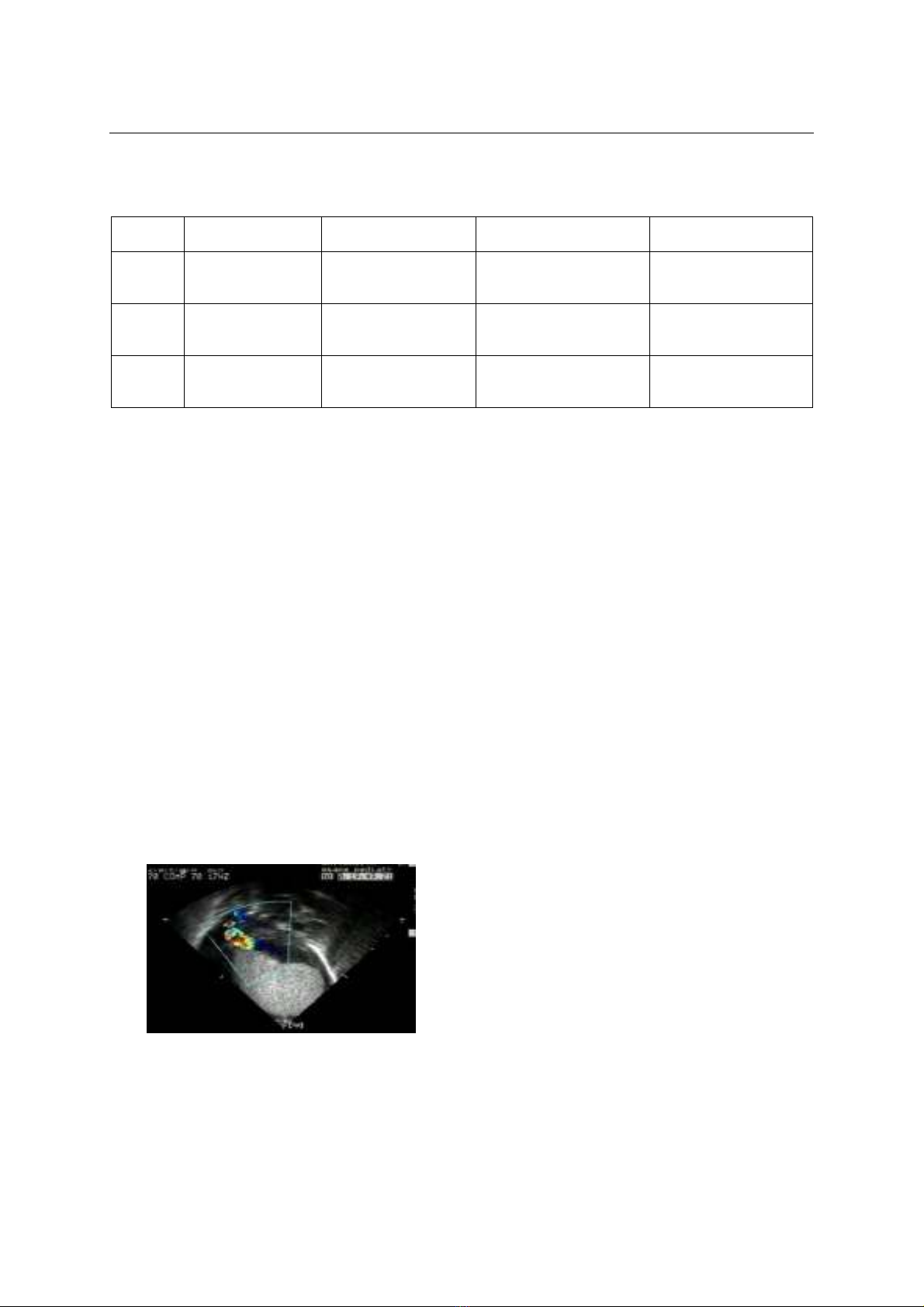
Kết quả
: Khi
chẩn đoán chúng tôi phát hiện 5 khối u, chủ yếu nằm
khu trú ở vách liên thất, tâm thất phải và tâm thất
trái. Chỉ 1 trẻ với sự hiện diện của khối u này gây tắc
nghẽn đáng kể. Trong quá trình theo dõi, chúng tôi
quan sát thấy sự thoái triển của u cơ vân ở tim cả về
số lượng và kích thước trong tất cả 3 bệnh nhân. 1
bệnh nhân bị rối loạn nhịp tim và dẫn đến Hội chứng
Wolf-Parkinson-White. Liên quan với bệnh xơ cứng củ,
chúng tôi đã chẩn đoán bệnh xơ cứng củ dựa trên lâm
sàng ở 3 trẻ.
Kết luận:
U cơ vân ở tim là khối u có
tiên lượng thuận lợi vì bệnh thường không gây ra triệu
chứng và khối u thường thoái triển về số lượng và kích
thước. Tuy nhiên, do thường có mối liên quan phức
tạp với bệnh xơ cứng củ và các rối loạn thần kinh, tiên
1Trung tâm Nhi, Bệnh viện Trung ương Huế
Chịu trách nhiệm chính: Trần Kiêm Hảo
Email: trankiemhaobvh@yahoo.com
Ngày nhận bài: 3.2.2020
Ngày phản biện khoa học: 23.3.2020
Ngày duyệt bài: 27.3.2020
lượng có thể không thuận lợi.
Từ khóa:
U cơ vân ở tim, Bệnh xơ cứng củ, Động kinh
SUMMARY
RHABDOMYOMAS IN CHILDREN WITH
TUBEROUS SCLEROSIS COMPLEX
Background:
Rhabdomyomas are the most
common type of cardiac tumors in children.
Anatomically, they can be considered as hamartomas.
They are usually randomly diagnosed antenatally or
postnatally sometimes presenting in the neonatal
period with haemodynamic compromise or severe
arrhythmias although most neonatal cases remain
asymptomatic. Typically rhabdomyomas are multiple
lesions and usually regress spontaneously but are
often associated with tuberous sclerosis complex
(TSC), an autosomal dominant multisystem disorder
caused by mutations in either of the two genes, TSC1
or TSC2. Diagnosis of tuberous sclerosis is usually
made on clinical grounds and eventually confirmed by
a genetic test by searching for TSC genes mutations.
Methods:
We report our experience on 3 cases
affected with rhabdomyomas and diagnosed from
January 2015 to December 2019, focusing on the
cardiac outcome and on association with the signs of
tuberous sclerosis complex. We performed
echocardiography using initially a Philips Sonos 2500
with a 7,5/5 probe. We investigated the family history,
brain, skin, kidney and retinal lesions, development of
seizures, and neuropsychiatric disorders.
Results:
At
diagnosis we detected 5 masses localized in
interventricular septum, right ventricle and left
ventricle. During follow-up we observed a reduction of
rhabdomyomas in terms of both number and size in all
patients. One patient had an arrhythmia and led to
Wolf-Parkinson-White Syndrome. For all patients the
arrhythmia spontaneously totally disappeared or was
reduced gradually. With regarding to association with
tuberous sclerosis, we diagnosed tuberous sclerosis
clinically in 3 babies.
Conclusion:
Rhabdobyomas are
tumors with favorable prognosis because they
frequently do not cause symptoms and they often
regress in numbers and size. Nevertheless, due to
frequent association with tuberous sclerosis complex
and the resulting neurological impairment, the