TẠP CHÍ Y häc viÖt nam tẬP 546 - th¸ng 1 - sè 3 - 2025
169
Key Concepts and Perioperative Implications.
Journal of cardiothoracic and vascular anesthesia.
Dec 2019;33(12):3486-3495. doi:10.1053/
j.jvca.2019.01.030
2. Boersma SN, Maes S, Joekes K, Dusseldorp
E. Goal processes in relation to goal attainment:
predicting health-related quality of life in
myocardial infarction patients. Journal of health
psychology. Nov 2006;11(6):927-41. doi:10.1177/
1359105306069095
3. Wang W, Thompson DR, Ski CF, Liu M.
Health-related quality of life and its associated
factors in Chinese myocardial infarction patients.
Eur J Prev Cardiol. Mar 2014;21(3):321-9.
doi:10.1177/2047487312454757
4. Mai VQ, Sun S, Van Minh H, et al. An EQ-5D-
5L value set for Vietnam. Quality of Life Research.
2020;29(7):1923-1933.
5. Siriyotha S, Pattanaprateep O,
Srimahachota S, Sansanayudh N,
Thakkinstian A, Limpijankit T. Factors
associated with health-related quality of life in
patients undergoing percutaneous coronary
intervention: Thai PCI registry. Original Research.
2023- November-08 2023;10doi:10.3389/fcvm.
2023.1260993
6. Shan L, Saxena A, McMahon R. A systematic
review on the quality of life benefits after
percutaneous coronary intervention in the elderly.
Cardiology. 2014;129(1): 46-54. doi:10.1159/
000360603
7. Yan BP, Chan LLY, Lee VWY, et al. Sustained
3-Year Benefits in Quality of Life After
Percutaneous Coronary Interventions in the
Elderly: A Prospective Cohort Study. Value in
health: the journal of the International Society for
Pharmacoeconomics and Outcomes Research. Apr
2018; 21(4): 423-431. doi:10.1016/ j.jval.
2017.10.004
8. Weintraub WS, Spertus JA, Kolm P, et al.
Effect of PCI on quality of life in patients with
stable coronary disease. The New England journal
of medicine. Aug 14 2008;359(7):677-87. doi:
10.1056/NEJMoa072771
CASE LÂM SÀNG: UNG THƯ DẠ DÀY HAI VỊ TRÍ, BA LOẠI TẾ BÀO
Lê Minh Sơn1, Nguyễn Minh An2, Trần Tiến Quyết3
TÓM TẮT41
Đặt vấn đề: Trong y văn có 5 trường ung thư dạ
dày thể biểu mô tuyến đồng thời với u lympho không
Hodgkin tế bào B lớn lan tỏa, nhưng chưa có báo cáo
nào thông báo về ca bệnh có sự kết hợp ung thư biểu
mô thần kinh nội tiết tế bào lớn kết hợp với 2 loại tế
bào trên trong cùng một cơ quan. Ca lâm sàng:
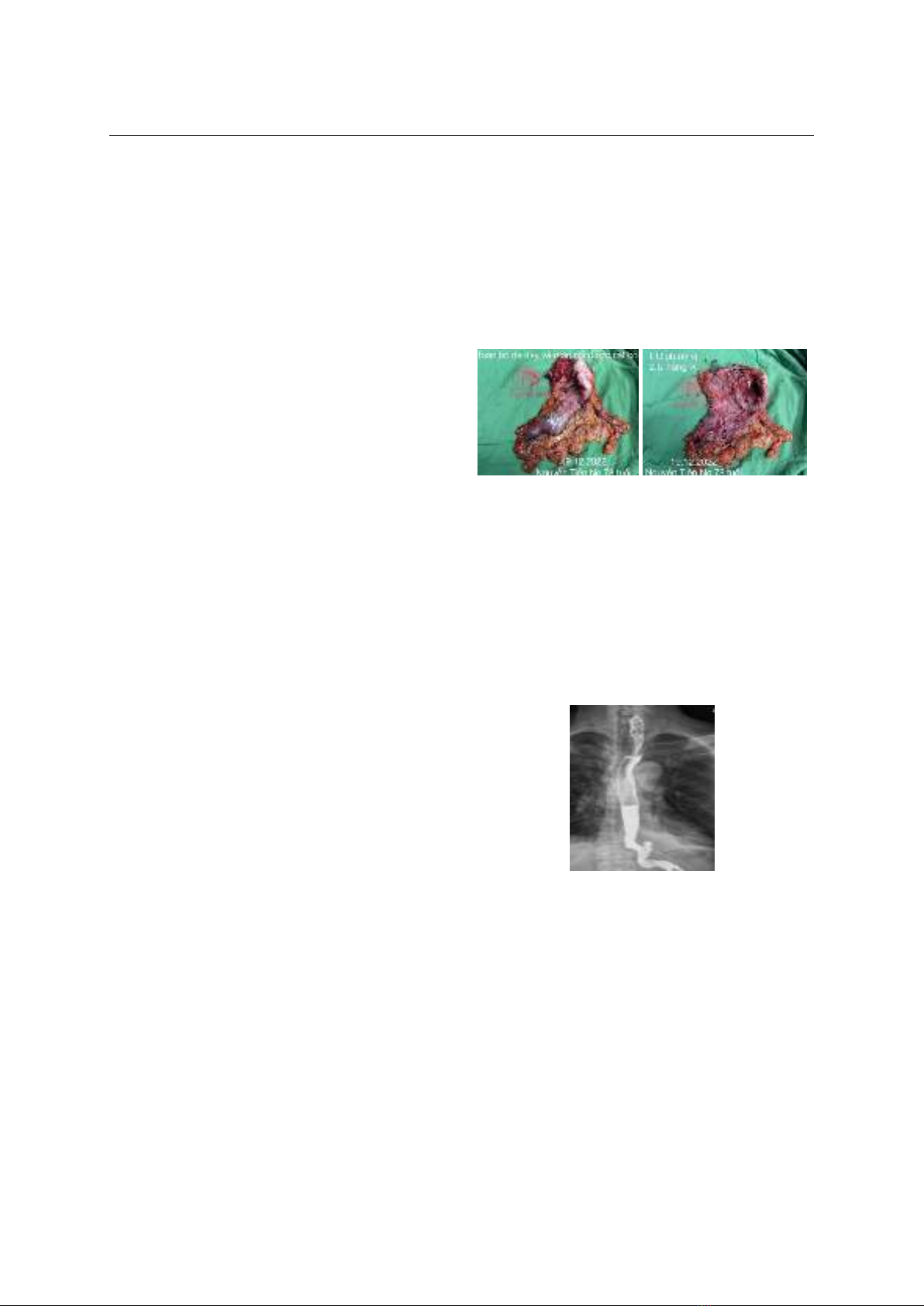
Bệnh nhân nam 78 tuổi, vào viện vì phân đen và khối
sa lồi vùng hậu môn khi đại tiện. Lâm sàng có da niêm
nhợt, trĩ hỗn hợp độ III. Nội soi dạ dày để tầm soát
nguyên nhân thiếu máu khác: cách góc tâm vị 3cm
phía bờ cong lớn, tổn thương 10*8cm, bờ gồ cao,
cứng, đáy loét phủ giả mạc, bờ cong nhỏ có tổn
thương 3*4cm, bờ cứng dễ chảy máu. Kết quả sinh
thiết ung thư biểu mô tuyến. Bệnh nhân được phẫu
thuật nội soi cắt toàn bộ dạ dày, nạo vét hạch D2+, ra
viện sau 10 ngày hậu phẫu không biến chứng. Hóa mô
miễn dịch: tổn thương trên u lympho không Hodgkin
lan tỏa tế bào B lớn, tổn thương dưới ung thư biểu mô
tuyến biệt hóa vừa, có ổ biệt hóa ung thư biểu mô
thần kinh nội tiết tế bào lớn. Sau 1 đợt hóa chất phác
đồ TS-1, bệnh nhân mắc viêm phổi bệnh viện và tử
vong sau 5 tháng phẫu thuật. Kết luận: Xuất huyết
tiêu hóa thấp do bệnh trĩ thường gặp, đôi khi nhầm
lẫn với các bệnh lý ung thư đường tiêu hóa cần được
1Bệnh viện Đa khoa Xanh Pôn
2Trường Cao đẳng Y tế - Hà Nội
3Bệnh viện Đa khoa Xanh Pôn
Chịu trách nhiệm chính: Lê Minh Sơn
Email: lmsxanhpon1@gmail.com
Ngày nhận bài: 21.10.2024
Ngày phản biện khoa học: 22.11.2024
Ngày duyệt bài: 27.12.2024
khám xét kỹ lưỡng, ung thư dạ dày 3 loại tế bào, 2 vị
trí hiếm gặp, cần được phối hợp điều trị nhiều chuyên
khoa.
Từ khóa:
ung thư đồng thời hai loại tế bào,
ung thư biểu mô tuyến dạ dày, u lympho không
Hodgkin tế bào B lớn lan tỏa.
SUMMARY
CLINICAL CASE: GASTRIC CANCER WITH
TWO LOCATIONS, THREE CELL TYPES
Background: In the literature, there are 5 cases
of gastric adenocarcinoma with diffuse large B-cell
non-Hodgkin lymphoma, but there have been no
reports of a case of large cell neuroendocrine
carcinoma combined with the above two cell types in
the same organ. Case: A 78-year-old male patient
was admitted to the hospital because of melena and
protrusion in the anal canal during defecation.
Clinically there is pale skin, mixed hemorrhoids grade
III. Gastroscopy to screen for other causes of anemia:
3cm from the cardial angle on greater curvature,
10*8cm, high, hard edges, pseudomembranous
ulcerated bottom, 3*4cm lesions on the lesser
curvature, hard margin easy to bleed. Biopsy results of
adenocarcinoma. The patient underwent laparoscopic
total gastrectomy, and D2+ lymph node dissection,
and was discharged after 10 days of surgery without
complications. Immunohistochemistry: upper lesions
diffuse large B-cell non-Hodgkin lymphoma, lower
lesions moderately differentiated adenocarcinoma.
After 1 course of chemotherapy with TS-1 regimen,
the patient was diagnosed with hospital-acquired
pneumonia and died 5 months after surgery.
Conclusion: Low gastrointestinal bleeding due to
hemorrhoids is common, sometimes confused with
gastrointestinal cancers that need to be carefully
examined, gastric cancer with 3 cell types, and 2