
RESEARCH Open Access
Prognostic Impact of MiR-155 in Non-Small Cell
Lung Cancer Evaluated by in Situ Hybridization
Tom Donnem
1,2*
, Katrine Eklo
3,4
, Thomas Berg
3,4
, Sveinung W Sorbye
3,4
, Kenneth Lonvik
3,4
, Samer Al-Saad
3,4
,
Khalid Al-Shibli
3,5
, Sigve Andersen
1,2
, Helge Stenvold
1,2
, Roy M Bremnes
1,2
, Lill-Tove Busund
3,4
Abstract
Background: In recent years, microRNAs (miRNAs) have been found to play an essential role in tumor
development. In lung tumorigenesis, targets and pathways of miRNAs are being revealed, and further translational
research in this field is warranted. MiR-155 is one of the miRNAs most consistently involved in various neoplastic
diseases. We aimed to investigate the prognostic impact of the multifunctional miR-155 in non-small cell lung
cancer (NSCLC) patients.
Methods: Tumor tissue samples from 335 resected stage I to IIIA NSCLC patients were obtained and tissue
microarrays (TMAs) were constructed with four cores from each tumor specimen. In situ hybridization (ISH) was
used to evaluate the expression of miR-155.
Results: There were 191 squamous cell carcinomas (SCCs), 95 adenocarcinomas (ACs), 31 large cell carcinomas and
18 bronchioalveolar carcinomas. MiR-155 expression did not have a significant prognostic impact in the total
cohort (P = 0.43). In ACs, high miR-155 expression tended to a significant negative prognostic effect on survival in
univariate analysis (P = 0.086) and was an independent prognostic factor in multivariate analysis (HR 1.87, CI 95%
1.01 - 3.48, P = 0.047). In SCC patients with lymph node metastasis, however, miR-155 had a positive prognostic
impact on survival in univariate (P = 0.034) as well as in multivariate (HR 0.45, CI 95% 0.21-0.96, P = 0.039) analysis.
Conclusions: The prognostic impact of miR-155 depends on histological subtype and nodal status in NSCLC.
Introduction
Lung cancer is the leading cause of cancer-related mor-
tality in both men and women [1]. Despite several new
treatment achievements, the consistently poor 5-year
survival for lung cancer patients underscores the need
for novel modalities for early detection, prognostification
and targeted therapies [1,2].
MicroRNAs (miRNAs) are approximately 19-22
nucleotides single stranded RNAs playing crucial roles
in regulating gene expression by either inducing
mRNA degradation or inhibiting translation [3,4].
These non-coding RNAs can simultaneously regulate
hundreds to thousands of their target genes or up to
one third of the genome, thereby controlling a wide
range of biological functions including apoptosis, pro-
liferation and differentiation [3,5].
To date miR-155 is one of the miRNAs most consis-
tently involved in neoplastic diseases in both hemato-
poietic malignancies (i.e. Hodgkin’s lymphoma, some
types of Non Hodgkin’s lymphoma, AML and CML)
and solid tumors (e.g. breast, colon, cervical, thyroid,
pancreatic and lung cancer) [6-16]. MiR-155 is also
involved in other biological processes like hematopoiesis,
inflammation and immunity [6]. The frequently detected
up-regulation of miR-155 in malignant cells indicates a
major role as an oncogene, however, a possible tumor
suppression function has also been suggested [17]. In
non-small cell lung cancer (NSCLC), miR-155 has so far
been considered as an oncogene and been associated
with a poor prognosis [13,16], though a recent large
scale study did not find miR-155 to have any prognostic
or predictive impact [18].
NSCLC classification according to histology and nodal
status are two of the most important determinants for
NSCLC treatment strategies [13,19]. However, a consid-
erable variability in prognosis has been observed for
* Correspondence: tom.donnem@uit.no
1
Department of Oncology, University Hospital of North Norway, Tromso,
Norway
Full list of author information is available at the end of the article
Donnem et al.Journal of Translational Medicine 2011, 9:6
http://www.translational-medicine.com/content/9/1/6
© 2011 Donnem et al; licensee BioMed Central Ltd. This is an Open Access article distributed under the terms of the Creative
Commons Attribution License (http://creativecommons.org/licenses/by/2.0), which permits unrestricted use, distribution, and
reproduction in any medium, provided the original work is properly cited.

subsets of patients with the same clinical features. Con-
sequently, the clinical incorporation of predictive and
prognostic molecular biomarkers with traditional cancer
staging should improve the management of patients
with NSCLC.
Squamous cell carcinomas (SCCs) and adenocarci-
nomas (ACs) are the major histological subtypes of
NSCLC. During recent years, treatment responses
and side effects by novel therapies have been corre-
lated to NSCLC subgroups according to histology,
gender, ethnicity and smoking status. The vascular
endothelial growth factor (VEGF) monoclonal anti-
body, bevacizumab, is only given to non-SCCs due to
the risk of fatal bleeding in SCCs [20]. Further, muta-
tions in epidermal growth factor receptor (EGFR) and
response to EGFR tyrosine kinase inhibitors appear
related to ACs, female gender, Asian ethnicity and
non-smokers, and the new antifolate agent peme-
trexed appears to have better response in non-SCC
patients and females [21,22]. Consequently, ACs and
SCCs are increasingly recognized as different diseases
instead of one.
In an unselected NSCLC cohort of 335 patients [23]
weaimedtoexplore,usingin situ hybridization on a
high throughput platform, possible prognostic roles by
miR-155 in all NSCLC cases and subgroups according
to histology and stage.
Patients and Methods
Patients and Clinical Samples
Primary tumor tissues from anonymized patients diag-
nosed with NSCLC pathologic stage I to IIIA at the
University Hospital of Northern Norway (UNN) and
Nordland Central Hospital (NLCH) from 1990 through
2004 were used in this retrospective study. In total, 371
patients were registered from the hospital database. Of
these, 36 patients were excluded from the study due to:
(i) Radiotherapy or chemotherapy prior to surgery (n =
10); (ii) Other malignancy within five years prior to
NSCLC diagnosis (n = 13); (iii) Inadequate paraffin-
embedded fixed tissue blocks (n = 13). Adjuvant che-
motherapy was not introduced in Norway during this
period (1990 - 2004). Thus, 335 patients with complete
medical records and adequate paraffin-embedded tissue
blocks were eligible.
This report includes follow-up data as of November
30, 2008. The median follow-up of survivors was 86
(range 48-216) months. The tumors were staged accord-
ing to the new 7th edition of TNM in Lung Cancer and
histologically subtyped and graded according to the
World Health Organization guidelines [19,24]. Regard-
ing N-status, ipsilateral peribronchial or hilar nodes and
intrapulmonary nodes are defined as N1, while N2
includes ipsilateral mediastinal or subcarinal nodes.
The term N+ (lymph node metastasis present) includes
both N1 and N2. The National Data Inspection Board
and The Regional Committee for Research Ethics
approved the study.
Microarray Construction
All lung cancer cases were histologically reviewed by
two pathologists (S.A.S. and K.A.S.) and the most
representative areas of viable tumor cells were care-
fully selected. The TMAs were assembled using a tis-
sue-arraying instrument (Beecher Instruments, Silver
Springs, MD). The Detailed methodology has been
previously reported [23]. Briefly, we used a 0.6 mm
diameter stylet, and the study specimens were routi-
nely sampled with four replicate core samples (differ-
ent areas) of tumor tissue. In addition normal lung
tissue localized distant from the primary tumor, and
one slide with normal lung tissue samples from 20
patients without a cancer diagnosis were stained. Mul-
tiple 4-μm sections were cut with a Micron micro-
tome (HM355S) and used for in situ hybridization
analysis.
In Situ Hybridization (ISH)
In situ hybridization was performed following the proto-
col developed by Nuovo et al. [25], with some minor
adjustments. Digoxigenin (DIG) labeled locked nucleic
acid (LNA) modified probes for miR-155 (hsa-miR-155),
positive control (U6, hsa/mmu/rno) and negative control
(scramble-miR) were purchased from Exiqon, Vedbek,
Denmark.
Briefly, we placed 4 μm sections of the TMA blocks in a
heater at 59°C over night to attach cores to the silane-
coated slide. Sections were deparaffinised with xylene (2 ×
5 min), rehydrated with ethanol (100 - 50 - 25% for 5 min
each), and treated with DEPC water for 1 min. Protease
treatment was performed with pepsin solution (1.3 mg/ml)
(Dako, Glostrup, Denmark) at 37°C for 50 min. Following a
postfixation step in 4% paraformaldehyde (PFA), hybridiza-
tion of the LNA-probe was carried out in a Hybrite (Abbott
Laboratories, IL) at 60°C for 5 min and 37°C over night
(12-18 h). Low-stringency post-hybridization wash done at
4°C in SSC with 2% BSA for 5 min, followed by incubation
with anti-DIG/alkaline phosphate conjugate antibodies
(Enzo Diagnostics, NY) in a heater at 37°C for 30 min. The
blue color was developed by incubation of the slide with
nitroblue tetrazolium and bromchloroindolyl phosphate
(NBT/BCIP) (Enzo Diagnostics, NY) at 37°C. The colori-
metric reaction was monitored visually and stopped by pla-
cing the slides in water when background coloring started
Donnem et al.Journal of Translational Medicine 2011, 9:6
http://www.translational-medicine.com/content/9/1/6
Page 2 of 9
to appear on the negative control (scrambled probe), vary-
ing from 15-30 min. The slides were counterstained with
nuclear fast red (Enzo Diagnostics, NY) to visualize the
nuclei, before cover glass mounting.
Scoring of ISH
The ARIOL imaging system (Genetix, San Jose, CA)
was used to scan the TMA slides of ISH staining. The
slides were loaded in the automated loader (Applied
Imaging SL 50) and specimens were scanned at low
(1.25×) and high resolution (20×) using the Olympus
BX 61 microscope with an automated platform (Prior).
Representative and viable tissue sections were scored
manually semiquantitatively for cytoplasmic staining
on computer screen. The dominant staining intensity
in tumor cells was scored as: 0 = negative; 1 = weak; 2
= intermediate; 3 = strong (Figure 1). In case of dis-
agreement (score discrepancy >1), the slides were re-
examined and a consensus was reached by the obser-
vers. In most cores there was a mixture of stromal
cells and tumor cells. By morphological criteria only
tumor cells were scored staining intensity.
All samples were anonymized and independently scored
by one experienced pathologist and one technician (S.W.S.
and K.E.). When assessing a variable for a given core, the
observers were blinded to the scores of the other observer
and to outcome. Mean score for each case was calculated
from all four cores and both examiners. The median
miR-155 expression value was used as cut-off.
Statistics
All statistical analyses were done using the statistical
package SPSS (Chicago, IL), version 17. The Chi-
square test and Fishers Exact test were used to exam-
ine the association between molecularmarkerexpres-
sion and various clinicopathological parameters. The
ISH scores from each observer were compared for
interobserver variability by use of a two-way random
effect model with absolute agreement definition. The
intraclass correlation coefficient (reliability coefficient)
was obtained from these results. Plots of the disease-
specific survival (DSS) according to marker expression
were drawn using Kaplan-Meier method, and statisti-
cal significance between survival curves was assessed
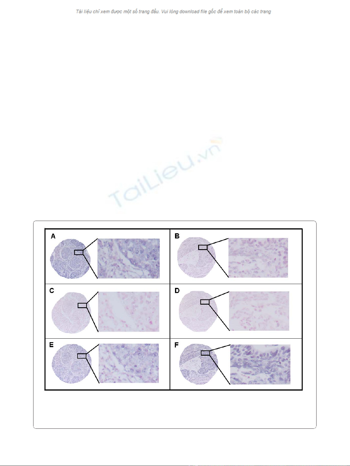
Figure 1 In situ hybridization (ISH) analysis of NSCLC representing strong and weak intensities for tumor cell miR-155 expression.
Negative (scramble-miR) and positive (U6) controls from the same tissue area are shown. Strong miR-155 staining (A) with corresponding
negative (C) and positive (E) controls to the left. Weak miR-155 staining (B) with corresponding negative (D) and positive (F) controls to the right.
ISH positive signals (miR-155 and U6) stain blue, while nuclei stain red.
Donnem et al.Journal of Translational Medicine 2011, 9:6
http://www.translational-medicine.com/content/9/1/6
Page 3 of 9

Table 1 Prognostic Clinicopathologic Variables as Predictors for Disease-Specific Survival in 335 NSCLC Patients
(Univariate Analyses; Log-rank Test)
Characteristic Patients (n) Patients (%) Median survival (months) 5-Year survival (%) P
Age 0.34
≤65 years 156 47 83 55
>65 years 179 53 NR 60
Sex 0.20
Female 82 25 190 63
Male 253 75 83 56
Smoking 0.23
Never 15 5 19 43
Current 215 64 NR 60
Former 105 31 71 54
Performance status 0.013
ECOG 0 197 59 NR 63
ECOG 1 120 36 64 52
ECOG 2 18 5 25 33
Weight loss 0.71
<10% 303 90 127 58
>10% 32 10 98 57
Histology 0.028
SCC 191 57 NR 66
Adenocarcinoma 95 34 47 41
LCC 31 9 98 56
BAC 18 NR 71
Differentiation <0.001
Poor 138 41 47 47
Moderate 144 43 190 64
Well 53 16 NR 68
Surgical procedure 0.004
Lobectomy + Wedge* 243 73 190 61
Pneumonectomy 92 27 37 47
Pathological stage <0.001
I 157 47 190 71
II 136 41 61 51
IIIa 42 12 17 23
Tumor status <0.001
1 85 25 190 74
2 188 56 84 57
362192536
Nodal status <0.001
0 232 69 190 66
176233543
2 27 8 18 18
Surgical margins 0.29
Free 307 92 190 58
Not free 28 8 47 47
Vascular infiltration <0.001
No 284 85 190 58
Yes 51 15 27 32
NR, not reached.
* Wedge, n = 10.
Abbreviations: SCC; squamous cell carcinoma; LCC, large-cell carcinoma; BAC, bronchioalveolar carcinoma.
Donnem et al.Journal of Translational Medicine 2011, 9:6
http://www.translational-medicine.com/content/9/1/6
Page 4 of 9

by the log rank test. DSS was determined from the
date of surgery to the time of lung cancer death. The
multivariate analysis was carried out using the Cox
proportional hazards model. Variables with P < 0.1
fromtheunivariateanalysis were entered into the Cox
regression analysis. The significance level used was
P<0.05.
Results
Clinicopathological Variables
Demographic, clinical, and histopathological variables are
shown in Table 1. The median age was 67 (range, 28-85)
years and the majority of patients were male (75%). The
NSCLC tumors comprised 191 squamous cell carcinomas
(SCCs), 95 adenocarcinomas (ACs), 31 large cell carcino-
mas and 18 bronchioloalveolar carcinomas. Due to nodal
metastasis or non-radical surgical margins, 59 (18%)
patients received adjuvant radiotherapy.
Interobserver variability
Interobserver scoring agreement was tested for miR-155.
The scoring agreement was good (r = 0.91, P < 0.001).
Expression of miR-155 and Correlations
MiR-155 was expressed in the cytoplasm of most neo-
plastic tumor cells and to a lesser extent expressed in
the cytoplasm of normal epithelial cells in lung tissue.
Based on morphological criteria, inflammatory cells
(macrophages, lymphocytes, granulocytes and plasma
cells), pneumocytes and fibroblasts, normal as well as
tumor associated, showed variable and in general
reduced cytoplasmic expression compared to tumor
cells.
There were no significant correlations between miR-
155 expression and any of the clinicopathological vari-
ables in the total material or in histological subgroups.
There was a tendency (P = 0.076) towards higher fre-
quency of high miR-155 expression in SCCs (52.4%) than
ACs (40.4%). From our large database with expression
data on different ligands, receptors and downstream pro-
teins related to angiogenesis, hypoxia, epithelial-
mesenchymal transition (EMT) as well as immunologic
markers [23,26-33], the strongest association was found
between miR-155 and phosphatase and tensin homolo-
gue (PTEN). There was an inverse correlation between
miR-155 and PTEN expression, r = - 0.23, P < 0.001
(Table 2).
Univariate Analysis
Survival analyses according to clinicophatological
variables are shown Table 1. Performance status (P =
0.013), histology (P = 0.028), histological differentiation
(P < 0.001), surgical procedure (P < 0.004), pathologi-
cal stage (P < 0.001), T-stage (P < 0.001), N-stage (P <
0.001) and vascular infiltration (P < 0.001) were all sig-
nificant prognostic indicators for DSS. DSS according
to miR-155 expression is shown in Table 3 and Figure
2 and 3. In the total material (P = 0.43) and in the
SCC subgroup (P = 0.88), miR-155 expression showed
no significant prognostic impact. High miR-155
expressiontendedtoanegativeprognosticroleinACs
(P = 0.086).
In SCC patients with lymph node metastasis, high
miR-155 expression appeared as a favorable prognostic
factor (P = 0.034) while none of the clinicopathological
variables were significant associated with DSS.
Multivariate Cox Proportional Hazards Analysis
In the overall material, performance status (P = 0.008),
histology (P = 0.001), pathological T-stage (P > 0.001),
N-stage (P < 0.001), histological differentiation (P =
0.02) and vascular infiltration (P = 0.002) appeared as
independent prognostic factors.
Results of miR-155 expression in multivariate analysis
are presented in Table 3. For SCCs patients, N-stage
(P = 0.001), histological differentiation (P = 0.011) and
vascular infiltration (P = 0.037) were independent prog-
nostic factors. In the SCC subgroup with nodal metasta-
sis, high miR-155 expression was an independent
significant positive prognostic factor (HR 0.45, CI 95%
0.21-0.96, P = 0.039) while none of the clinicopathologi-
cal variables had independent prognostic impact.
For ACs patients, N-stage (P = 0.001), performance
status (P = 0.001), vascular infiltration (P = 0.012) and
miR-155 expression (HR 1.87, CI 95% 1.01 - 3.48, P =
0.047) were independent prognostic factors.
Discussion
We present the first large-scale study combining
high-throughput TMA and in situ hybridization to
evaluate the prognostic impact of miR-155 expression.
In this unselected population of surgically resected
NSCLC patients, high miR-155 expression was an
independent negative prognostic factor in ACs, while
high miR-155 expression was an independent favor-
able prognosticator in SCC patients with regional
nodal metastasis.
Table 2 Crosstab showing the inverse correlation
between miR-155 and phosphatase and tensin
homologue (PTEN)
PTEN Total
Low expression High expression
miR-155 Low expression 119 40 159
High expression 144 13 157
Total 263 53 316
Spearman correlation, r = - 0.23, P < 0.001.
Donnem et al.Journal of Translational Medicine 2011, 9:6
http://www.translational-medicine.com/content/9/1/6
Page 5 of 9


![Bộ Thí Nghiệm Vi Điều Khiển: Nghiên Cứu và Ứng Dụng [A-Z]](https://cdn.tailieu.vn/images/document/thumbnail/2025/20250429/kexauxi8/135x160/10301767836127.jpg)
![Nghiên Cứu TikTok: Tác Động và Hành Vi Giới Trẻ [Mới Nhất]](https://cdn.tailieu.vn/images/document/thumbnail/2025/20250429/kexauxi8/135x160/24371767836128.jpg)


