
RESEARC H Open Access
Adenovirus-mediated delivery of bFGF small
interfering RNA reduces STAT3 phosphorylation
and induces the depolarization of mitochondria
and apoptosis in glioma cells U251
Jun Liu
1,2
, Xinnv Xu
3
, Xuequan Feng
4
, Biao Zhang
5
and Jinhuan Wang
2*
Abstract
Glioblastoma multiforme (GBM) carries a dismal prognosis primarily due to its aggressive proliferation in the brain
regulated by complex molecular mechanisms. One promising molecular target in GBM is over-expressed basic
fibroblast growth factor (bFGF), which has been correlated with growth, progression, and vascularity of human
malignant gliomas. Previously, we reported significant antitumor effects of an adenovirus-vector carrying bFGF
small interfering RNA (Ad-bFGF-siRNA) in glioma in vivo and in vitro. However, its mechanisms are unknown. Signal
transducer and activator of transcription 3 (STAT3) is constitutively active in GBM and correlates positively with the
glioma grades. In addition, as a specific transcription factor, STAT3 serves as the convergent point of various
signaling pathways activated by multiple growth factors and/or cytokines. Therefore, we hypothesized that the
proliferation inhibition and apoptosis induction by Ad-bFGF-siRNA may result from the interruption of STAT3
phosphorylation. In the current study, we found that in glioma cells U251, Ad-bFGF-siRNA impedes the activation
of ERK1/2 and JAK2, but not Src, decreases IL-6 secretion, reduces STAT3 phosphorylation, decreases the levels of
downstream molecules CyclinD1 and Bcl-xl, and ultimately results in the collapse of mitochondrial membrane
potentials as well as the induction of mitochondrial-related apoptosis. Our results offer a potential mechanism for
using Ad-bFGF-siRNA as a gene therapy for glioma. To our knowledge, it is the first time that the bFGF knockdown
using adenovirus-mediated delivery of bFGF siRNA and its potential underlying mechanisms are reported.
Therefore, this finding may open new avenues for developing novel treatments against GBM.
Keywords: bFGF, STAT3, IL-6, Glioblastoma multiforme
1. Introduction
Glioblastoma multiforme (GBM) is the most common
primary malignant brain tumor in adults. Despite tech-
nological advances in surgical resection followed by the
application of combined radiotherapy and chemother-
apy, GBM patients have a median overall survival of
nearly one year [1,2]. A wide variety of genetic altera-
tions that are frequently found in GBM are known to
promote the malignant phenotype, including the abnor-
mal activation of the PI3K-AKT and Ras-Raf-MEK-
MAPK signaling pathways, the suppression of p53,
retinoblastoma protein, and PTEN, as well as the ampli-
fication and/or alteration of epidermal growth factor
receptor (EGFR) and vascular endothelial growth factor
receptor (VEGFR) [3-5]. Basic fibroblast growth factor
(bFGF), a heparin-binding polypeptide growth factor,
exerts mitogenic and angiogenic effects on human astro-
cytic tumors in an autocrine way [6]. Overexpression of
bFGF, but not of fibroblast growth factor receptor1, in
the nucleus correlates with the poor prognosis of glio-
mas [7]. Thus, bFGF may be a promising target for
novel therapeutic approaches in glioma. Previously, we
reported that adenovirus-mediated delivery of bFGF
small interfering RNA (Ad-bFGF-siRNA) showed antitu-
mor effects and enhanced the sensitivity of glioblastoma
* Correspondence: wangjinhuanfch@yahoo.com.cn
2
Department of Neurosurgery, Tianjin Huan Hu Hospital(122
#
Qixiangtai
Road, Hexi District), Tianjin (300060), China
Full list of author information is available at the end of the article
Liu et al.Journal of Experimental & Clinical Cancer Research 2011, 30:80
http://www.jeccr.com/content/30/1/80
© 2011 Liu et al; licensee BioMed Central Ltd. This is an Open Access article distributed under the terms of the Creative Commons
Attribution License (http://creativecommons.org/licenses/by/2.0), which permits unrestricted use, distribution, and reproduction in
any medium, provided the original work is properly cited.

cells to chemotherapy in glioma cell U251 [8,9]. How-
ever, the major mechanisms involved remain unknown.
Recently, the signal transducer and activator of tran-
scription3 (STAT3) signaling pathway, which is constitu-
tively activated in a variety of human neoplasms [10], such
as leukemia, head and neck cancer, melanoma, breast can-
cer, prostate cancer, and glioma, has become a focal point
of cancer research. In GBM, abnormally activated STAT3
activates a number of downstream genes to regulate multi-
ple behaviors of tumor cells, such as survival, growth,
angiogenesis, invasion, and evasion of immune surveil-
lance. This aberrant STAT3 activation correlates with the
tumor grades and clinical outcomes [11]. STAT3 can be
activated by IL-6-family cytokines in the classic IL-6/JAK
pathway [12,13] and by the growth factors EGF, FGF, and
platelet-derived growth factor (PDGF) in target cells
expressing receptor tyrosine kinases [14]. The oncoprotein
Src can also directly activate STAT3 [15]. Given the fact
that bFGF can activate the STAT3 pathway in many cell
types, we investigated in this study whether the antitumor
effects of Ad-bFGF-siRNA correlate with the reduced acti-
vation of the STAT3 signaling pathway to further our cur-
rent understanding of the underlying mechanisms of Ad-
bFGF-siRNA-induced growth suppression and apoptosis
of glioma cells.
2. Materials and methods
2.1 Cell Culture and Adenovirus Infection
The human glioblastoma cell line U251 was cultured in
Dulbcco’s modified Eagle medium (DMEM) supplemen-
ted with 10% heat inactivated fetal bovine serum (FBS),
100 U/ml of penicillin, and 100 μg/ml of streptomycin
in a humidified atmosphere containing 5% CO
2
at 37°C.
All media and serum were purchased from Gibcol. Nor-
mal human astrocytes (NHA) were obtained and main-
tained in specific growth medium AGM bullet kit from
Clonetics-BioWhittaker (Walkersville, MD, USA).
U251 cells (2 × 10
5
) in serum-free DMEM were
infected with Ad-bFGF-siRNA at 100 MOI or an adeno-
virus vector expressing green fluorescent protein (Ad-
GFP) or null (Ad-null) as mock controls at 100 MOI.
Cells treated with DMSO were used as the controls. 8 h
later, the virus-containing medium was removed and
replaced with fresh DMEM containing 10% FBS. Cells
were further incubated for 24, 48, or 72 h, respectively.
Cells were then lysed and total protein was extracted.
2.2 Western Blot
Western blot analysis was performed as previously
described [8,9]. Briefly, the treated and untreated U251
cells were lysed in M-PER Reagent (Thermo Co, Ltd)
containing the halt protease and phosphatase inhibitor
cocktail. Protein (30 μg/lane), quantified with the BCA
protein assay kit (Pierce, Fisher Scientific), was separated
by 8-12% SDS-PAGE and transferred to PVDF mem-
branes. The membranes were blocked with 5% non-fat
dry milk in TBST (for non-phosphorylated proteins) or
5% BSA in TBST (for phosphorylated proteins) for 1 h
and then incubated with primary antibodies overnight at
4°C. After washing, the membranes were incubated with
secondary antibodies conjugated to horseradish peroxi-
dase (1:5000) for 1 h at room temperature and devel-
oped by an ECL kit (Thermo Co., Ltd.)
2.3 Antibodies and regents
The primary antibodies were obtained from Santa Cruz
(Beijing China) (bFGF, pJAK2 (Tyr1007/1008), STAT3,
pSTAT3 (Ser727), CyclinD1, Caspase3, Cytochrome C,
Bcl-xl, Bax, and Beta-actin). Other antibodies were form
Genemapping (Tianjin China) (JAK2, pSTAT3 (Tyr705),
anti-Src, anti-pSrc (Tyr419), anti-ERK1/2, anti-pERK1/2
(Thr202/Tyr204)). Human recombinant IL-6 was pur-
chased from Sigma (Beijing China).
2.4 ELISA Analysis of IL-6 Release
The U251 cells were infected as above and collected
from 0-24, 24-48, or 48-72 h periods IL-6 secretion was
determined using a human IL-6 ELISA kit (4A Biotech,
Beijing, China). The results were read using a microplate
reader at 450 nm. A standard curve prepared from
recombinant IL-6 was used to calculate the IL-6 produc-
tion of the samples.
2.5 Measurement of mitochondrial transmembrane
potential (ΔΨm)
Mitochondrial transmembrane potential (ΔΨm) was
measured with the mitochondrial membrane potential
assay kit with JC-1 (Beyotime, Shanghai, China). Cells
were infected with Ad-bFGF-siRNA at 100 MOI for 8 h
in 6-well plates, incubated in fresh DMEM for 72 h, and
collected and resuspended in fresh medium. Cells were
then incubated at 37°C for 20 min with 0.5 mL of JC-1
working solution. After that, the staining solution was
removed by centrifugation at 600 g for 3-4 min and
cells were washed twice with JC-1 staining 1 × buffer.
Finally, cells were resuspended in 0.6 mL of buffer. At
least 10,000 cells were analyzed per sample on the
FACScalibermachine(BDBiosciences,SanJose,CA,
USA). Additionally, ΔΨm was also observed by fluores-
cence microscopy. Briefly, untreated and treated cells
were cultured in 6-well plates, stained with 1.0 mL of
JC-1 working solution at 37°C for 20 min, washed twice
with JC-1 staining 1 × buffer, and then observed using a
fluorescence microscope at 200× (Olympus, Japan).
2.6 Statistical analysis
Results were analyzed using SPSS software 13.0 and
compared using one-way analysis of variance (ANOVA).
Liu et al.Journal of Experimental & Clinical Cancer Research 2011, 30:80
http://www.jeccr.com/content/30/1/80
Page 2 of 7
Data were presented as mean ± standard deviation (SD)
of three independent experiments. P< 0.05 was consid-
ered statistically significant
3. Results
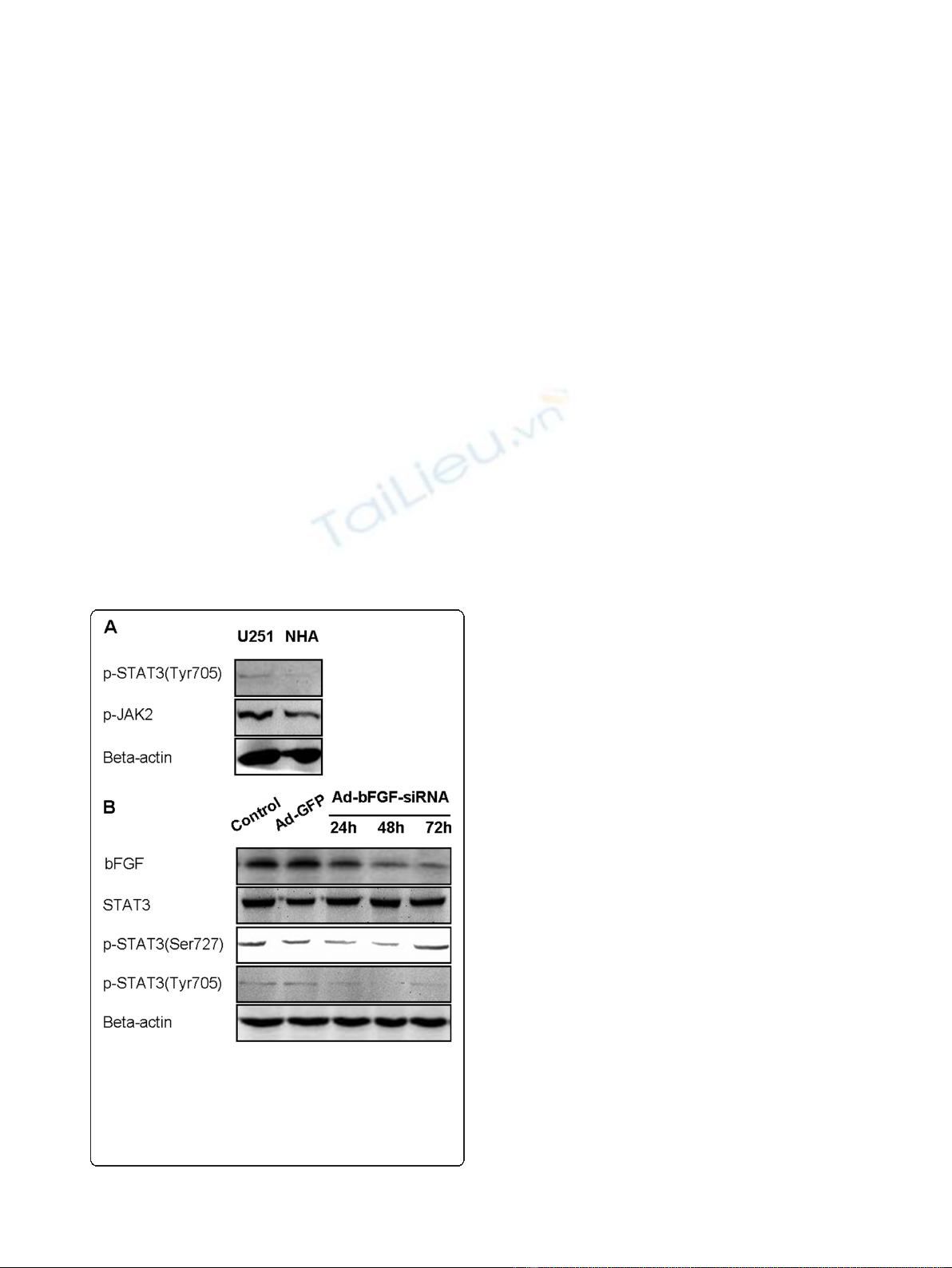
3.1 Ad-bFGF-siRNA reduces STAT3 phosphorylation at
Ser727 and Tyr705 in a time-dependent manner in U251
cells
First, to investigate whether STAT3 and upstream
kinases JAK1/2 are activated in U251 cells, we per-
formed western blot and showed a higher expression of
pSTAT3 Tyr705 and pJAK2 in the glioblastoma cell line
U251 than in NHA (Figure 1A). The level of pJAK1 was
not significantly elevated in U251 cells (data not shown).
Next, we knocked down bFGF using Ad-bFGF-siRNA,
and the decrease in bFGF protein levels was confirmed
by western blot (Figure 1B). Then, we examined whether
Ad-bFGF-siRNA treatment affects STAT3 phosphoryla-
tion. STAT3 is fully activated when both of its two con-
served amino acid residues Tyr705 and Ser727 are
phosphorylated [16]. For this propose, we extracted total
proteins from DMSO, Ad-GFP, and Ad-bFGF-siRNA
treatment groups at 24, 48, and 72 h time points and
examined the levels of total and phosphorylated STAT3
by western blot. The total STAT3 expression remained
similar among three groups across different time points
(Figure 1B). Interestingly, the expression of pSTAT3
Ser727 moderately decreased at 24 and 48 h and then
restored to the control level at 72 h. Furthermore, com-
pared with the levels under the control and Ad-GFP
treatment, the level of pSTAT3 Tyr705 under Ad-bFGF-
siRNA treatment was markedly decreased at all three
time points, even to an undetectable level at 48 h point.
Thus, these findings suggested that Ad-bFGF-siRNA
interferes with the activation of STAT3 in a time-depen-
dent manner and this decrease in pSTAT3 could not be
explained by a constitutional decrease in total STAT3.
3.2 Ad-bFGF-siRNA reduces the activation of upstream
kinases of the STAT3 signaling pathway and decreases
the levels of downstream molecules
STAT3 is regulated by upstream kinases, including
extracellular signal-regulated kinases (ERKs), JAKs, and
non receptor tyrosine kinases, including Ret, Src, and
the Bcl-Abl fusion protein [17]. Therefore, to better
understand how the upstream cascade of STAT3 is
affected by Ad-bFGF-siRNA in U251 cells, we examined
the phosphorylation of ERK1/2, JAK2, and Src under
Ad-bFGF-siRNA treatment.
Interestingly, despite similar protein levels of total
ERK1/2, when infected with Ad-bFGF-siRNA, the level
of pERK1/2 decreased at 24 and 48 h compared with
the levels in the Ad-GFP and control groups and
increased to the control level at 72 h (Figure 2A). Simi-
larly, while no change in total JAK2 was observed, the
level of pJAK2 decreased at 24, 48, and 72 h time points
(Figure 2A). In contrast, after bFGF knockdown, the
total and phosphorylated Src decreased at 48 h in a
similar manner, indicating that the phosphorylation/acti-
vation of Src is probably not affected by bFGF knock-
down (Figure 2A).
To further explore the inhibition of STAT3 phosphor-
ylation by Ad-bFGF-siRNA,weexaminedthelevelsof
two downstream targets of STAT3: CyclinD1, which
regulates cell cycle, and Bcl-xl, which is an important
apoptosis-suppressor and is usually down-regulated in
apoptotic cells. As shown in Figure 2B, at the 72 h time
point, the levels of both CyclinD1 and Bcl-xl in the Ad-
bFGF-siRNA group were significantly decreased com-
pared with the levels in the Ad-GFP and control groups.
3.3 Correlation between pSTAT3 down-regulation and IL-
6 secretion induced by Ad-bFGF-siRNA
GBM cells secrete IL-6 both in an autocrine and local-
crine way, and this IL-6 secretion is responsible for the
persistent activation of STAT3 in GBM [18]. To exam-
ine whether Ad-bFGF-siRNA inhibits STAT3
Figure 1 Ad-bFGF-siRNA reduces STAT3 phosphorylation in
U251 cells. (A) Western blot analysis revealed that the levels of
pSTAT3 (Tyr705) and pJAK2 are higher in U251 cells than in normal
human astrocytes (NHA). (B) Ad-bFGF-siRNA (MOI = 100) reduces
STAT3 phosphorylation (both Tyr705 and Ser727) in a time-
dependent manner in U251 cells. Total STAT3 expression remains
stable.
Liu et al.Journal of Experimental & Clinical Cancer Research 2011, 30:80
http://www.jeccr.com/content/30/1/80
Page 3 of 7
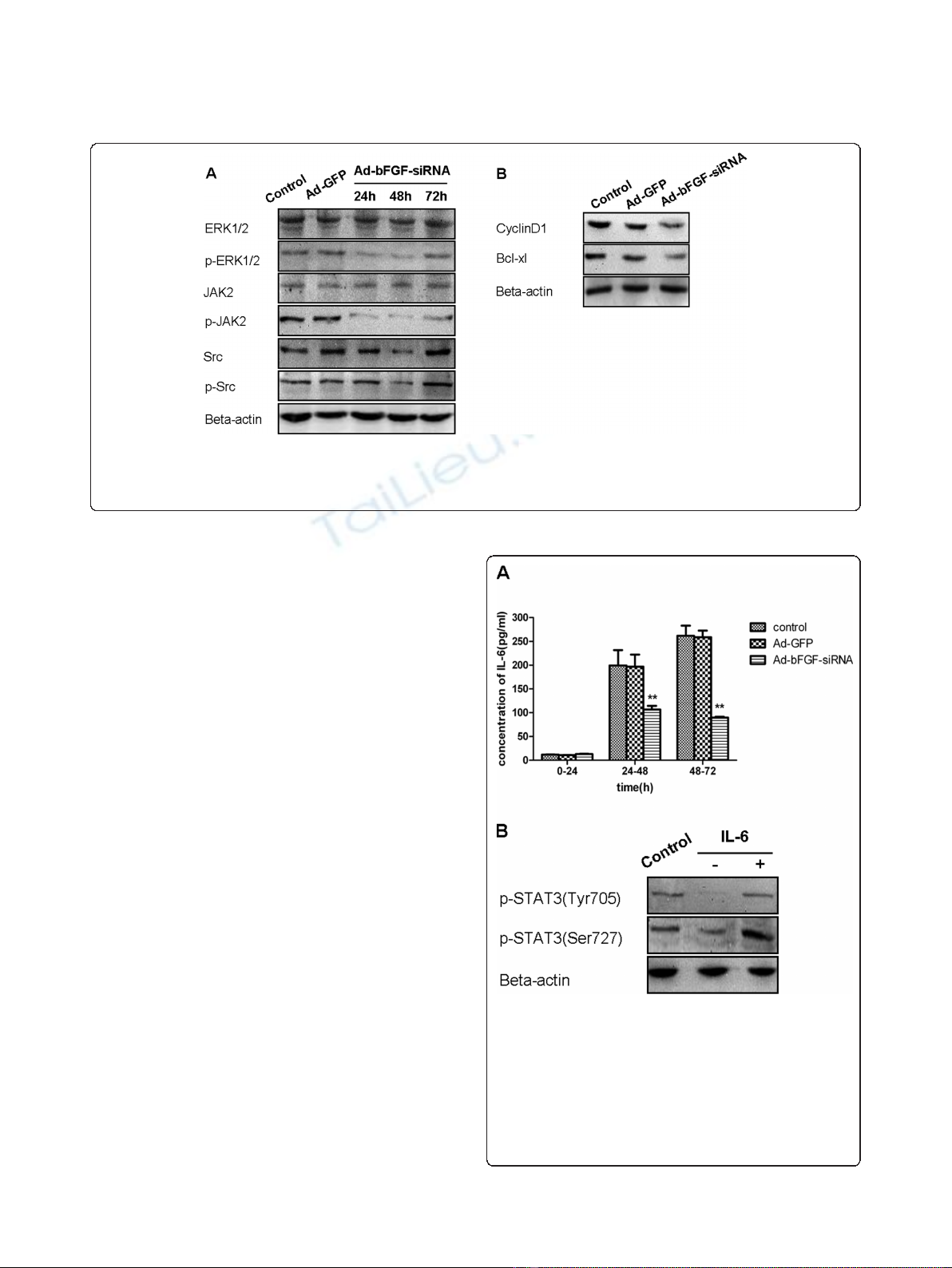
phosphorylation by reducingIL-6secretion,wetested
the IL-6 level in the supernatant of U251 cells. The level
of IL-6 was very low during the first 24 h and no signifi-
cant difference was observed between the three groups
(concentration in pg/mL: control: 11.93 ± 0.34; Ad-GFP:
10.92 ± 0.14; and Ad-bFGF-siRNA: 13.15 ± 0.74) (Figure
3A). During 24-72 h, the IL-6 level in the control and
Ad-GFP groups increased markedly (24-48 h: control:
199.46 ± 32.11 and Ad-GFP: 196.99 ± 25.24; 48-72 h:
control: 261.74 ± 21.47 and Ad-GFP: 258.50 ± 14.21)
(Figure 3A). In contrast, the IL-6 level in the Ad-bFGF-
siRNA group, although increased from that of the first
24 h, was significantly lower than that of the control
and Ad-GFP groups (p < 0.0001; 24-48 h: 106.66 ± 7.70;
48-72 h: 89.87 ± 1.82) (Figure 3A). In conclusion, Ad-
bFGF-siRNA inhibits IL-6 cytokine expression in a
time-dependent manner.
To explore whether exogenous IL-6 can rescue Ad-
bFGF-siRNA-inhibited STAT3 activation, U251 cells
infected for 48 h were treated with serum-free DMEM
in the presence or absence of recombinant IL-6 (100
ng/ml) for 24 h. Cells treated with DMSO for 72 h were
used as a negative control. As shown in Figure 3B, the
phosphorylation of STAT3 at both Tyr705 and Ser727
was elevated after stimulated with IL-6 for 24 h.
3.4 Ad-bFGF-siRNA induces depolarization of
mitochondria and apoptosis in U251 cells
Given the central role of mitochondria in orchestrating
the apoptotic processes, we assessed the mitochondrial
transmembrane potential (ΔΨm) after bFGF knockdown
by Ad-bFGF-siRNA using JC-1 staining. JC-1 forms high
orange-red fluorescent J-aggregates (FL-2 channel) at
Figure 2 Ad-bFGF-siRNA reduces the activation of upstream molecules and the expression of downstream molecules of STAT3 in
U251 cells. (A) Ad-bFGF-siRNA (MOI = 100) reduces the phosphorylation/activation of ERK1/2 and JAK2 in a time-dependent manner in U251
cells. Total ERK1/2 and JAK2 expression remains stable. Total and phosphorylated Src decreases at 48 h in a similar manner. (B) Ad-bFGF-siRNA
(MOI = 100) reduces the expression of CyclinD1 and Bcl-xl at 72 h time point.
Figure 3 Ad-bFGF-siRNA reduces IL-6 secretion in U251 cells.
(A) ELISA analysis showed that IL-6 secretion in the Ad-bFGF-siRNA
group (MOI = 100) was lower than that in the control and Ad-GFP
groups during both 24-48 h and 48-72 h periods. **: p < 0.0001.
Data are presented as mean ± SD, n = 3. (B) U251 cells infected
with Ad-bFGF-siRNA for 48 h were treated with serum-free DMEM
in the presence or absence of recombinant IL-6 (100 ng/ml) for 24
h. Cells treated with DMSO for 72 h served as controls. The
phosphorylation of STAT3 at both Tyr705 and Ser727 is elevated
after stimulated with IL-6 for 24 h.
Liu et al.Journal of Experimental & Clinical Cancer Research 2011, 30:80
http://www.jeccr.com/content/30/1/80
Page 4 of 7
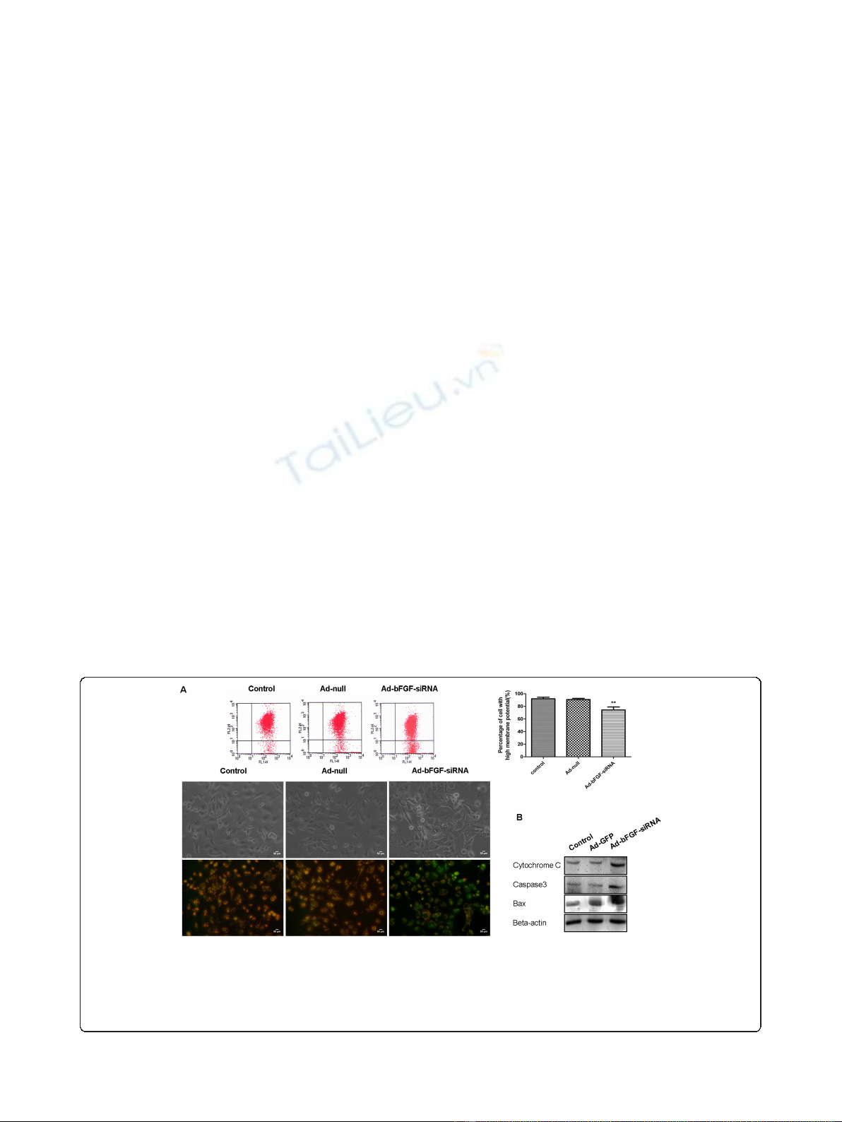
hyperpolarized membrane potentials and weak green
fluorescent monomers (FL-1 channel) at depolarized
membrane potentials. The results showed that the con-
trol and Ad-Null cells exhibited high orange-red fluores-
cence and weak green fluorescence (Figure 4A),
indicating hyperpolarized mitochondria. In contrast,
after treated with Ad-bFGF-siRNA (MOI = 100) for 72
h, an increased subpopulation of cells displayed
decreased orange-red fluorescence, suggesting the col-
lapse of mitochondrial membrane potentials. The ratio
of cells with high membrane potentials in the Ad-bFGF-
siRNA group (90.87 ± 1.84%) decreased significantly
from that in the control and Ad-Null groups (92.12 ±
2.50% and 74.42 ± 4.66%, respectively; p < 0.0005)
Furthermore, to reveal whether apoptosis is triggered
by Ad-bFGF-siRNA, we examined the levels of three
important players in apoptosis: Cytochrome C, Cas-
pase3, and Bax. As shown in Figure 4B, the level of
Cytochrome C, Caspase3, and Bax was markedly higher
in the Ad-bFGF-siRNA group than in the control and
Ad-GFP groups, confirming the activation of apoptosis
under Ad-bFGF-siRNA treatment.
4. Discussion
Recent studies have demonstrated that over-activation of
STAT3 is observed in several human malignant tumors
and cell lines, including glioblastoma [19,20]. Abnormal
and constitutive activation of STAT3 may be responsible
for glioma progression through regulating the expres-
sion of target genes, such as CyclinD1, Bcl-xl, IL-10, and
VEGF, whereas functional inactivation of STAT3 by
dominant-negative STAT3 mutants inhibits proliferation
and induce apoptosis of glioma [21]. Since STAT3 is
activated by cytokine receptor-associated tyrosine
kinasesorgrowthfactorreceptorintrinsictyrosine
kinases, besides antagonizing the function of relevant
kinases or receptors, targeting the over-expressed
ligands that inappropriately stimulate the activation of
STAT3 is also a promising strategy for glioma [22].
In this study, we provided evidence that Ad-bFGF-
siRNA can inhibit the phosphorylation of STAT3 by
down regulating the activation of ERK1/2 and JAK2, but
not Src signaling transduction (Figure 1 and 2). This
inhibition of STAT3 phosphorylation/activation subse-
quently down-regulates downstream substrates of
STAT3 and induces mitochondria-related apoptosis in
U251 cells (Figure 2 and 4). Importantly, the aberrant
expression of IL-6 in GBM cells is also interrupted by
Ad-bFGF-siRNA (Figure 3), which could be a potential
mechanism for Ad-bFGF-siRNA to serve as a targeted
therapy for glioma in vitro and in vivo.
bFGF exerts functions via its specific binding to the
high affinity transmembrane tyrosine kinase receptors
[23] and the low affinity FGF receptors (FGFR1-4) [24].
The binding of bFGF by FGFRs causes dimerization and
autophosphorylation of receptors and subsequently acti-
vates serine-threonine phosphorylation kinases such as
Raf, which triggers the classic Ras-Raf-MEK-MAPK
(ERK) signaling pathway [25]. As a central component
of the MAPK cascade, over-activated ERK1/2 contri-
butes to malignant transformation [26]. After ERK1/2 is
phosphorylated and dimerized, it translocates into the
Figure 4 Ad-bFGF-siRNA reduces the mitochondrial transmembrane potential (ΔΨm) and induces apoptosis in U251 cells.(A)
Cytofluorimetric analysis using JC-1 staining demonstrated that Ad-bFGF-siRNA treatment (MOI = 100) induces depolarization of mitochondria.
Percentages of cells with high ΔΨm (%) are shown in each column. Data are represented as mean ± SD of three replicates (**: P < 0.0005).
Changes in ΔΨm were also detected by fluorescence microscopy. Magnification: 200×. Scale bar: 50 μm. Normal cells that have high ΔΨm show
punctuate yellow fluorescence. Apoptotic cells show diffuse green fluorescence because of the decrease in mitochondrial membrane potential.
(B) Western blot analysis revealed that Ad-bFGF-siRNA (MOI = 100 for 72 h) increases the expressions of Cytochrome C, Caspase3, and Bax.
Liu et al.Journal of Experimental & Clinical Cancer Research 2011, 30:80
http://www.jeccr.com/content/30/1/80
Page 5 of 7


%20--%3e%3cdefs%3e%3cstyle%3e%20.st0%20{%20fill:%20%23fff;%20}%20.st1%20{%20fill:%20%237800fa;%20}%20%3c/style%3e%3c/defs%3e%3cpath%20class='st1'%20d='M117.78,12.18H43.11c2.9,3.47,4.65,7.94,4.65,12.82,0,5.6-2.3,10.66-6.01,14.29h76.02l7.22-13.56-7.22-13.56Z'/%3e%3cg%3e%3cpath%20class='st0'%20d='M53.58,26.17h-.59v-1.46h.59v-4.96h2.83c1.78,0,2.67.94,2.67,2.82v5.76c0,1.87-.89,2.81-2.67,2.81h-2.83v-4.96ZM55.36,21.37v3.34h1.1v1.46h-1.1v3.34h1.01c.61,0,.91-.37.91-1.1v-5.93c0-.74-.3-1.1-.91-1.1h-1.01Z'/%3e%3cpath%20class='st0'%20d='M65.99,31.14h-1.8l-.31-2.07h-2.19l-.31,2.07h-1.64l1.82-11.39h2.62l1.82,11.39ZM65.28,18.04c-.25.46-.51.77-.75.94-.21.15-.47.22-.79.22-.26,0-.57-.07-.92-.22l-.38-.15c-.14-.05-.26-.07-.37-.07-.3,0-.53.18-.71.54l-.91-.68c.25-.46.51-.77.75-.94.21-.14.48-.21.79-.21.26,0,.57.07.92.21l.38.15c.14.05.26.07.37.07.3,0,.53-.18.71-.54l.91.68ZM61.91,27.52h1.73l-.87-5.76-.87,5.76Z'/%3e%3cpath%20class='st0'%20d='M74.53,26.89v1.52c0,1.91-.89,2.86-2.67,2.86s-2.67-.95-2.67-2.86v-5.93c0-1.91.89-2.86,2.67-2.86s2.67.95,2.67,2.86v1.11h-1.69v-1.22c0-.75-.31-1.12-.93-1.12s-.93.37-.93,1.12v6.15c0,.74.31,1.11.93,1.11s.93-.37.93-1.11v-1.63h1.69Z'/%3e%3cpath%20class='st0'%20d='M81.4,31.14h-1.8l-.31-2.07h-2.19l-.31,2.07h-1.64l1.82-11.39h2.62l1.82,11.39ZM75.9,19.2l1.52-1.91h1.71l1.51,1.91h-1.61l-.76-.95-.75.95h-1.61ZM77.32,27.52h1.73l-.87-5.76-.87,5.76ZM83.1,15.99l-1.76,1.91h-1.26l1.17-1.91h1.86Z'/%3e%3cpath%20class='st0'%20d='M84.86,19.75c1.78,0,2.67.94,2.67,2.82v1.48c0,1.87-.89,2.81-2.67,2.81h-.85v4.28h-1.79v-11.39h2.64ZM84.01,21.37v3.86h.85c.58,0,.87-.36.87-1.08v-1.71c0-.71-.29-1.07-.87-1.07h-.85Z'/%3e%3cpath%20class='st0'%20d='M93.51,19.75c1.78,0,2.67.94,2.67,2.82v1.48c0,1.87-.89,2.81-2.67,2.81h-.85v4.28h-1.79v-11.39h2.64ZM92.66,21.37v3.86h.85c.58,0,.87-.36.87-1.08v-1.71c0-.71-.29-1.07-.87-1.07h-.85Z'/%3e%3cpath%20class='st0'%20d='M98.8,31.14h-1.79v-11.39h1.79v4.88h2.03v-4.88h1.83v11.39h-1.83v-4.88h-2.03v4.88Z'/%3e%3cpath%20class='st0'%20d='M105.36,24.55h2.46v1.62h-2.46v3.34h3.09v1.63h-4.88v-11.39h4.88v1.63h-3.09v3.18ZM108.17,17.29l-1.76,1.91h-1.26l1.17-1.91h1.86Z'/%3e%3cpath%20class='st0'%20d='M112.2,19.75c1.78,0,2.67.94,2.67,2.82v1.48c0,1.87-.89,2.81-2.67,2.81h-.85v4.28h-1.79v-11.39h2.64ZM111.35,21.37v3.86h.85c.58,0,.87-.36.87-1.08v-1.71c0-.71-.29-1.07-.87-1.07h-.85Z'/%3e%3c/g%3e%3ccircle%20class='st1'%20cx='25'%20cy='25'%20r='20'/%3e%3cpath%20class='st0'%20d='M32.78,19.27c2.92,0,4.43,2.55,5.28,5.33l.71,2.17c.14.38-.33.75-.71.75h-5.61c.19-.33.24-.71.09-1.08l-.75-2.45c-.43-1.32-.99-2.64-1.79-3.77.75-.57,1.65-.94,2.78-.94h0ZM25,18.38c3.25,0,4.9,2.78,5.89,5.89l.76,2.45c.14.42-.33.8-.8.8h-11.69c-.42,0-.94-.38-.8-.8l.75-2.45c.99-3.11,2.64-5.89,5.89-5.89h0ZM25,11.35c1.74,0,3.11,1.37,3.11,3.11s-1.37,3.11-3.11,3.11-3.11-1.41-3.11-3.11,1.41-3.11,3.11-3.11h0ZM17.27,19.27c1.08,0,1.98.38,2.73.94-.8,1.13-1.37,2.45-1.74,3.77l-.8,2.45c-.14.38-.05.75.09,1.08h-5.56c-.42,0-.9-.38-.75-.75l.71-2.17c.9-2.78,2.41-5.33,5.33-5.33h0ZM17.27,12.91c1.51,0,2.78,1.27,2.78,2.83s-1.27,2.83-2.78,2.83-2.83-1.27-2.83-2.83,1.27-2.83,2.83-2.83h0ZM32.78,12.91c1.56,0,2.78,1.27,2.78,2.83s-1.23,2.83-2.78,2.83-2.83-1.27-2.83-2.83,1.27-2.83,2.83-2.83h0ZM27.07,28.56v.09c0,.57-.24,1.08-.61,1.46h0v.05c-.38.33-.9.57-1.46.57s-1.08-.24-1.46-.61h0c-.38-.38-.61-.9-.61-1.46v-.09h1.41v.09c0,.19.05.38.19.47v.05c.09.09.28.19.47.19s.38-.09.47-.19v-.05c.14-.09.24-.28.24-.47t-.05-.09h1.41ZM30.99,28.56v.09c0,1.65-.66,3.16-1.74,4.24-1.08,1.08-2.59,1.79-4.24,1.79s-3.16-.71-4.24-1.79l-.05-.05c-1.04-1.08-1.7-2.55-1.7-4.2v-.09h1.41v.09c0,1.27.47,2.4,1.27,3.25h.05c.85.85,1.98,1.37,3.25,1.37s2.4-.52,3.25-1.37c.85-.8,1.37-1.98,1.37-3.25v-.09h1.37ZM34.99,28.56v.09c0,2.78-1.13,5.28-2.92,7.07-1.79,1.79-4.29,2.92-7.07,2.92s-5.23-1.13-7.07-2.92c-1.79-1.79-2.92-4.29-2.92-7.07v-.09h1.41v.09c0,2.4.94,4.53,2.5,6.08,1.56,1.56,3.72,2.5,6.08,2.5s4.52-.94,6.08-2.5c1.56-1.56,2.5-3.68,2.5-6.08v-.09h1.41Z'/%3e%3c/svg%3e)