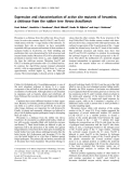
Dermaseptin DA4, although closely related to dermaseptin B2, presents chemotactic and Gram-negative selective bactericidal activities Constance Auvynet1,2,*, Pierre Joanne2,(cid:2), Julie Bourdais3, Pierre Nicolas2,(cid:2), Claire Lacombe2,4,(cid:3) and Yvonne Rosenstein1
1 Departamento de Medicina Molecular y Bioprocesos, Instituto de Biotecnologia, Universidad Nacional Auto´ noma de Me´ xico, Col Chamilpa, Cuernavaca, Morelos, Mexico 2 FRE 2852, Peptidome de la peau des amphibiens, CNRS ⁄ Universite´ Paris–Pierre et Marie Curie, Paris, France 3 Independent scholar, Cuernavaca, Morelos, Mexico 4 UFR Sciences et Technologie, Universite´ Paris 12–Val de Marne, Cre´ teil, France
Keywords antimicrobial peptide; chemotaxis; dermaseptin; frog skin; peptide–membrane interactions
Correspondence Y. Rosenstein, Departamento de Medicina Molecular y Bioprocesos, Instituto de Biotecnologia, Universidad Nacional Auto´ noma de Me´ xico, Avenida Universidad 2001, Col Chamilpa, Cuernavaca, Morelos 62270, Mexico Fax: +52 73172388 Tel: +52 5 55 66 22 76 63 E-mail: yvonne@ibt.unam.mx C. Lacombe, Laboratoire des Biomole´ cules, Universite´ Pierre et Marie Curie-CNRS-ENS, 4 Place Jussieu, 75252 Paris cedex 05, France Fax: +33 1 44 27 55 64 Tel: +33 1 44 27 51 59 E-mail: claire.lacombe@upmc.fr
Antimicrobial peptides participate in innate host defense by directly elimi- nating pathogens as a result of their ability to damage the microbial mem- brane and by providing danger signals that will recruit innate immune cells to the site of infection. Dermaseptin DA4 (DRS-DA4), a new antimicrobial peptide of the dermaseptin superfamily, was identified based on its chemo- tactic properties, contrasting with the currently used microbicidal proper- ties assessment. The peptide was isolated and purified by size exclusion HPLC and RP-HPLC from the skin of the Mexican frog, Pachymedu- sa dacnicolor. MS and amino acid sequence analyses were consistent with the structure GMWSKIKNAGKAAKAAAKAAGKAALGAVSEAM. CD experiments showed that, unlike most antimicrobial peptides of the derm- aseptin superfamily, DRS-DA4 is not structured in the presence of zwitteri- onic lipids. DRS-DA4 is a potent chemoattractant for human leukocytes and is devoid of hemolytic activity; in addition, bactericidal tests and mem- brane perturbation assays on model membranes and on Escherichia coli and Staphylococcus aureus strains have shown that the antibacterial effects of DRS-DA4 and permeabilization of the inner membrane are exclusively selective for Gram-negative bacteria. Interestingly, despite high sequence homology with dermaseptin S4, dermaseptin B2 was not able to induce directional migration of leukocytes, and displayed a broader bactericidal spectrum. A detailed structure–function analysis of closely related peptides with different capabilities, such as DRS-DA4 and dermaseptin B2, is criti- cal for the design of new molecules with specific attributes to modulate immunity and/or act as microbicidal agents.
Present addresses *INSERM UMR-S 945 Immunite´ et Infec- tion, Universite´ Pierre et Marie Curie, Paris, France (cid:2)Biogene` se des Signaux Peptidiques (BIOSIPE), ER3-UPMC, Universite´ Pierre et Marie Curie, Paris, France
Abbreviations DDK, dermadistinctin K; DiSC3(5), 3,3¢-dipropylthiadicarbocyanine iodide; DMPC, 1,2-dimyristoyl-sn-glycero-3-phosphatidylcholine; DMPG, 1,2-dimyristoyl-sn-glycero-3-phosphatidylglycerol; DRS-B2, dermaseptin B2; DRS-DA3, dermaseptin DA3; DRS-DA4, dermaseptin DA4; DRS-L1, dermaseptin L1; DRS-S1, dermaseptin S1; DRS-S9, dermaseptin S9; DSC, differential scanning calorimetry; ERK, extracellular signal-regulated kinase; fMLP, formyl-methionyl-leucyl-phenylalanine; FPR, formyl peptide receptor; FPRL-1, formyl peptide receptor-like 1; Gal-ONp, 2-nitrophenyl b-D-galactopyranoside; GPCR, G-protein-coupled receptor; ITC, isothermal titration calorimetry; LUV, large unilamellar vesicle; MAPK, mitogen-activated protein kinase; MIC, minimum inhibitory concentration; MLV, multilamellar liposome vesicle; PMN, polymorphonuclear; PTX, pertussis toxin; SDF1-a, stromal cell-derived factor 1a; TFA, trifluoroacetic acid.
FEBS Journal 276 (2009) 6773–6786 ª 2009 The Authors Journal compilation ª 2009 FEBS
6773
C. Auvynet et al.
Chemotactic and antimicrobial DRS-DA4
(cid:3)Laboratoire des Biomole´ cules, Universite´ Pierre et Marie Curie-CNRS-ENS, Paris cedex 05, France
(Received 12 August 2009, accepted 21 September 2009)
doi:10.1111/j.1742-4658.2009.07392.x
Introduction
the interface of
but no or little hemolytic activity [8]. The microbicidal activity of these lysine-rich linear polycationic peptides, most of which are composed of 24–34 amino acids structured as an amphipathic a-helix in polar solvents, is thought to result from the interaction of the amphi- pathic a-helical structure with the membrane bilayer of target microorganisms.
Most peptides belonging to the dermaseptin super- family have been identified primarily on the basis of their antimicrobial activity. However, additional bio- logical functions have been recognized that may, or may not, be directly associated with pathogen clearance. For instance, adenoregulin [dermaseptin B2 (DRS-B2)] was first identified as a peptide able to stimulate binding of agonists to A1-adenosine receptors [9], and was further shown to enhance the binding potency of several GPCR agonists [10]. Frog skin insulintropic peptide (FSIP), also a member of this superfamily, significantly stimu- lates insulin release in glucose-responsive BRIN-BD 11 cells [11], and dermaseptin S1 (DRS-S1) has been reported to stimulate the microbicidal activity of poly- morphonuclear (PMN) leukocytes [12] and dermaseptin S9 (DRS-S9) to chemoattract PMN leukocytes [13].
At innate and adaptive immunity, antimicrobial peptides have been shown to enhance the overall immune response [1]. The majority of these peptides are cationic, with a net charge of +2 to +7, and contain up to 50% hydrophobic amino acids. This amphipathic design, consisting of spatially separated hydrophobic and charged regions, is believed to allow the insertion of the peptide into microbial membranes. Until recently, direct antimicrobial activity against bacteria, fungi, parasites and viruses was considered to be the primary function of antimicrobial peptides. However, there is now increasing evidence that anti- microbial peptides are multifunctional molecules of fundamental importance in host defense, modulating the innate and adaptive immune systems. In addition to microbicidal activity, a large number of antimicro- bial peptides, such as the human cathelicidin LL-37 and the defensins, have been found to modulate the immune response by directing the migration of immune cells to the site of injury, as well as by activating leuko- cytes and promoting cytokine release, wound repair, angiogenesis, and neutralization of microbial products [2,3]. In particular, the chemotactic activity of antimi- crobial peptides is mostly mediated through G-protein- coupled receptors (GPCRs) such as the CC-chemokine receptor 6, the formyl peptide receptor (FPR), and the formyl peptide receptor-like 1 (FPRL-1) [4–7].
Frog skin is a rich source of antimicrobial peptides, with more than half of the peptides described to date having been isolated from South American Hylidae or European, Asian or North American Ranidae; the peptides are involved in the defense of the frog against predation or invading microorganisms. More than 80 antimicrobial peptides have been isolated from only 12 species of the Phyllomedusinae subfamily, belonging to the genera Agalychnis, Hylomantis, Pachymedusa, and Phyllomedusa. Among them, the dermaseptins, a super- family of structurally and functionally related peptides produced by the Hylidae family, have potent micro- bicidal activity at micromolar concentrations against a wide range of microorganisms (Gram-positive and Gram-negative bacteria, fungi, yeasts, and protozoa),
We report herein the isolation and characterization of a new dermaseptin-related peptide, GMWSKIKNA GKAAKAAAKAAGKAALGAVSEAM, named derm- aseptin DA4 (DRS-DA4), according to the new derm- aseptin nomenclature [14]. DRS-DA4 was obtained by fractioning the skin exudate of Pachymedusa dacnicol- or, and was first identified on the basis of its chemo- tactic properties rather than on the classic assessment its antimicrobial activity. Interestingly, although of DRS-DA4 was found to share strong sequence homol- ogy with DRS-B2, it has distinct biological activities. DRS-DA4, but not DRS-B2, induced human leukocyte migration and activation mainly through a GPCR, probably FPRL-1; moreover, it was devoid of hemo- lytic activity. Unlike DRS-B2, which is active on Gram-positive as well as on Gram-negative bacteria, DRS-DA4 only exhibited direct antibacterial activity on Gram-negative bacteria, together with perturbation the inner membrane against of Gram-negative of
FEBS Journal 276 (2009) 6773–6786 ª 2009 The Authors Journal compilation ª 2009 FEBS
6774
C. Auvynet et al.
Chemotactic and antimicrobial DRS-DA4
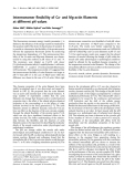
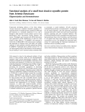
RP-HPLC on a semipreparative column (Fig. 1B). A peak with a retention time of 38.7 min was found to have strong chemotactic activity, inducing the direc- tional migration of human leukocytes (Fig. 1B, insert). The sequence of the purified fraction determined by tandem MS (experimental monoisotopic mass of the protonated peptide: 3063.26) and Edman sequencing gave unequivocally the sequence GMWSKIKNAGKA AKAAAKAAGKAALGAVSEAM. According to the new dermaseptin nomenclature [14], this peptide was named DRS-DA4.
bacteria. The identification of a novel antimicrobial peptide on the basis of its immunomodulatory capacity broadens the panel of activities of these peptides within Hylidae frog genera. In addition, our analysis the biophysical characteristics and properties of of these peptides contributes to our current knowledge regarding the different activities of antimicrobial peptides, and opens the possibility of using them as templates for the design of new molecules with specific attributes to modulate immunity and ⁄ or act as micro- bicidal agents.
Results
Isolation, purification and structure of DRS-DA4
DRS-DA4 was purified to homogeneity from P. dacni- color skin exudates by a two-step protocol. Specifically, 1.1 mL of skin secretions recovered by gently squeez- ing the parotoid glands of a single living frog was first fractionated on a Sephadex G-50 column (Fig. 1A), and each fraction was tested for chemotactic activity. The chemotactic fraction III was further purified by
To confirm the sequence and to demonstrate that the biological activities of the purified natural peptide reflected its intrinsic properties, DRS-DA4 was synthe- sized by the solid-phase method. After HPLC purifica- tion on a semipreparative column, synthetic DRS-DA4 was indistinguishable from the natural product, eluting exactly at the same position (38.7 min) as the natural corresponding product and giving the same monoi- sotopic mass to charge ratio (3063.28) by MALDI-TOF MS (data not shown). Further characterization of conformational and biological properties was the performed with the synthetic peptide.
A
0.9
0.8
V0
)
0.7
Fraction II
0.6
Fraction III
0.5
Fraction I
0.4
0.3
0.2
m n 0 8 2 ( e c n a b r o s b A
0.1
0.0
0
10
20
30
40
50
60
70
Fraction number
B
A c e t o n i t r i l
e (
%
)
Fig. 1. (A) Profile of fractionation of the P. dacnicolor skin exudates on a Sephadex G-50 column. The absorbance at 280 nm is represented as a solid line. (B) RP-HPLC separation of the recovered fraction III using a semipreparative column. Elution was achieved with a 0–60% linear gradient of solvent (dotted line). The arrow points to the elution position of synthetic DRS-DA4 under the same conditions. The absorbance at 220 nm is represented as a solid line. Insert: neutrophil migration induced in response to the peak indicated by the arrow. The peak solution was diluted 1 ⁄ 10 from column 6 to column 1. RPMI medium was used as negative control (column 0) and fMLP (100 nM) as positive control.
FEBS Journal 276 (2009) 6773–6786 ª 2009 The Authors Journal compilation ª 2009 FEBS
6775
C. Auvynet et al.
Chemotactic and antimicrobial DRS-DA4
A
B
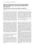
Fig. 2. (A) Amino acid sequences of DRS-DA4 and DRS-B2, together with some antimicrobial peptides with nearest sequences. Identical amino acids between these peptides are underlined. (B) Helical wheel projection of the DRS-DA4, DRS-B2, DDK, and DRS-L1. Hexagonal and round backgrounds refer to basic and acid amino acids respectively, pentagonal backgrounds to hydrophilic residues, and squares to hydrophobic residues.
A protein database was
screened for
similar sequences, using the embl-ebi fasta 3 program, and this revealed that DRS-DA4 belonged to the derm- aseptin superfamily. A sequence alignment showed
that the antimicrobial peptides dermaseptin DA3 [DRS-DA3 (PD-33)] [15], DRS-B2 [9], dermadistinctin isolated from K (DDK) Phyllomedusa P. dacnicolor,
[16] and ARP-AC1 [17], Phyllomedusa bicolor,
FEBS Journal 276 (2009) 6773–6786 ª 2009 The Authors Journal compilation ª 2009 FEBS
6776
C. Auvynet et al.
Chemotactic and antimicrobial DRS-DA4
and Agalychnis callidryas,
A
84.8% identity
B
distincta, respectively, presented the highest sequence homology with DRS- DA4 (87.9% identity and 93.9% similarity for DRS-DA3, and 97% similarity for DRS-B2, and 84.8% identity and 93.9% similarity for DDK; Fig. 2A). Both, DRS-DA3 and DRS-B2 have six positive charges (Lys), and only one negative charge (Glu). The resulting total overall net charge for DRS-DA4 is +5, whereas DRS-B2 has an overall net charge of +4, owing to the carboxyamidated end, three negative charges (Glu) and the extra positive charge of the N-terminus. Figure 2A shows dermaseptin L1 (DRS-L1) [18] from the lemur leaf frog Hyloman- tis lemur, which showed only 48.6% identity and 22.4% similarity but whose biological properties led us to make a comparison. The helical wheel projection of Edmundson showed the partial amphipathic character of the helix obtained for DRS-DA4, with hydrophobic residues on one face of the helix and polar or charged residues on the opposite face, corresponding to a hydrophobic sector that subtends a radial angle of 160(cid:2) (Fig. 2B).
DRS-DA4 induces directional migration of human leukocytes
triplicate wells. Similar
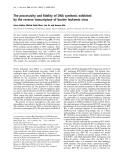
(A) Fig. 3. DRS-DA4 is a potent chemoattractant of leukocytes. Neutrophil migration induced in response to DRS-DA4 or fMLP (100 nM) as a positive control and medium as a negative control. Addition of the same concentrations of DRS-DA4 to the upper and lower wells of the chemotaxis chamber abolished the chemotactic effect. (B) Monocyte migration in response to DRS-DA4, DRS-B2 or SDF1-a (1 nM) as positive control. The data shown represent the average cell migration of results were obtained from three different experiments.
rather
Like natural DRS-DA4, synthetic DRS-DA4 induced the migration of human neutrophils (Fig. 3A) and monocytes (Fig. 3B), with a typical bell-shaped dose– response curve. Maximum activity was observed at a concentration of 10 lm for both cell types. Addition of various concentrations of DRS-DA4 to the upper wells of the Boyden chamber abolished the migration of the cells, suggesting that DRS-DA4 induced che- motactic movement than enhanced random movement (Fig. 3A). As described previously [13], DRS-B2 did not induce the migration of leukocytes within the range of concentrations tested for DRS- DA4.
of the cells with 1.14 nm fMLP, a concentration at which fMLP is considered to be an FPR agonist, did not inhibit the motility of the cells in response to DRS-DA4 or fMLP (100 nm). As expected, preincuba- tion of neutrophils with fMLP (1.14 nm) prior to the chemotaxis assay inhibited the migration induced by fMLP (1.14 nm) (Fig. 4A).
a
through
seven-helix
Like formyl-methionyl-leucyl-phenylalanine (fMLP), most chemotactic antimicrobial peptides induce cell transmembrane migration Gia-protein-coupled receptor such as FPR or FPRL-1 [19]. Pretreatment with pertussis toxin (PTX) – a specific inhibitor of Gia-protein-coupled receptors – prior to the onset of the chemotactic assay partially inhibited the motility of neutrophils induced by DRS-DA4. As expected, neutrophils preincubated with PTX before exposure to fMLP failed to migrate (Fig. 4A).
Preincubation of neutrophils for 30 min with 100 nm fMLP, an FPRL-1 agonist, inhibited by up to 50% the migration of the cells in response to a gradient of DRS-DA4 or fMLP (100 nm). However, preincubation
Stimulation of a GPCR by chemotactic agonist ligands can lead to the activation of mitogen-activated protein kinase (MAPK) pathways in target cells [20]. Incubation of neutrophils with DRS-DA4 (10 lm) or fMLP (100 nm) for 5 or 10 min induced the phos- phorylation of the extracellular signal-regulated kinase (ERK)1 ⁄ 2 MAPK, but not that of the p38 MAPK (Fig. 4B). Preincubation with PTX or PD98059 (a specific inhibitor of MEK1 and MEK2) before the addition of DRS-DA4 (10 lm) or fMLP (100 nm) inhibited the phosphorylation of ERK1 ⁄ 2 but induced
FEBS Journal 276 (2009) 6773–6786 ª 2009 The Authors Journal compilation ª 2009 FEBS
6777
C. Auvynet et al.
Chemotactic and antimicrobial DRS-DA4
A
suggest that DRS-DA4 chemotactic activity is medi- ated through a GPCR, probably FPRL-1, and that it is coupled to the ERK1 ⁄ 2 MAPK pathway.
DRS-DA4, but not DRS-B2, is only microbicidal for Gram-negative bacteria
B
PTX – PD98059 –
10 ’ 10 ’ 5 ’ 10 ’ 10 ’ 10 ’ 10 ’ 10 ’ 10 ’ 10 ’ – – + – + – + – + – –
– + + –
– + +
pErk1/2
Erk1/2
pp38
p38
DRS-DA4 exhibited good to moderate antimicrobial activity against all of the Gram-negative strains tested, as no colony was counted when the peptide–Escherichia coli (both E. coli strains tested) or peptide–Pseudo- monas aeruginosa mixtures were incubated on LB agar plates overnight, indicative of a bactericidal effect. How- ever, no microbicidal activity was detected against any of the Gram-positive strains tested, at concentrations up to 100 lm. In comparison, DRS-B2 presented strong antimicrobial activities against both Gram-negative and Gram-positive strains (Table 1). No hemolytic or apop- totic activities were observed on rat blood cells or leuko- cytes with DRS-DA4 or DRS-B2, respectively, for concentrations up to 100 lm (data not shown).
C
DRS-DA4 interacts preferentially with 1,2-dimyristoyl-sn-glycero-3-phosphatidylglycerol (DMPG) vesicles mimicking Gram-negative bacteria
fMLP (1.14 nM), or
When we used the membrane potential-sensitive dye 3,3¢-dipropylthiadicarbocyanine iodide [DiSC3(5)] to assess the ability of the peptide to damage, and thus depolarize, prokaryotic membranes [21,22], we found that addition of DRS-DA4 resulted in concentration- independent increases in DiSC3(5) fluorescence within the range 10–50 lm, indicative of equivalent membrane depolarization for both strains tested, i.e. E. coli and Staphylococcus epidermidis (Table 1). Thus, in contrast to its Gram-negative-specific microbicidal activity, DRS-DA4 was able to depolarize the membranes of Gram-positive and Gram-negative bacteria.
Fig. 4. DRS-DA4-induced neutrophil migration through a seven- transmembrane GPCR. (A) DRS-DA4-induced chemotaxis is medi- ated through a GPCR. Neutrophils were preincubated with medium, PTX, fMLP (100 nM) prior to challenge with DRS-DA4, fMLP (1.14 nM) or fMLP (100 nM). (B) ERK1 ⁄ 2 phosphor- ylation is induced in response to DRS-DA4. Human neutrophils were treated with medium, DRS-DA4, or fMLP, with or without preincubation with PTX and ⁄ or PD98059. The same membrane was stripped and blotted with antibody against ERK1 ⁄ 2 or antibody against p38. Similar results were obtained from three separate (C) ERK1 ⁄ 2 phosphorylation is necessary for DRS- experiments. DA4-induced. Human neutrophils were preincubated with PD98058 (ERK1 ⁄ 2 inhibitor) or SB202190 (p38 inhibitor) prior to the chemo- taxis assay, as described in (A). The data shown are representative of two independent experiments.
the phosphorylation of p38. Moreover, pretreatment of neutrophils with PD98059 led to decreased migra- tion in response to DRS-DA4 or fMLP (100 nm), whereas SB202190, a specific inhibitor of the p38 MAPK, did not (Fig. 4C). All together, these results
To further characterize the bactericidal activity of DRS-DA4, we measured its ability to damage the bacterial membrane. Permeabilization of the inner mem- brane can be assessed by monitoring the b-galactosidase substrate 2-nitrophenyl b-d-galactopyranoside (Gal- ONp). In E. coli strain ML35p and in Staphylococ- cus aureus strain ST1036, which lack lac permease, Gal- ONp entry is blocked by the integrity of the inner mem- brane; if Gal-ONp crosses this barrier, it can be cleaved by the cytoplasmic b-galactosidase, resulting in a color change from clear to yellow, reflecting membrane dam- age. As shown in Fig. 5A, the time needed to obtain the maximum Gal-ONp hydrolysis in E. coli increased as the concentration of DRS-DA4 decreased. On the other hand, no permeabilization of S. aureus ST1065 was
FEBS Journal 276 (2009) 6773–6786 ª 2009 The Authors Journal compilation ª 2009 FEBS
6778
C. Auvynet et al.
Chemotactic and antimicrobial DRS-DA4
Table 1. Antimicrobial activities of DRS-DA4 and DRS-B2. Bacterial strains were considered to be resistant (R) when their growth was not inhibited by peptide concentrations up to 100 lM. The data shown correspond to the MIC (lM). E. coli 363 ATCC 1175 or S. epidermidis BM 3302 transmembrane potential changes were induced by DRS-DA4 and assessed with the DiSC3(5) probe: membrane depolarization was monitored by an increase in fluorescence after the addition of peptide at the MIC. Triton X-100 was used to fully collapse the membrane potential. B, bactericidal; ND, not determined.
Antimicrobial activity (MIC)
DRS-DA4
Membrane depolarization assay
DRS-B2
Gram-negative
Gram-positive
E. coli ML35p E. coli 363 ATCC 11775 Ps. aeruginosa ATCC 27853 En. faecalis ATCC 29212 S. aureus ATCC 25923 S. epidermidis BM 3302
5, B 5, B 40, B R R R
ND 0.39, Ba 3.1, Ba ND 0.7, Ba ND
ND 55 ± 9% ND ND ND 50 ± 5%
a See ref [26].
observed for concentrations of DRS-DA4 up to 100 lm (Fig. 5B and data not shown). These results indicate that the bacterial membrane of Gram-negative strains is one of the main targets of DRS-DA4.
during the lipid fusion resulting from the insertion of the peptide molecules. The main transition was totally abolished at 1 : 20 peptide ⁄ lipid ratios, indicating total disorganization of lipid DMPG bilayer (Fig. 5C).
Like
those of DMPG, aqueous dispersions of DMPC showed two endothermic transitions, a pretran- sition occurring at 13.1 (cid:2)C with an enthalpy of about 1.1 kcalÆmol)1, and a main transition at 23.9 (cid:2)C with an enthalpy of 11 kcalÆmol)1, which was again within the range previously published [24,25]. At low con- centrations of DRS-DA4 (1 : 100 peptide ⁄ lipid ratio), no modifications of the thermogram were observed. The pretransition was then gradually abolished as the peptide/lipid ratio increased. At greater ratios only (1 : 20 peptide ⁄ lipid ratio), DRS-DA4 induced some changes to the DMPC melting profile, indicating an interaction with this type of vesicle, whereas the main transition peak was still present, without modification of its temperature.
enthalpy
for
((cid:2) 40%). The
CD spectra of DRS-DA4 (Fig. 5E) revealed that DRS-DA4 adopted an a-helix conformation in the presence of DMPG vesicles, whereas it remained as a random coil in aqueous solution and in the presence of DMPC liposomes, whatever the peptide/lipid ratio (data not shown). In the presence of DMPG vesicles, the spectrum of DRS-DA4 showed a profile with minima at 208 and 220 nm, suggesting a major contri- conformational bution of a-helix propensity of DRS-DA4 contrasts with that of DRS- B2, which has been found to be structured as an a-helix (55%), whatever the lipidic composition of the vesicles [26].
Discussion
Hundreds of antimicrobial peptides have been isolated from frogs, but very few studies dealing with their immunomodulatory capacities have been published.
Differential scanning calorimetry (DSC) is a power- ful, nondisturbing thermodynamic technique that is useful for the study of lipid–protein interactions in model membranes as well as for the evaluation of anti- microbial peptide interactions with lipid bilayer model membranes [23]. We evaluated the effect of various concentrations of DRS-DA4 on the pretransition and the main transition of the zwitterionic 1,2-dimyristoyl-sn- glycero-3-phosphatidylcholine (DMPC) and anionic DMPG membranes used as models for eukaryotic and respectively. DSC prokaryotic plasma membranes, thermograms illustrating the effect of the incorporation of increasing quantities of DRS-DA4 on the thermo- tropic phase behavior of multilamellar liposome vesi- cles (MLVs) of DMPG or DMPC are shown in Fig. 5C,D. In the absence of the peptide, DMPG exhibited two endothermic events: a less energetic pre- transition near 12.8 (cid:2)C, arising from the conversion of the lamellar phase to the rippled gel phase, and a sec- ond, more energetic, main transition at 23.2 (cid:2)C, result- ing from the conversion of the rippled gel phase to the lamellar liquid-crystalline phase. These results, together the pretransition with the values ((cid:2) 1 kcalÆmol)1) and the main transition (8.8 kcalÆ mol)1), are comparable with previous data [24,25]. The incorporation of DRS-DA4 into DMPG MLVs signifi- cantly altered their thermotropic phase behavior. The presence of the peptide abolished the pretransition, even at the lowest concentration tested (peptide/lipid ratio 1 : 100), which is within the same range of order as the minimum inhibitory concentration (MIC) for Gram-negative strains. Increasing concentrations of DRS-DA4 broadened the DMPG main phase transi- tion peak, probably because of a loss of cooperativity
FEBS Journal 276 (2009) 6773–6786 ª 2009 The Authors Journal compilation ª 2009 FEBS
6779
C. Auvynet et al.
Chemotactic and antimicrobial DRS-DA4
A
B
2.0
2.0
)
1.5
1.5
20 µM 10 µM 5 µM 1.2 µM 0.3 µM Control
1.0
1.0
m n 5 0 4 ( e c n a b r o s b A
0.5
0.5
0.0
0.0
0
2000
4000
6000
8000 10 000 12 000 14 000
0
2000
4000
8000
10 000
12 000
Time (s)
6000 Time (s)
C
D
30
30
25
25
20
20
DMPC
DMPG
15
15
10
10
1/100
1/100
) 1 – l o m · k · l a c k ( p C
) 1 – l o m · k · l a c k ( p C
5
5
0
0
1/20
1/20
5
10
15
20
25
30
35
40
5
10
15
20
25
30
35
40
Temperature (°C)
Temperature (°C)
16
E
14
12
DMPG DMPC Buffer
10
8
6
4
) 1 – m c · 1 –
M (
2
0
–2
–4
–6
190
200
210
220
230
240
250
260
Wavelength (nm)
Fig. 5. The bactericidal capacity of DRS-DA4 is linked to its interaction with DMPG. (A, B) Kinetics of bacterial membrane leakage of E. coli ML35p (A) and S. aureus ST1065 (B) after treatment with increasing concentrations of DRS-DA4. The membrane leakage was followed by measuring Gal-ONp hydrolysis at 405 nm. (C, D) DSC heating thermograms illustrating the effects of DRS-DA4 on the thermotropic phase behavior of DMPG (C) and DMPC (D) MLVs. The top scan corresponds to the lipid alone, and the peptide ⁄ lipid molar ratios of the lower scans are indicated. Thermodynamic parameters are given in Table 2. (E) CD spectra of DRS-DA4 in buffer, DMPG and DMPC with a peptide ⁄ lipid molar ratio of 1 : 50. The data shown are representative of three experiments.
The first antimicrobial peptide shown to exhibit immu- nological properties was DRS-S1, a 34 amino acid cationic antimicrobial peptide, which stimulates the microbicidal activity of PMN leukocytes [12]. A few other frog peptides, temporin A, rana-6 and pLR-like
peptides and, recently, another dermaseptin-related peptide, DRS-S9, have been shown to be microbicidal as well as immunomodulatory [13,27–29]. Here, we report the isolation, by screening for chemotactic activ- ity, of a new member of the dermaseptin superfamily,
FEBS Journal 276 (2009) 6773–6786 ª 2009 The Authors Journal compilation ª 2009 FEBS
6780
C. Auvynet et al.
Chemotactic and antimicrobial DRS-DA4
DRS-DA4, from the defensive skin secretion of the Mexican leaf frog P. dacnicolor.
through different
extent of helical
the
We showed that DRS-DA4 triggered the in vitro directional migration of human neutrophils and mono- cytes with a typical bell-shaped dose–response curve, and a maximal response at a concentration of 10 lm. Our results suggested that the chemotactic effect was mediated by the low-affinity Gia-protein-coupled recep- tor FPRL-1, recently reported to interact with many different ligands, such as fMLP, the a-helical antimi- crobial peptides LL-37 and temporin A, and the amy- loidogenic peptides Ab1–42 and, presumably, DRS-S9 [13,27,30,31] (Fig. 4B). Whether FPRL-1 interacts with its various agonists functional domains remains to be investigated. In addition, as only human leukocytes were used in this study, it remains to be discovered whether DRS-DA4 is also capable of recruiting frog leukocytes. Indeed, frog pep- tides are secreted at the outer surface of the skin, and it is presently not known whether they also enter the blood circulation or inner tissues to act on leukocytes or to modulate other biological functions.
residues on the opposite face, leading us to propose that the amphipathic a-helix structure is an important feature of these membrane-permeating peptides. Secon- dary structure prediction methods and CD spectros- copy have also shown that dermaseptins contain 45–90% helix in structure-promoting solvents [16,26, 34,39]. We illustrated here by CD (Fig. 5E) that DRS- DA4 was randomly coiled in water and, unexpectedly, also in the presence of DMPC vesicles. In contrast, DRS-DA4 adopted an a-helical structure in the pres- ence of DMPG vesicles, consistent with its cidal action against prokaryotic cells. Thus, DRS-DA4 fits the model proposed by Khandelia et al. [35], in which the composition of the target membrane (zwitterionic or anionic) modulates content induced in antimicrobial peptides. CD results are con- sistent with data from the calorimetric tests and iso- thermal titration calorimetry (ITC) experiments with DMPC large unilamellar vesicles (LUVs) (data not shown), again suggesting very weak or no interaction with this lipid. In contrast, strong perturbations of the pretransition and the main phase transition (Fig. 5C and Table 2) were recorded in the presence of DMPG, suggesting that electrostatic interactions participate in the peptide–lipid interaction, and that DRS-DA4 is able to penetrate the acyl chain region.
A highly cationic charge such as that of DRS-DA4 favors the accumulation of peptides on negatively charged DMPG bilayers via electrostatic interactions, suggesting that the bactericidal activity of DRS-DA4 towards Gram-negative bacteria results from the pref- erential binding of the peptide to the negatively charged lipopolysaccharides of the outer membrane, the subsequent membrane damage occurs and that through hydrophobic interactions with the inner target membrane, which is rich in neutral phosphatidyletha- nolamine. This hypothesis is supported by a study showing that helical amphipathicity prevails over hydrophobicity in interfacial binding, underlining the
As described for other agonists [20], the interaction of DRS-DA4, presumably with FPRL-1, led to the activation of the ERK1 ⁄ 2 MAPK pathway, but not to that of the p38 MAPK pathway. The chemotaxis data suggested that ERK phosphorylation, but not that of p38, was necessary for the chemotactic process. Inter- estingly, inhibition of FPRL-1 by PTX has been reported to activate the p38 pathway [32]. Consistent with this, inhibition of ERK1 ⁄ 2 phosphorylation by a specific inhibitor (PD98059) activated the p38 MAPK pathway in response to DRS-DA4, suggesting that DRS-DA4 may additionally bind to a non-GPCR receptor and, through a p38-dependent pathway, con- trol other cell functions such as degranulation or cyto- kine ⁄ chemokine gene expression and release. This is in accordance with studies indicating that inhibition of one signaling pathway could activate another one [27,33].
Table 2. Thermodynamic parameters obtained by DSC for the interaction of DRS-DA4 with MLVs of either DMPG or DMPC. –, no pretransition observed.
Pretransition
Transition
Lipid
Peptide ⁄ lipid ratio
DH (kcalÆmol)1)
DH (kcalÆmol)1)
T ((cid:2)C)
T ((cid:2)C)
DMPG
DMPC
0 1 : 100 1 : 20 0 1 : 100 1 : 20
12.8 – – 13.0 14.1 13.5
1 – – 1.1 1.2 0.1
23.2 23.3 23.9 23.9 24.0 23.9
9.6 10.6 4.3 11.8 10.2 5.0
The 32 amino acid DRS-DA4 exhibits the typical characteristics of the dermaseptin superfamily: it is a linear, Lys-rich cationic peptide with the conserved Trp at position 3. A blast search revealed high sequence homology of DRS-DA4 with DRS-DA3 (PD-33), also isolated from P. dacnicolor, and, more surprisingly, with DRS-B2 isolated from Ph. bicolor, DDK from Ph. distincta, and ARP-AC1 from A. cal- lidryas [17] (Fig. 2A). However, unlike these other pep- tides, DRS-DA4 is not carboxyamidated. Modeling revealed antimicrobial peptides as idealized helices their highly amphipathic nature, with hydrophobic residues on one face of the helix and polar or charged
FEBS Journal 276 (2009) 6773–6786 ª 2009 The Authors Journal compilation ª 2009 FEBS
6781
C. Auvynet et al.
Chemotactic and antimicrobial DRS-DA4
lytic activity.
data show that, like plasticins, DRS-DA4 was able to depolarize the membrane of Gram-positive (S. epider- midis ST1065) as well as of Gram-negative (E. coli ML35p) strains of bacteria, but it was toxic only for Gram-negative strains (Table 1).
shown the
localized in highly curved regions of
importance of amphipathicity as a driving force for cell In addition, conformational constraints and appropriate positioning of aromatic residues for the formation of hydrophobic clusters have been shown to be critical for antimicrobial activ- ity and selectivity [36]. Moreover, the presence of regions with different order and polarity within the membrane has existence of domains enriched in phosphatidylethanolamine or phosphatidyl- glycerol, the bacterial membrane [37]. Segregation of the membrane lipid components, leading to clustering of anionic lip- ids through an induced lateral-phase separation, and the subsequent perturbation of existing domains of the membrane has been proposed as a mechanism contrib- uting to the antimicrobial activity of numerous antimi- crobial peptides. In agreement with this, as they have significant amounts of both anionic and zwitterionic lipids, most Gram-negative bacteria are more suscepti- ble to this membrane-disrupting mechanism. Consis- tent with this, and as reported for oligo-acyl lysine [38], we found that Gram-negative bacteria were killed by DRS-DA4.
In contrast to the situation with clinically used anti- biotics, resistance to natural antimicrobial peptides is not frequent, raising interest in the use of antimicro- to fight antibiotic-resistant microbes. bial peptides Moreover, recent data have suggested that antimicro- bial peptides participate actively in preventing the appearance of resistant mutants, and are thus the last infections line of defense dealing with persistent [41]. Our data showing the antibacterial potency of DRS-DA4, resulting from its ability to interact with anionic model membranes and to induce directional locomotion of mammalian cells through a receptor, presumably FPLR-1, highlight the multifunctionality of antimicrobial peptides as antibiotics and immuno- modulatory molecules [3,42,43]. Modification of DRS- DA4 to enhance its direct bactericidal effect and to extend its antibacterial activity to Gram-positive strains, without compromising its immunomodulatory potency, could be achieved, as for dermaseptin S4, through acylation [44], or, as for magainin 2 analogs, amidation [45]. This new class of peptides will be use- ful for therapeutic application purposes.
and DDK. However, despite
Experimental procedures
Frogs
Male and female specimens of P. dacnicolor were captured in the state of Morelos (Mexico) and housed in a nonsterile environment, in a large plastic container covered by a fence. phyllodendron, potos and dracena were used as perches, and a water bowl was provided for nocturnal baths. Once a week, the frogs were fed with crickets.
Purification of the peptide
by size first
Fresh skin exudate was recovered by gently squeezing the latero-dorsal portion of a frog skin, resuspended in de-ion- ized water, and centrifuged for 15 min at 400 g. The super- exclusion natant was fractionated chromatography on a Sephadex G-50 fine column (60 · 0.75 cm) eluted with 10% acetic acid. Absorbance was monitored at 280 nm. Three main fractions were obtained and tested for their chemotactic activity. Fraction III was further fractionated by RP-HPLC on a semipreparative col- umn (Nucleosil 5 lm C18, 250 · 10 mm), using a solvent system composed of water containing 0.1% trifluoroacetic acid (TFA) as solvent A, and acetonitrile containing 0.07%
It has been suggested that an uninterrupted section of five hydrophobic residues, as identified on the heli- cal wheel, is sufficient for good antimicrobial activity, with reduced hemolysis [39]. This is the case for DRS-DA4 strong sequence homology with DRS-B2, DRS-DA4 exhibits distinct biological activities. Contrasting with the wide microbicidal spectrum of DRS-B2, DRS-DA4’s bacte- ricidal capacity was selective for Gram-negative bacte- ria (Table 1), and, whereas DRS-B2 did not induce leukocyte motility, DRS-DA4, at equivalent concentra- tions, was a potent chemotactic agent for PMN leuko- cytes (Fig. 4B). A comparison of the helical wheel projections of DRS-B2, DDK, ARP-AC1 and DRS-L1 (a dermaseptin with a similar selectivity for Gram- negative bacteria [18]) revealed that, if the last three residues – which are rarely involved in the a-helix formation – are disregarded, these peptides exhibit an amphipathic distribution (Fig. 2B). The main informa- tion provided by the Edmundson projection is that DRS-DA4 and DRS-L1, the two peptides that are active only on Gram-negative strains, do not expose a negative residue on the apolar face, as do DRS-B2 and DDK, which have a Glu at position 31, or ARP-AC1, with an Asp at position 27. In agreement with this, the position of acidic residues seems to be a critical para- meter for the antibacterial activity against Staphylo- coccus strains [40]. However, the ability of a peptide to depolarize the cytoplasmic membrane does not neces- sarily correlate with bactericidal activity. Indeed, our
FEBS Journal 276 (2009) 6773–6786 ª 2009 The Authors Journal compilation ª 2009 FEBS
6782
C. Auvynet et al.
Chemotactic and antimicrobial DRS-DA4
TFA as solvent B. The column was eluted at 5 mLÆmin)1 with a 0–60% linear gradient of solvent B for 65 min.
ESI-MS, and peptide sequencing by ESI-MS/MS and Edman degradation
dye DiSC3(5) (kex = 622 nm, kem = 670 nm) after addition of the peptides, on a Perkin-Elmer LS 50B spectrophoto- meter with spectro winlab software (Perkin-Elmer, Cour- tabeouf, France). Full dissipation of the membrane potential was obtained by adding Triton X-100 (final concentration, 0.1%). The membrane potential-dissipating activity of the peptides was calculated as follows: percentage membrane depolarization = 100 · [(Fp ) F0) ⁄ (Fg ) F0)], where F0 is the stable fluorescence value after addition of the KCl, Fp is the fluorescence value 10 min after addition of the peptide, and Fg is the fluorescence signal after addition of Triton X-100. ESI-MS and tandem MS (MS ⁄ MS) experiments were per- formed on a Q-TOF II mass spectrometer (Waters, Guyan- court, France) equipped with its standard ESI source and operated under the control of masslynx 3.5 software (Waters) [46].
Solid-phase peptide synthesis and purification
Peptide-induced permeabilization of the cytoplasmic membrane of E. coli
Permeabilization of the cytoplasmic membrane of E. coli ML35p (generously provided by S. Rebuffat, Laboratoire Mole´ cules de Communication et Adaptation des micro- organismes, Museum National d’Histoire Naturelle, Paris) or of S. aureus ST1065 by DRS-DA4 was assayed by mea- suring the b-galactosidase activity with the chromogenic substrate Gal-ONp. Bacteria were grown in 10% LB broth, washed twice with NaCl ⁄ Pi (0.15 m phosphate, 0.2 m NaCl, pH 7.4), and diluted in 10% LB in NaCl ⁄ Pi to an absor- bance of 0.5 at 630 nm. The assay was carried out in a 96-well microtiter plate. Aliquots (15 lL) of bacterial sus- pension were mixed with 2.5 mm Gal-ONp and incubated with various concentrations of peptide. The hydrolysis of Gal-ONp was monitored by measuring the absorbance at 405 nm of released o-nitrophenol with a Fluostar Galaxy (BMG LabTech, Champigny-sur-Marne, France).
Antimicrobial assays and hemolytic activity
Synthesis and purification of the peptides were performed by the platform ‘Inge´ nierie des Prote´ ines et Synthe` se Peptidique of IFR83’ (UPMC, Paris, France). The synthesis of DRS-DA4 and DRS-B2 was carried out by a solid-phase Fastmoc chemistry procedure on an Applied Biosystems 433A Automated Peptide Synthesizer (Applera, Courtab- oeuf, France). Resins and Fmoc-protected amino acids were purchased from Merck Chemicals (Novabiochem, Notting- ham, UK) and solvents from Carlo Erba, SDS Val de Reuil, France (Aix-en-Provence, France), as previously described [47]. Fmoc-Met-NovasynTGA (100–200 mesh) was used for DRS-DA4. Briefly, synthesis products were cleaved from the resin with a mixture of TFA (94%), H2O (2.5%), ethanedithiol (2.5%), and tri-isopropylsilan (1%), precipitated in ether, centrifuged at 5000 g for 10 min, and lyophilized. Peptides were purified by semipreparative HPLC (C18 reverse-phase column, PrepLC 25 mm module, 250 · 100 mm, 15 mm particle; Waters) with a Waters 1252 Binary HPLC pump (flow rate 8 mLÆmin)1). Purity was assessed by MALDI-TOF MS (Voyager DE P60; Applied Biosystems, Courtaboeuf, France).
Membrane polarization assay
The Gram-negative bacterial strains E. coli ATCC ML35p, E. coli ATCC 11775, Ps. aeruginosa ATCC 27853, S. aureus ATCC 25923, S. aureus ST1065 (generously given by M. Falord and T. Msadek, Biology of Gram Positive Patho- gens, CNRS URA 2172, Institut Pasteur, Paris), S. epide- rmidis BM3302 (generously given by O. Chesneau, Unite´ Membranes Bacte´ riennes CNRS URA 2172, Institut Pasteur, Paris) and Enterococcus faecalis ATCC 29212 were used. Strains were cultured and antimicrobial assays were performed as described previously [13]. The hemolytic activity of the peptide was determined using fresh rat erythrocytes, as previously described [47].
CD spectroscopy
FEBS Journal 276 (2009) 6773–6786 ª 2009 The Authors Journal compilation ª 2009 FEBS
6783
The CD spectra of the peptide were recorded on a Jobin- Yvon CD6 dichrograph linked to a PC microprocessor, as previously described [13]. Spectra were scanned at room temperature in a quartz-optical cell with a 1 mm path length. Spectral measurements were obtained at wavelengths The cytoplasmic membrane depolarization was determined with the membrane potential-sensitive dye DiSC3(5) (Molec- ular Probes–Invitrogen, Cergy Pontoise, France) and E. coli 363 ATCC 11775 or S. epidermidis BM3302, as described by Zhu et al. [48]. Briefly, bacteria were harvested in the mid- logarithm phase by centrifugation at 1000 g 10 min at 4 (cid:2)C, washed twice in 5 mm glucose and 5 mm Hepes (pH 7.2), resuspended to an absorbance of 0.2 at 630 nm in the same buffer, and incubated with 1 lm DiSC3(5) at 37 (cid:2)C until a stable reduction of fluorescence was achieved; once the dye uptake was maximal, as indicated by a stable reduction in fluorescence due to the quenching of the accumulated dye in the bacteria, 0.1 m KCl was added to maintain a high mem- brane potential gradient. Membrane depolarization was monitored by measuring the change in the intensity of fluorescence emission of the membrane potential-sensitive
C. Auvynet et al.
Chemotactic and antimicrobial DRS-DA4
some experiments, neutrophils were preincubated with PTX (200 ngÆmL)1) (Sigma-Aldrich, St Louis, MO, USA), fMLP (1.14 nm), fMLP (100 nm), PD98059 (30 lm) (Calbiochem) or SB202190 (10 lm) (Calbiochem, Los Angeles, CA, USA) for 30 min at 37 (cid:2)C, prior to evaluation of their migration towards DRS-DA4. of 190–260 nm with a bandwidth of 1 nm. Typically, five scans were accumulated and averaged. The CD spectrum of the peptide was measured in phosphate buffer (10 mm phos- phate, pH 7.0), and in the presence of DMPC or DMPG vesicles, at a peptide ⁄ lipid molar ratio of 1 : 50. The relative (m)1 cm)1) at a-helix content was estimated from De 220 nm, according to Zhong and Johnson [49].
Cell activation and immunoblot
DSC and ITC
(1 : 1),
Purified human neutrophils (2 · 106 per sample) resuspended in RPMI-1640 were incubated with DRS-DA4 (10 lm), fMLP (100 nm) or chemotaxis medium for the indicated peri- ods of time at 37 (cid:2)C in 5% CO2. In some cases, neutrophils were pretreated for 30 min at 37 (cid:2)C with PTX and ⁄ or with PD98059. Phosphorylation of MAPK ERK1 ⁄ ERK2 and p38 were detected with an antibody against phospho-ERK1 ⁄ 2 (Santa Cruz Biotechnology, Santa Cruz, CA, USA) and an antibody against phospho-p38 (Cell Signaling Technology, Inc., Danvers, MA, USA) by chemiluminescence with ECL (Amersham Biosciences Inc., Piscataway, NJ, USA). Membranes were reprobed for ERK1 ⁄ 2 (Santa Cruz Bio- technology) and p38 (Santa Cruz Biotechnology).
Acknowledgements
One milligram of DMPC or DMPG (Avanti Polar Lipids) was dissolved in 100 lL of chloroform or 100 lL of chloro- form ⁄ methanol respectively. Lipid films were obtained by evaporating solvents under a nitrogen stream, desiccated for several hours under vacuum at 40 (cid:2)C, and resuspended in NaCl ⁄ Pi to obtain MLVs at a final concentra- tion of 1 mgÆmL)1. The peptide was then added to this solu- tion to achieve lipid ⁄ peptide ratios of 100 : 1, 50 : 1, or 20 : 1. NaCl ⁄ Pi and this mixture were then degassed under vacuum for 15 min before being loaded into the reference cell of a Nano-Differential Scanning Calorimeter III (Calorime- try Sciences Corp., Lindon, UT, USA) and in the sample cell, respectively. Scans were recorded between 0 and 40 (cid:2)C at scan rates of 0.5 (cid:2)CÆmin)1 for the heating and 1 (cid:2)CÆmin)1 for the cooling. MLVs were used rather than LUVs for DSC experiments, because they exhibit much more cooperative lipid phase transitions. As the thermotropic phase behavior was not completely reproducible, we performed at least 10 scans. Thermogram analysis, using the last scans once equi- librium had been reached, was performed with cpcalc, the software provided with the calorimeter. Final figures were plotted with origin 6.0 (Microcal, Inc., Northampton, MA, USA).
Cell preparation and chemotaxis assays
This work was supported by grants from the Ministe` re des Affaires Etrange` res (France), the Centre National de la Recherche Scientifique (CNRS), the Universite´ Pierre et Marie Curie, the Secretaria de Relaciones Exteriores (Mexico), and CONACyT (Mexico). The authors are grateful to C. El Amri (Universite´ Pierre et Marie Curie, UR4-Enzymologie Mole´ culaire et Fonc- tionnelle, Paris, France) for help with ITC experiments and fruitful discussions, R. Thai (Laboratoire de Bio- chimie des Prote´ ines, CEA, Saclay, France) for peptide sequencing, F. Bruston (Universite´ Denis Diderot, Unite´ EA4413, Paris, France) for helpful advice, espe- cially in CD, A. Alagon, G. Corzo, H. Clement and E. Melchy (Instituto de Biotecnologica, Universidad Nac- ional Auto´ noma de Me´ xico, Me´ xico) for suggestions and technical help with HPLC purification, F. Abassi (Laboratoire de Biochimie, Faculte´ de Me´ decine de Sousse, Sousse, Tunisia) for help with antimicrobial assays, I. Alves (Laboratoire des Biomole´ cules, Univer- site´ Pierre et Marie Curie-CNRS-ENS, Paris, France) for the LUV preparation facilities, and A. Robin and S. Khedimi for technical assistance.
References
1 Hancock RE & Sahl HG (2006) Antimicrobial and
FEBS Journal 276 (2009) 6773–6786 ª 2009 The Authors Journal compilation ª 2009 FEBS
6784
host-defense peptides as new anti-infective therapeutic strategies. Nat Biotechnol 24, 1551–1557. Neutrophils and monocytes were obtained as previously described [13]. The chemotactic activity was tested on calcein- labeled human neutrophils or monocytes, using a 48-well (Neuro Probe, Gaithersburg, microchemotaxis chamber MD, USA) as described previously [13]. Briefly, different concentrations of DRS-DA4 or DRS-B2, each diluted in chemotaxis medium, were placed in the bottom wells of the chamber; fMLP (1.14 nm), fMLP (100 nm) or stromal cell- derived factor 1a (SDF1-a) (1 nm) was used as positive control, and chemotaxis medium as negative control. The chamber was incubated for 45 min (neutrophils) or 4 h (monocytes) at 37 (cid:2)C in a 5% CO2 humidified atmosphere. Cell migration was assessed by measuring the fluorescence of the lower face of the membranes with an Alpha Innotech image analyzer, using fluorchem 8800 software from Alpha Innotech. To differentiate between chemotaxis and chemokinesis, experiments were performed in which the same concentrations of DRS-DA4 present in the lower wells were added simultaneously to the upper wells. For
C. Auvynet et al.
Chemotactic and antimicrobial DRS-DA4
2 Zaiou M & Gallo RL (2002) Cathelicidins, essential gene-encoded mammalian antibiotics. J Mol Med 80, 549–561. 3 Zasloff M (2002) Antimicrobial peptides of multicellular 14 Amiche M, Ladram A & Nicolas P (2008) A consistent nomenclature of antimicrobial peptides isolated from frogs of the subfamily Phyllomedusinae. Peptides 29, 2074–2082. organisms. Nature 415, 389–395. 4 Yang D, Chertov O, Bykovskaia SN, Chen Q, Buffo 15 Wechselberger C (1998) Cloning of cDNAs encoding new peptides of the dermaseptin-family. Biochim Biophys Acta 1388, 279–283. 16 Batista CVF, Rosendo da Silva L, Sebben A, Scaloni
MJ, Shogan J, Anderson M, Schroder JM, Wang JM, Howard OM et al. (1999) Beta-defensins: linking innate and adaptive immunity through dendritic and T cell CCR6. Science 286, 525–528. 5 Chen X, Yang D, Shen W, Dong HF, Wang JM, A, Ferrara L, Paiva GR, Olamendi-Portugal T, Possani LD & Bloch CJ (1999) Antimicrobial peptides from the Brazilian frog Phyllomedusa distincta. Peptides 20, 679–686.
Oppenheim JJ & Howard MZ (2000) Characterization of chenodeoxycholic acid as an endogenous antagonist of the G-coupled formyl peptide receptors. Inflamm Res 49, 744–755.
17 Wang L, Zhou M, McClelland A, Reilly A, Chen T, Gagliardo R, Walker B & Shaw C (2008) Novel dermaseptin, adenoregulin and caerin homologs from the Central American red-eyed leaf frog, Agalychnis callidryas, revealed by functional peptidomics of defensive skin secretion. Biochimie 90, 1435–1441. 18 Conlon JM, Woodhams DC, Raza H, Coquet L, 6 Biragyn A, Ruffini PA, Leifer CA, Klyushnenkova E, Shakhov A, Chertov O, Shirakawa AK, Farber JM, Segal DM, Oppenheim JJ et al. (2002) Toll-like receptor 4-dependent activation of dendritic cells by beta- defensin 2. Science 298, 1025–1029.
Leprince J, Jouenne T, Vaudry H & Rollins-Smith LA (2007) Peptides with differential cytolytic activity from skin secretions of the lemur leaf frog Hylomantis lemur (Hylidae: Phyllomedusinae). Toxicon 50, 498–506. 19 Yang D, Chen Q, Chertov O & Oppenheim JJ (2000) Human neutrophil defensins selectively chemoattract naive T and immature dendritic cells. J Leukoc Biol 68, 9–14. 7 De Y, Chen Q, Schmidt AP, Anderson GM, Wang JM, Wooters J, Oppenheim JJ & Chertov O (2000) LL-37, the neutrophil granule- and epithelial cell-derived cath- elicidin, utilizes formyl peptide receptor-like 1 (FPRL1) as a receptor to chemoattract human peripheral blood neutrophils, monocytes, and T cells. J Exp Med 192, 1069–1074.
8 Nicolas P & Amiche M (2006) The dermaseptins. In Handbook of Biologically Active Peptides (Kastin AJ ed), pp. 295–304. Elsevier, San Diego, CA. 20 Selvatici R, Falzarano S, Mollica A & Spisani S (2006) Signal transduction pathways triggered by selective formylpeptide analogues in human neutrophils. Eur J Pharmacol 534, 1–11.
9 Daly JW, Caceres J, Moni RW, Gusovsky F, Moos M Jr, Seamon KB, Milton K & Myers CW (1992) Frog secretions and hunting magic in the upper Amazon: identification of a peptide that interacts with an adenosine receptor. Proc Natl Acad Sci USA 89, 10960–10963. 21 Wu M, Maier E, Benz R & Hancock RE (1999) Mecha- nism of interaction of different classes of cationic anti- microbial peptides with planar bilayers and with the cytoplasmic membrane of Escherichia coli. Biochemistry 38, 7235–7242. 22 Yang ST, Shin SY & Kim JI (2007) Interaction mode 10 Moni RW, Romero FS & Daly JW (1995) The
of a symmetric Trp-rich undecapeptide PST11-RK with lipid bilayers. FEBS Lett 581, 157–163. 23 Seto GW, Marwaha S, Kobewka DM, Lewis RN, amphiphilic peptide adenoregulin enhances agonist binding to A1-adenosine receptors and [35S]GTP gamma S to brain membranes. Cell Mol Neurobiol 15, 465–493.
11 Marenah L, Shaw C, Orr DF, McClean S, Flatt PR & Abdel-Wahab YH (2004) Isolation and characterisation of an unexpected class of insulinotropic peptides in the skin of the frog Agalychnis litodryas. Regul Pept 120, 33–38. Separovic F & McElhaney RN (2007) Interactions of the Australian tree frog antimicrobial peptides aurein 1.2, citropin 1.1 and maculatin 1.1 with lipid model membranes: differential scanning calorimetric and Fourier transform infrared spectroscopic studies. Biochim Biophys Acta 1768, 2787–2800.
24 Alves ID, Goasdoue N, Correia I, Aubry S, Galanth C, Sagan S, Lavielle S & Chassaing G (2008) Membrane interaction and perturbation mechanisms induced by two cationic cell penetrating peptides with distinct charge distribution. Biochim Biophys Acta 1780, 948–959. 12 Ammar B, Perianin A, Mor A, Sarfati G, Tissot M, Nicolas P, Giroud JP & Roch-Arveiller M (1998) Dermaseptin, a peptide antibiotic, stimulates micro- bicidal activities of polymorphonuclear leukocytes. Biochem Biophys Res Commun 247, 870–875. 13 Auvynet C, El Amri C, Lacombe C, Bruston F,
FEBS Journal 276 (2009) 6773–6786 ª 2009 The Authors Journal compilation ª 2009 FEBS
6785
25 Andrushchenko VV, Vogel HJ & Prenner EJ (2007) Interactions of tryptophan-rich cathelicidin anti- microbial peptides with model membranes studied by Bourdais J, Nicolas P & Rosenstein Y (2008) Structural requirements for antimicrobial versus chemoattractant activities for dermaseptin S9. FEBS J 275, 4134–4151.
C. Auvynet et al.
Chemotactic and antimicrobial DRS-DA4
37 Epand RM & Epand RF (2009) Lipid domains in differential scanning calorimetry. Biochim Biophys Acta 1768, 2447–2458. bacterial membranes and the action of antimicrobial agents. Biochim Biophys Acta 1788, 289–294. 38 Epand RM, Rotem S, Mor A, Berno B & Epand RF
(2008) Bacterial membranes as a predictor of antimicro- bial potency. J Am Chem Soc 130, 14346–14352. 26 Galanth C, Abbassi F, Lequin O, Ayala-Sanmartin J, Ladram A, Nicolas P & Amiche M (2009) Mechanism of antibacterial action of dermaseptin B2: interplay between helix–hinge–helix structure and membrane cur- vature strain (dagger). Biochemistry 48, 313–327.
39 Blondelle SE & Houghten RA (1992) Design of model amphipathic peptides having potent antimicrobial activities. Biochemistry 31, 12688–12694. 40 Joanne P, Falord M, Chesneau O, Lacombe C, Castano
27 Chen Q, Wade D, Kurosaka K, Wang ZY, Oppenheim JJ & Yang D (2004) Temporin A and related frog antimicrobial peptides use formyl peptide receptor-like 1 as a receptor to chemoattract phagocytes. J Immunol 173, 2652–2659.
S, Desbat B, Auvynet C, Nicolas P, Msadek T & El Amri C (2009) Comparative study of two plasticins: specificity, interfacial behavior, and bactericidal activity. Biochemistry 48, 9372–9383. 41 Haine ER, Moret Y, Siva-Jothy MT & Rolff J (2008)
Antimicrobial defense and persistent infection in insects. Science 322, 1257–1259.
28 Salmon AL, Cross LJ, Irvine AE, Lappin TR, Dathe M, Krause G, Canning P, Thim L, Beyermann M, Rothemund S et al. (2001) Peptide leucine arginine, a potent immunomodulatory peptide isolated and structurally characterized from the skin of the Northern Leopard frog, Rana pipiens. J Biol Chem 276, 10145–10152. 42 Finlay BB & Hancock RE (2004) Can innate immunity be enhanced to treat microbial infections? Nat Rev Microbiol 2, 497–504. 43 Mookherjee N & Hancock RE (2007) Cationic host
29 Graham C, Richter SC, McClean S, O’Kane E, Flatt PR & Shaw C (2006) Histamine-releasing and anti- microbial peptides from the skin secretions of the dusky gopher frog, Rana sevosa. Peptides 27, 1313–1319. defence peptides: innate immune regulatory peptides as a novel approach for treating infections. Cell Mol Life Sci 64, 922–933. 44 Porat Y, Marynka K, Tam A, Steinberg D & Mor A
30 Yang D, Chertov O & Oppenheim JJ (2001) The role of mammalian antimicrobial peptides and proteins in awakening of innate host defenses and adaptive immu- nity. Cell Mol Life Sci 58, 978–989. (2006) Acyl-substituted dermaseptin S4 derivatives with improved bactericidal properties, including on oral microflora. Antimicrob Agents Chemother 50, 4153–4160.
31 Carretero M, Escamez MJ, Garcia M, Duarte B, Holguin A, Retamosa L, Jorcano JL, Rio MD & Larcher F (2008) In vitro and in vivo wound healing- promoting activities of human cathelicidin LL-37. J Invest Dermatol 128, 223–236. 45 Juretic D, Chen HC, Brown JH, Morell JL, Hendler RW & Westerhoff HV (1989) Magainin 2 amide and analogues. Antimicrobial activity, membrane depolar- ization and susceptibility to proteolysis. FEBS Lett 249, 219–223. 46 Favreau P, Cheneval O, Menin L, Michalet S, Gaertner
32 Lattin J, Zidar DA, Schroder K, Kellie S, Hume DA & Sweet MJ (2007) G-protein-coupled receptor expression, function, and signaling in macrophages. J Leukoc Biol 82, 16–32. 33 Heit B, Tavener S, Raharjo E & Kubes P (2002) An
H, Principaud F, Thai R, Menez A, Bulet P & Stocklin R (2007) The venom of the snake genus Atheris contains a new class of peptides with clusters of histidine and glycine residues. Rapid Commun Mass Spectrom 21, 406–412. 47 Vanhoye D, Bruston F, El Amri S, Ladram A, Amiche intracellular signaling hierarchy determines direction of migration in opposing chemotactic gradients. J Cell Biol 159, 91–102. 34 Lequin O, Bruston F, Convert O, Chassaing G &
M & Nicolas P (2004) Membrane association, electro- static sequestration, and cytotoxicity of Gly-Leu-rich peptide orthologs with differing functions. Biochemistry 43, 8391–8409. 48 Zhu WL, Song YM, Park Y, Park KH, Yang ST, Nicolas P (2003) Helical structure of dermaseptin B2 in a membrane-mimetic environment. Biochemistry 42, 10311–10323.
35 Khandelia H, Ipsen JH & Mouritsen OG (2008) The impact of peptides on lipid membranes. Biochim Biophys Acta 1778, 1528–1536. 36 Wessolowski A, Bienert M & Dathe M (2004) Kim JI, Park IS, Hahm KS & Shin SY (2007) Substi- tution of the leucine zipper sequence in melittin with peptoid residues affects self-association, cell selectivity, and mode of action. Biochim Biophys Acta 1768, 1506–1517.
FEBS Journal 276 (2009) 6773–6786 ª 2009 The Authors Journal compilation ª 2009 FEBS
6786
Antimicrobial activity of arginine- and tryptophan-rich hexapeptides: the effects of aromatic clusters, D-amino acid substitution and cyclization. J Pept Res 64, 159–169. 49 Zhong L & Johnson WC Jr (1992) Environment affects amino acid preference for secondary structure. Proc Natl Acad Sci USA 89, 4462–4465.