R E V I E W A R T I C L E
a-enolase: a promising therapeutic and diagnostic tumor target Michela Capello, Sammy Ferri-Borgogno, Paola Cappello and Francesco Novelli
Department of Medicine and Experimental Oncology, Center for Experimental Research and Medical Studies (CeRMS), San Giovanni Battista Hospital, University of Turin, Italy
Keywords a-enolase; cancer; immune response; post-translational modifications; tumor-associated antigen
Correspondence F. Novelli, Center for Experimental Research and Medical Studies (CeRMS), San Giovanni Battista Hospital, Via Cherasco 15, 10126 Turin, Italy Fax: +39 011 633 6887 Tel: +39 011 633 4463 E-mail: franco.novelli@unito.it
(Received 5 November 2010, revised 19 January 2011, accepted 21 January 2011)
a-enolase (ENOA) is a metabolic enzyme involved in the synthesis of pyru- vate. It also acts as a plasminogen receptor and thus mediates activation of plasmin and extracellular matrix degradation. In tumor cells, EMOA is it is upregulated and supports anaerobic proliferation (Warburg effect), expressed at the cell surface, where it promotes cancer invasion, and is sub- jected to a specific array of post-translational modifications, namely acety- lation, methylation and phosphorylation. Both ENOA overexpression and its post-translational modifications could be of diagnostic and prognostic value in cancer. This review will discuss recent information on the biochemical, proteomics and immunological characterization of ENOA, particularly its ability to trigger a specific humoral and cellular immune response. In our opinion, this information can pave the way for effective new therapeutic and diagnostic strategies to counteract the growth of the most aggressive human disease.
doi:10.1111/j.1742-4658.2011.08025.x
Introduction
tissues [1,7–9]. The monomer of ENOA consists of a smaller N-terminal domain (residues 1–133) and a lar- ger C-terminal domain (residues 141–431). In eukarya, enzymatically active enolase consists of a dimeric form in which two subunits face each other in an antiparal- lel manner [1,10]; some eubacterial enolases, by con- trast, are octameric [11]. Enolase can form homo- or heterodimers, such as aa, ab, bb, ac and cc [1].
and b-enolase,
Enolase is a metalloenzyme that catalyzes the dehydra- tion of 2-phospho-d-glycerate to phosphoenolpyruvate in the second half of the glycolytic pathway. In the reverse reaction (anabolic pathway), which occurs dur- ing gluconeogenesis, the enzyme catalyzes the hydra- tion of phosphoenolpyruvate to 2-phospho-d-glycerate [1,2]. Enolase is found from archaebacteria to mam- mals, and its sequence is highly conserved [3]. In mam- mals, three genes, ENO1, ENO2 and ENO3 encode for the enzyme, a-enolase (ENOA), three isoforms of c-enolase respectively, with high sequence identity [4–6]. The expression of these iso- forms is tissue specific: ENOA is present in almost all adult tissues, b-enolase is expressed in muscle tissues and c-enolase is found in neurons and neuroendocrine
Apart from its enzymatic activity, in many prokary- otic and eukaryotic cells, ENOA is expressed on the cell surface, where it acts as a plasminogen receptor promoting cell migration and cancer metastasis [12– 23]. Moreover, ENO1 can be translated into a 37 kDa protein, c-myc promoter-binding protein (MBP-1), by using an alternative start codon [24]. MBP-1 lacks the
Abbreviations EGFR, epidermal growth factor receptor; ENOA, a-enolase; ERK, extracellular signal-regulated kinase; MBP-1, c-myc promoter-binding protein; MHC, major histocompatibility complex; MMP, matrix metalloproteinase; PAI-1, plasminogen activator inhibitor-1; PTM, post- translational modification; TAA, tumor-associated antigen; tPA, tissue-type plasminogen activator; uPA, urokinase-type plasminogen activator; uPAR, urokinase-type plasminogen activator receptor.
FEBS Journal 278 (2011) 1064–1074 ª 2011 The Authors Journal compilation ª 2011 FEBS
1064
M. Capello et al.
a-enolase in tumor diagnosis and therapy
first 96 residues of ENOA and localizes in the nucleus, where it binds to the c-myc P2 promoter and acts as a transcription repressor, leading to tumor suppression [25–27]. ENOA associates with MBP-1 in the tran- scriptional regulation of the oncogene c-myc [28].
ENOA is a surface plasminogen-binding receptor in tumors
receptor
signal-regulated kinase
In breast, lung and pancreatic neoplasia, ENOA is localized on the surface of cancer cells [29–31], whereas in melanoma and nonsmall cell lung carcinoma cells it can also be secreted by exosomes [32,33]. How ENOA is displayed on the cell surface remains unknown. Many glycolytic enzymes and cytosolic proteins that lack N-terminal signal peptide reach the surface of eukaryotic cells [34]. In mammal cells, some export routes of unconventional protein secretion have been postulated: membrane blebbing, membrane flip-flop, endosomal recycling or a plasma membrane trans- porter [35]. One possibility is that phosphoinositides recruit ENOA and translocate it to the cell surface [36]. It is not known if surface ENOA is also present as a monomer. As the monomeric form is catalytically inefficient it could be available to interact with other proteins that mediate its transport to the cell surface [37]. However, in breast cancer cells, surface ENOA maintains its catalytic activity, suggesting that cell sur- face localization does not affect this function [31].
its inhibitor a2-antiplasmin and enhancement of the proteolytic activity of cell-bound plasmin [13,51]. Pro- teolysis mediated by cell-associated plasmin contributes to both physiological processes, such as tissue remodel- ing and embryogenesis, and to pathophysiological processes, such as cell invasion, metastasis and inflam- matory response [19,45]. A noteworthy positive corre- lation exists between elevated levels of plasminogen activation and malignancy [46,52]. Higher expression levels of uPA and ⁄ or plasminogen activator inhibitor-1 (PAI-1) in tumor tissues correlate with aggressiveness and poor prognosis. ENOA takes part, together with (uPAR), urokinase plasminogen activator integrins and some cytoskeletal proteins, in a multipro- tein complex, called metastasome, responsible for adhesion, migration and proliferation in ovarian can- cer cells [53]. In human follicular thyroid carcinoma cells, retinoic acid causes a decrease in ENOA levels that coincides with their reduced motility [54], and cell surface ENOA is enhanced in breast cancer cells ren- dered superinvasive following paclitaxel treatment [55]. In pancreatic cancer patients, deregulated expression of many proteins involved in the plasminogen pro-fibri- nolytic cascade (annexin A2, PAI-2, uPA, uPAR, MMP- 1 and MMP-10) correlates with survival [56–59]. In the same tumor, tPA activates a mitogenic signal mediated by extracellular (ERK)-1 ⁄ 2 through epidermal growth factor receptor (EGFR) and annexin A2 [60,61]. These proteins probably form a complex that also includes ENOA, as it has been pulled down with annexin A2, cytokeratin 8 and tPA in raft membrane fractions of pancreatic cancer cells [62].
ENOA is a tumor-associated antigen (TAA)
Interaction of
Cell surface ENOA is one of the many plasminogen- binding molecules that include actin [38], gp330 [39], cytokeratin 8 [40], histidine-proline rich glycoprotein [41], glyceraldehyde-3-phosphate dehydrogenase [42], annexin II [43], histone H2B [44] and gangliosides [14]. ENOA and most of these proteins have C-terminal lysines predominantly responsible for plasminogen acti- vation [45]. the plasminogen lysine- binding sites with ENOA is dependent upon recognition of ENOA C-terminal lysines K420, K422 and K434 [14]. In view of the surface potential of the human ENOA crystal structure, an additional plasminogen binding site that includes K256 has been proposed [10].
TAAs are self-proteins that can trigger multiple spe- cific immune responses in the autologous host [63]. Activation of the immune system against TAAs occurs at an early stage of tumorigenesis, as illustrated by the detection of high titers of autoantibodies in patients with early-stage cancer [64], and correlates with the progression of malignant transformation [65]. It is not entirely clear how TAAs are able to trigger humoral responses, especially as many of those discovered so far are intracellular proteins, but are thought to be altered in a way that renders the proteins immunogenic [66,67]. Several hypotheses have been proposed: these self-proteins could be overexpressed, mutated, misfold- ed, aberrantly degraded or localized so that autoreac- tive immune responses in cancer patients are induced [65,68,69]. Moreover TAAs that have undergone post- translational modifications (PTMs) (e.g. glycosylation,
Binding with ENOA lysyl residues leads to activa- tion of plasminogen to plasmin by the proteolytic action of either tissue-type (tPA) or urokinase-type (uPA) plasminogen activators [19,46]. Plasmin is a ser- ine protease with a broad spectrum substrate, includ- ing fibrin, extracellular matrix components (laminin, fibronectin) and proteins involved in extracellular matrix degradation (matrix metalloproteinases, such as MMP3) [47–50]. Binding of plasminogen to the cell surface has profibrinolytic consequences: enhancement of plasminogen activation, protection of plasmin from
FEBS Journal 278 (2011) 1064–1074 ª 2011 The Authors Journal compilation ª 2011 FEBS
1065
M. Capello et al.
a-enolase in tumor diagnosis and therapy
phosphorylation, acetylation, oxidation and proteolytic cleavage) may be perceived as foreign by the immune system [66–68]. The immune response against such immunogenic epitopes of TAAs induces the production of autoantibodies as serological biomarkers for cancers [70]. Both its overexpression in tumors and its ability to induce a humoral and ⁄ or cellular immune response in cancer patients classify ENOA as a true TAA.
Results from a bioinformatic study support a correla- tion between ENOA expression and tumorigenicity [52,77]. Moreover, ENOA’s enzymatic activity may also be increased in breast tumor tissue, especially in metastatic sites [82,83]. Increased ENOA expression can influence chemotherapy treatments, as shown in estrogen receptor-positive breast tumors, where it induces tamoxifen resistance [78], and in colorectal car- cinoma cells, where it is overexpressed after 5-fluoro- uracil administration [87].
ENOA expression is increased in tumors
ENOA PTMs in tumors
lines (Table 2)
PTMs are common mechanisms that control signal transduction, protein-protein interaction and transloca- tion [105,106]. Reversed-phase liquid chromatography, nanospray tandem mass spectrometry has been used to characterize ENOA PTMs in several cancer and normal cell (http://www.uniprot.org/ uniprot/P06733) [107–115].
The overexpression of ENOA is associated with tumor development through a process known as aerobic gly- colysis or the Warburg effect [71]. Warburg observed that cancer cells consume more glucose than normal cells and generate ATP by converting pyruvate to lac- tic acid, even in the presence of a normal oxygen sup- ply [72]. The mechanism of the Warburg effect was uncertain until the recent identification of upregulation of glycolytic enzymes by hypoxia-inducible factor. When a solid tumor exceeds 1 mm3, its cells face hyp- oxic stress due to slow angiogenesis [73,74]. Because the ENO1 promoter contains a hypoxia responsive ele- ment [75,76], ENOA is upregulated at the mRNA and ⁄ or protein level in several tumors, including brain [77], breast [78–83], cervix [77,84,85], colon [77,86,87], eye [77], gastric [77,88,89], head and neck [90,91], kid- ney [77], leukemia [92], liver [77,93,94], lung [77,95–99], muscle [77], ovary [77,100], pancreas [29,77,101,102], prostate [77,103], skin [104] and testis [77] (Table 1).
Acetylation, methylation and phosphorylation are the main PTMs (Table 2). Acetylation was found in cervix and colon cancer, leukemia, normal pancreatic ducts and tumoral pancreatic cells. Fourteen acetylated lysine residues are common to leukemia, pancreatic cancer and normal pancreas, and one of them is the only acetylated residue in cervix tumor. Three acetyla- tions are common to both leukemia and pancreatic cancer, whereas three are specific for normal and tumoral pancreatic cells. However, six specific acety- lated lysines were found in pancreatic cancer cells, and
Table 1. Expression of ENOA, the immune response to it and clinical correlations in cancer.
Cancer
ENOA enhanced expression
Immune response to ENOA
Clinical correlations
Ab [69,125]
DP, DFI, M [69,78]
Ab (79%) [91,123,124], T [131,132]
OS, PFS [91]
Ab (33–86%) [120,121]
Ab (7–80%) [30,69,96,99,126–129]
M [93,94] DP, OS, PFS [69,99]
Ab (62%) [119], T [29]
OS, PFS [119]
Ab (38–100%) [104,122]
Brain Breast Cervix Colon Eye Gastric Head and neck Kidney Leukemia Liver Lung Muscle Ovary Pancreas Prostate Skin Testis
m [77] m (68%), p, e (100%) [78–83] m, p [77,84,85] m, p [77,86,87] m [77] m (73%), p [77,88,89] m (68%), p [90,91] m [77] p (> 50%) [92] m, p (17–80%) [77,93,94] m, p (79–100%) [77,95–99] m [77] m, p [77,100] m (100%), p (82–90%) [29,77,101,102] m, p (100%) [77,103] m [104] m [77]
Percentages indicate the reported frequencies of enhanced ENOA mRNA, protein and enzymatic activity or the frequencies of anti-ENOA Ig. m, mRNA; p, protein; e, enzymatic activity; Ab, antibody production; T, T cell response; DP, disease progression; DFI, disease-free interval; M, malignancy; OS, overall survival; PFS, progression-free survival.
FEBS Journal 278 (2011) 1064–1074 ª 2011 The Authors Journal compilation ª 2011 FEBS
1066
M. Capello et al.
a-enolase in tumor diagnosis and therapy
n
i
i
i
e u d s e r
h c a e
/ g r o . t o r p n u . w w w
f o
i
4 1 f e r # 3 3 7 6 0 P / t o r p n u
5 1 1
0 1 1
9 0 1 – 7 0 1
4 1 1 – 2 1 1
5 1 1
/ / : p t t h
1 1 1
e c n e r e f e R
n o i t i s o p
e h t
1 8 2
o t
3 6 2
, 0 4
0 9 3
7 8 2
7 8 2
r e f e r
n o i t i s o P
9 1 4
, 7 3
, 1 4
, 4 4
, 4 4
, 4 5 2
2 7
9 1 4
7 5
3 6
l
s r e b m u n
i
i
e u d s e R
r e S
r e S
r h T
r y T
r y T
n o i t a y r o h p s o h P
r e S
r h T
r e S
r y T
r e S
; e n s o r y t
, r y T
6 1 4
i
, 7 8 1
, 6 8 2
, 0 0 3
, 7 6 1
3 8 3
, 0 5 2
, 5 1 4
, 7 7 3
, 1 0 1
, 4 7 2
, 9 9 2
; e n n o e r h t
, 1 0 1
, 8 7 3
, 5 2 2
, 4 1 4
, 5 7 3
, 6 9
, 6 9
, 6 6 2
, 4 7 2
, r h T
, 0 0 3
, 2 2 2
, 7 7 3
, 3 9 2
, 8 8
, 8 8
, 9 0 2
, 9 0 2
, 6 8
, 9 9 2
, 9 1 2
, 5 7 3
, 0 5 2
; e n i r e s
, 3 0 2
, 6 8 , 8 4
, 8 4
, 3 0 2
, 7 9 2
, 0 1 2
, 2 5 3
, 9 1 2
6 1 4
, r e S
, 1 9
, 5 4
, 5 4
, 1 9
, 4 9 2
, 7 8 1
, 3 9 2
, 0 1 2
, 5 1 4
8 7 3
i
, 3 2
, 1 2
n o i t i s o P
, 1 2
, 3 2
; e n s y
l
, s y L
l
i
l
l
p s A
u G
e u d s e R
n o i t a y h t e M
p s A
u G
l
; e t a m a t u g
l
, u G
, 3 9 1
, 3 9 1
, 3 0 1
, 8 2 2
, 5 8 2
0 2 4
, 1 8 2
, 3 4 3
, 6 2 1
, 2 9
, 6 2 1
; e t a t r a p s a
, 9 8
, 1 2 2
, 1 8 2
, 6 0 4
, 6 5 2
, 5 3 3
, 9 8
, 2 9
, 1 8
, p s A
, 2 0 2
, 2 6 2
, 8 5 3
, 3 3 2
0 2 4
, 1 8 2
, 1 8
, 9 8
, 0 8
, 0 8
, 1 8
, 3 9 1
, 6 5 2
, 3 4 3
, 8 2 2
, 6 0 4
, 3 3 2
0 2 4
, 1 7
. s e u s s i t
, 1 7
, 0 8
, 4 6
, 6 2 1
, 9 3 2
, 5 3 3
, 1 2 2
, 3 4 3
, 8 2 2
, 6 0 4
, 4 6
, 1 7
, 0 6
, 5 0 1
, 3 3 2
, 0 3 3
, 9 9 1
, 5 8 2
, 2 0 2
, 8 5 3
r e c n a c
, 8 2
, 4 6
1 7
, 5
2
n o i t i s o P
d n a
l
l
i
a m r o n
n
s y L
s y L
s y L
s y L
r e S
n o i t a y t e c A
e u d s e R
i
. e c n e u q e s
i
d c a
s M T P
i
i
y e n d k
r e c n a c
i
o n m a
A O N E
i
i
s a e r c n a p
a m o n c r a c
l
. 2
r e c n a c
r e c n a c
i
e p y t
A O N E
l
l l
c i t a e r c n a P
g n u L
a m e k u e L
x v r e C
n o o C
e C
e h t
a m r o N
c n o y r b m E
e l b a T
FEBS Journal 278 (2011) 1064–1074 ª 2011 The Authors Journal compilation ª 2011 FEBS
1067
M. Capello et al.
a-enolase in tumor diagnosis and therapy
three in leukemia. The only acetylated serine identified is specific for colon cancer (Table 2).
In pancreatic
Methylation has been assessed in normal and tumor- al pancreas only. Twenty-four aspartate and glutamate residues were found in both cell types. However, five aspartates and five glutamates are specifically methy- lated only in pancreatic cancer (Table 2).
In many cancer patients, including pancreatic [119], leukemia [120,121], melanoma [104,122], head and neck [91,123,124], breast [69,125] and lung [30,69,96,99, 126–129], ENOA has been shown to induce autoanti- cancer body production (Table 1). patients, autoantibodies to ENOA are directed against two upregulated isoforms phosphorylated in Ser 419 [115,119] (Table 2). Protein phosphorylation increases the affinity of peptides for MHC molecules that can be recognized by T cells [130].
Phosphorylation is the PTM that displays the most specific pattern in each cell line. Two serine and one threonine residues were specifically found in cervix cancer, one threonine and one serine in embryonic kid- ney, three serines and two threonines in leukemia; whereas two tyrosine residues were found in both leu- kemia and lung cancer and one serine in both tumoral and normal pancreas.
In pancreatic cancer, ENOA elicits a CD4+ and CD8+ T cell response both in vitro and in vivo [29]. Anti-MHC class I Ig inhibited the cytotoxic activity of ENOA-stimulated CD8+ T cell against pancreatic tumor cells, but no MHC class I restricted peptide of ENOA has been identified so far. Moreover, in pancre- atic ductal adenocarcinoma patients, production of anti-ENOA IgG is correlated with the ability of T cells to be activated in response to the protein [29], thus confirming the induction of a T and B cell integrated antitumor activation against ENOA. In oral squamous cell carcinoma, an HLA-DR8-restricted peptide (amino acid residues 321–336) of human ENOA recognized by CD4+ T cell and able to confer cytotoxic susceptibility has been identified [131,132].
Clinical correlations
ENOA in tumor cells is subjected to more acetyla- tion, methylation and phoshorylation than in normal tissues, indicating that many PTMs are associated with cancer development and some are specific for each kind of tissue or cancer. This can reflect the specific activation of pro-mitogenic signaling pathways in tumor cells. In many cases, PTMs regulate the stability and functions of proteins; for example, in metabolic enzymes, acetylation acts as an on ⁄ off switch mecha- nism [116], whereas methylation on carboxylate side- chains enhances hydrophobicity by increasing the affin- ity of proteins for phospholipids [115]. We speculate that PTMs are important mechanisms in the regulation of ENOA functions, localization and immunogenicity.
ENOA induces a specific immune response in tumors
The diagnostic and prognostic value of ENOA expres- sion and production of autoantibodies to it has been illustrated in several tumors (Table 1). In breast can- cer, enhanced ENOA expression is correlated with greater tumor size, poor nodal status and a shorter dis- ease-free interval [78]. In head and neck and nonsmall cell lung cancer, patients with high ENOA expression had significantly poorer clinical outcomes than low expressers, including shorter overall- and progression- free survival [91,99]. In hepatocellular cancer, expres- sion of ENOA increased with tumor de-differentiation and correlated positively with venous invasion [93,94].
phosphorylated ENOA autoantibodies
Several TAAs induce the production of IgG autoanti- body in cancer patients via an integrated immune response triggered by CD4+ T cells, CD8+ T cells and B cells. TAAs released by secretion, shedding or tumor cell lysis are captured by antigen presenting cells, processed and presented by either major histocompatibility complex (MHC) class I or MHC class II molecules for priming and activation of CD8+ and CD4+ T cells, respectively. Uptake of antigen by B cells also occurs and is driven by membrane Ig, leading to MHC class II antigen presentation to CD4+ T cells. Activated CD4 + T cells, through the secretion of appropriate cytokines, trigger B cells to produce IgG against the same TAA [117], and CD8+ T cells to differentiate into TAA-spe- cific cytotoxic T lymphocytes. In vivo maintenance and survival of TAA-specific cytotoxic T lymphocytes is also dependent on cytokines released by CD4 + T cells [118]. This coordinated immune response suggests that IgGs against TAA are not only a diagnostic tool, but also allow the selection of TAAs for cancer immunotherapy.
In breast and lung cancer patients, anti-ENOA autoantibodies are decreased in the advanced stages of the disease [69]. In pancreatic cancer, detection of au- toantibodies against Ser 419 phosphorylated ENOA usefully complemented the diagnostic performance of serum CA19.9 levels up to 95%. The presence of this humoral response was also correlated with a longer progression-free survival upon gemcitabine treatment and overall survival, supporting the clinical significance of [119]. The concept that autoantibody levels can also function as markers for the diagnosis and prognosis of cancers has been extensively pursued [69,133].
FEBS Journal 278 (2011) 1064–1074 ª 2011 The Authors Journal compilation ª 2011 FEBS
1068
M. Capello et al.
a-enolase in tumor diagnosis and therapy
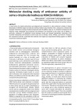
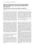
pancreas-specific Ser 419 phosphorylated ENOA is upregulated and induces the production of autoanti- bodies with diagnostic and prognostic value (Fig. 1).
Pancreatic ductal cell
Acknowledgements
Pancreatic cancer cell
Non-pancreatic cancer cell
The authors thank Dr W. Zhou for discussion on the role of post-translational modifications in the regulation of protein functions and Dr J. Iliffe who critically reviewed the manuscript. This work was supported in part by grants from the Associazione Italiana Ricerca sul Cancro (AIRC); Fondazione San Paolo (Special Project Oncology); Ministero della Salute: Progetto strategico, ISS-ACC, Progetto integrato Oncologia; Regione Piemonte: Ricerca Industriale e Sviluppo Precompetitivo (BIOPRO and ONCOPROT), Ricerca (BIOTHER), Industriale ‘Converging Technologies’ Progetti strategici su tematiche di interesse regionale o sovra regionale (IMMONC), Ricerca Sanitaria Finalizzata, Ricerca Sanitaria Applicata; Ribovax Biotechnologies (Geneva, Switzerland) and Fondazione Italiana Ricerca sul Cancro (FIRC).
References
Auto-antibodies against phosphorylated ENOA
Unphosphorylated ENOA
Phosphorylated ENOA
1 Pancholi V (2001) Multifunctional alpha-enolase: its role in diseases. Cell Mol Life Sci 58, 902–920. 2 Wold F (1971) Macromolecules: Structure and Function. Prentice-Hall, Englewood Cliffs, NJ. 3 Piast M, Kustrzeba-Wojcicka I, Matusiewicz M &
Fig. 1. Production of autoantibodies to phosphorylated ENOA in pancreatic cancer. ENOA is overexpressed in tumor cells compared with normal tissues and it is present on the surface of different cell types where it acts as a plasminogen receptor. ENOA is phosphor- ylated on Ser 419 only in pancreatic tissues, the overexpression of this post-translationally modified ENOA in tumor condition induces the production of autoantibodies with clinical relevance in pancre- atic cancer patients.
Banas T (2005) Molecular evolution of enolase. Acta Biochim Pol 52, 507–513.
4 Craig SP, Day IN, Thompson RJ & Craig IW (1990) Localisation of neurone-specific enolase (ENO2) to 12p13. Cytogenet Cell Genet 54, 71–73.
Conclusions
5 Feo S, Oliva D, Barbieri G, Xu WM, Fried M & Giall- ongo A (1990) The gene for the muscle-specific enolase is on the short arm of human chromosome 17. Genom- ics 6, 192–194.
6 Rider CC & Taylor CB (1975) Enolase isoenzymes. II. Hybridization studies, developmental and phylogenetic aspects. Biochim Biophys Acta 405, 175–187. 7 Fletcher L, Rider CC & Taylor CB (1976) Enolase
In several kinds of
isoenzymes. III. Chromatographic and immunological characteristics of rat brain enolase. Biochim Biophys Acta 452, 245–252. 8 Fletcher L, Rider CC, Taylor CB, Adamson ED, Luke
BM & Graham CF (1978) Enolase isoenzymes as markers of differentiation in teratocarcinoma cells and normal tissues of mouse. Dev Biol 65, 462–475. 9 Marangos PJ, Zis AP, Clark RL & Goodwin FK
Taken as a whole, these findings illustrate the multi- functional properties of ENOA in tumorigenesis, and its key implications in cancer proliferation, invasion and immune response. In cancer cells, ENOA is overex- pressed and localizes on their surface, where it acts as a key protein in tumor metastasis, promoting cellular metabolism in anaerobic conditions and driving tumor invasion through plasminogen activation and extracel- lular matrix degradation. It also displays a characteris- tic pattern of PTMs, namely acetylation, methylation and phosphorylation, that regulate protein functions and immunogenicity. tumor, patients develop an integrated response of CD4+, CD8+ T cells and B cells against ENOA, together with anti-ENOA autoantibodies in their sera. Clinical corre- lations propose ENOA as a novel target for cancer immunotherapy. In pancreatic cancer, for example, the
FEBS Journal 278 (2011) 1064–1074 ª 2011 The Authors Journal compilation ª 2011 FEBS
1069
(1978) Neuronal, non-neuronal and hybrid forms of enolase in brain: structural, immunological and func- tional comparisons. Brain Res 150, 117–133.
M. Capello et al.
a-enolase in tumor diagnosis and therapy
ically to alpha-enolase on rat neuronal plasma mem- brane. J Neurochem 63, 2048–2057. 23 Kim JW & Dang CV (2005) Multifaceted roles of 10 Kang HJ, Jung SK, Kim SJ & Chung SJ (2008) Struc- ture of human alpha-enolase (hENO1), a multifunc- tional glycolytic enzyme. Acta Crystallogr D Biol Crystallogr 64, 651–657.
11 Ehinger S, Schubert WD, Bergmann S, Hammersch- midt S & Heinz DW (2004) Plasmin(ogen)-binding alpha-enolase from Streptococcus pneumoniae: crystal structure and evaluation of plasmin(ogen)-binding sites. J Mol Biol 343, 997–1005. glycolytic enzymes. Trends Biochem Sci 30, 142–150. 24 Feo S, Arcuri D, Piddini E, Passantino R & Giallongo A (2000) ENO1 gene product binds to the c-myc pro- moter and acts as a transcriptional repressor: relation- ship with Myc promoter-binding protein 1 (MBP-1). FEBS Lett 473, 47–52. 12 Dudani AK, Cummings C, Hashemi S & Ganz PR
25 Ray R & Miller DM (1991) Cloning and characteriza- tion of a human c-myc promoter-binding protein. Mol Cell Biol 11, 2154–2161.
26 Subramanian A & Miller DM (2000) Structural analy- sis of alpha-enolase. Mapping the functional domains involved in down-regulation of the c-myc protoonco- gene. J Biol Chem 275, 5958–5965. 27 Lo Presti M, Ferro A, Contino F, Mazzarella C, (1993) Isolation of a novel 45 kDa plasminogen receptor from human endothelial cells. Thromb Res 69, 185–196. 13 Miles LA, Dahlberg CM, Plescia J, Felez J, Kato K & Plow EF (1991) Role of cell-surface lysines in plasmin- ogen binding to cells: identification of alpha-enolase as a candidate plasminogen receptor. Biochemistry 30, 1682–1691. 14 Redlitz A, Fowler BJ, Plow EF & Miles LA (1995)
The role of an enolase-related molecule in plasminogen binding to cells. Eur J Biochem 227, 407–415. 15 Pancholi V & Fischetti VA (1998) alpha-enolase, a Sbacchi S, Roz E, Lupo C, Perconti G, Giallongo A, Migliorini P et al. (2010) Myc promoter-binding protein-1 (MBP-1) is a novel potential prognostic marker in invasive ductal breast carcinoma. PLoS ONE 5, e12961.
novel strong plasmin(ogen) binding protein on the sur- face of pathogenic streptococci. J Biol Chem 273, 14503–14515.
28 Perconti G, Ferro A, Amato F, Rubino P, Randazzo D, Wolff T, Feo S & Giallongo A (2007) The kelch protein NS1-BP interacts with alpha-enolase ⁄ MBP-1 and is involved in c-Myc gene transcriptional control. Biochim Biophys Acta 1773, 1774–1785. 29 Cappello P, Tomaino B, Chiarle R, Ceruti P, 16 Sundstrom P & Aliaga GR (1992) Molecular cloning of cDNA and analysis of protein secondary structure of Candida albicans enolase, an abundant, immuno- dominant glycolytic enzyme. J Bacteriol 174, 6789– 6799.
17 Pal-Bhowmick I, Mehta M, Coppens I, Sharma S & Jarori GK (2007) Protective properties and surface localization of Plasmodium falciparum enolase. Infect Immun 75, 5500–5508. Novarino A, Castagnoli C, Migliorini P, Perconti G, Giallongo A, Milella M et al. (2009) An integrated humoral and cellular response is elicited in pancreatic cancer by alpha-enolase, a novel pancreatic ductal adenocarcinoma-associated antigen. Int J Cancer 125, 639–648.
18 Felez J, Chanquia CJ, Fabregas P, Plow EF & Miles LA (1993) Competition between plasminogen and tissue plasminogen activator for cellular binding sites. Blood 82, 2433–2441.
30 He P, Naka T, Serada S, Fujimoto M, Tanaka T, Hashimoto S, Shima Y, Yamadori T, Suzuki H, Hirashima T et al. (2007) Proteomics-based identifica- tion of alpha-enolase as a tumor antigen in non-small lung cancer. Cancer Sci 98, 1234–1240. 31 Seweryn E, Pietkiewicz J, Bednarz-Misa IS, Ceremuga I,
19 Lopez-Alemany R, Longstaff C, Hawley S, Mirshahi M, Fabregas P, Jardi M, Merton E, Miles LA & Felez J (2003) Inhibition of cell surface mediated plasminogen activation by a monoclonal antibody against alpha-eno- lase. Am J Hematol 72, 234–242. 20 Moscato S, Pratesi F, Sabbatini A, Chimenti D, Scav- Saczko J, Kulbacka J & Gamian A (2009) Localization of enolase in the subfractions of a breast cancer cell line. Z Naturforsch C 64, 754–758. 32 Mears R, Craven RA, Hanrahan S, Totty N,
uzzo M, Passatino R, Bombardieri S, Giallongo A & Migliorini P (2000) Surface expression of a glycolytic enzyme, alpha-enolase, recognized by autoantibodies in connective tissue disorders. Eur J Immunol 30, 3575– 3584. Upton C, Young SL, Patel P, Selby PJ & Banks RE (2004) Proteomic analysis of melanoma-derived exosomes by two-dimensional polyacrylamide gel electrophoresis and mass spectrometry. Proteomics 4, 4019–4031. 21 Wygrecka M, Marsh LM, Morty RE, Henneke I,
FEBS Journal 278 (2011) 1064–1074 ª 2011 The Authors Journal compilation ª 2011 FEBS
1070
33 Yu X, Harris SL & Levine AJ (2006) The regulation of exosome secretion: a novel function of the p53 protein. Cancer Res 66, 4795–4801. 34 Maxwell CA, McCarthy J & Turley E (2008) Cell- Guenther A, Lohmeyer J, Markart P & Preissner KT (2009) Enolase-1 promotes plasminogen-mediated recruitment of monocytes to the acutely inflamed lung. Blood 113, 5588–5598. 22 Nakajima K, Hamanoue M, Takemoto N, Hattori T, Kato K & Kohsaka S (1994) Plasminogen binds specif- surface and mitotic-spindle RHAMM: moonlighting or dual oncogenic functions? J Cell Sci 121, 925–932.
M. Capello et al.
a-enolase in tumor diagnosis and therapy
35 Nickel W (2005) Unconventional secretory routes: teinase expressed on the surface of invasive tumour cells. Nature 370, 61–65. direct protein export across the plasma membrane of mammalian cells. Traffic 6, 607–614.
50 Vassalli JD & Pepper MS (1994) Tumour biology. Membrane proteases in focus. Nature 370, 14–15. 51 Plow EF, Freaney DE, Plescia J & Miles LA (1986)
36 Lopez-Villar E, Monteoliva L, Larsen MR, Sachon E, Shabaz M, Pardo M, Pla J, Gil C, Roepstorff P & Nombela C (2006) Genetic and proteomic evidences support the localization of yeast enolase in the cell sur- face. Proteomics 6(Suppl 1), S107–S118. The plasminogen system and cell surfaces: evidence for plasminogen and urokinase receptors on the same cell type. J Cell Biol 103, 2411–2420.
52 Liu KJ & Shih NY (2007) The role of enolase in tissue invasion and metastasis of pathogens and tumor cells. J Cancer Mol 3, 45–48. 37 Pal-Bhowmick I, Krishnan S & Jarori GK (2007) Dif- ferential susceptibility of Plasmodium falciparum versus yeast and mammalian enolases to dissociation into active monomers. FEBS J 274, 1932–1945.
38 Dudani AK & Ganz PR (1996) Endothelial cell surface actin serves as a binding site for plasminogen, tissue plasminogen activator and lipoprotein(a). Br J Haema- tol 95, 168–178. 53 Saldanha RG, Molloy MP, Bdeir K, Cines DB, Song X, Uitto PM, Weinreb PH, Violette SM & Baker MS (2007) Proteomic identification of lynchpin urokinase plasminogen activator receptor protein interactions associated with epithelial cancer malignancy. J Prote- ome Res 6, 1016–1028.
54 Trojanowicz B, Winkler A, Hammje K, Chen Z, Seku- lla C, Glanz D, Schmutzler C, Mentrup B, Hombach- Klonisch S, Klonisch T et al. (2009) Retinoic acid-med- iated down-regulation of ENO1 ⁄ MBP-1 gene products caused decreased invasiveness of the follicular thyroid carcinoma cell lines. J Mol Endocrinol 42, 249–260. 55 Dowling P, Meleady P, Dowd A, Henry M, Glynn S & Clynes M (2007) Proteomic analysis of isolated mem- brane fractions from superinvasive cancer cells. Bio- chim Biophys Acta 1774, 93–101. 56 Crippa MP (2007) Urokinase-type plasminogen activa- 39 Kanalas JJ & Makker SP (1991) Identification of the rat Heymann nephritis autoantigen (GP330) as a receptor site for plasminogen. J Biol Chem 266, 10825–10829. 40 Hembrough TA, Li L & Gonias SL (1996) Cell-surface cytokeratin 8 is the major plasminogen receptor on breast cancer cells and is required for the accelerated activation of cell-associated plasminogen by tissue-type plasminogen activator. J Biol Chem 271, 25684–25691. 41 Borza DB & Morgan WT (1997) Acceleration of plas- minogen activation by tissue plasminogen activator on surface-bound histidine-proline-rich glycoprotein. J Biol Chem 272, 5718–5726. tor. Int J Biochem Cell Biol 39, 690–694. 57 Quemener C, Gabison EE, Naimi B, Lescaille G,
42 Winram SB & Lottenberg R (1996) The plasmin-bind- ing protein Plr of group A streptococci is identified as glyceraldehyde-3-phosphate dehydrogenase. Microbiol- ogy 142(Pt 8), 2311–2320.
Bougatef F, Podgorniak MP, Labarchede G, Lebbe C, Calvo F, Menashi S et al. (2007) Extracellular matrix metalloproteinase inducer up-regulates the urokinase- type plasminogen activator system promoting tumor cell invasion. Cancer Res 67, 9–15.
43 Kassam G, Choi KS, Ghuman J, Kang HM, Fitzpa- trick SL, Zackson T, Zackson S, Toba M, Shinomiya A & Waisman DM (1998) The role of annexin II tetra- mer in the activation of plasminogen. J Biol Chem 273, 4790–4799. 44 Das R, Burke T & Plow EF (2007) Histone H2B as a 58 Siren V, Salmenpera P, Kankuri E, Bizik J, Sorsa T, Tervahartiala T & Vaheri A (2006) Cell-cell contact activation of fibroblasts increases the expression of matrix metalloproteinases. Ann Med 38, 212–220. functionally important plasminogen receptor on macro- phages. Blood 110, 3763–3772. 45 Felez J (1998) Plasminogen binding to cell surfaces. Fibrinolysis Proteolysis 12, 183–189.
59 Smith R, Xue A, Gill A, Scarlett C, Saxby A, Clarkson A & Hugh T (2007) High expression of plasminogen activator inhibitor-2 (PAI-2) is a predictor of improved survival in patients with pancreatic adenocarcinoma. World J Surg 31, 493–502. 60 Hurtado M, Lozano JJ, Castellanos E, Lopez- 46 Andreasen PA, Egelund R & Petersen HH (2000) The plasminogen activation system in tumor growth, inva- sion, and metastasis. Cell Mol Life Sci 57, 25–40. 47 Lijnen HR, Van Hoef B, Lupu F, Moons L, Car-
Fernandez LA, Harshman K, Martinez AC, Ortiz AR, Thomson TM & Paciucci R (2007) Activation of the epidermal growth factor signalling pathway by tissue plasminogen activator in pancreas cancer cells. Gut 56, 1266–1274. meliet P & Collen D (1998) Function of the plasmino- gen ⁄ plasmin and matrix metalloproteinase systems after vascular injury in mice with targeted inactivation of fibrinolytic system genes. Arterioscler Thromb Vasc Biol 18, 1035–1045.
FEBS Journal 278 (2011) 1064–1074 ª 2011 The Authors Journal compilation ª 2011 FEBS
1071
48 Plow EF, Felez J & Miles LA (1991) Cellular regula- tion of fibrinolysis. Thromb Haemost 66, 32–36. 49 Sato H, Takino T, Okada Y, Cao J, Shinagawa A, 61 Ortiz-Zapater E, Peiro S, Roda O, Corominas JM, Aguilar S, Ampurdanes C, Real FX & Navarro P (2007) Tissue plasminogen activator induces pancre- atic cancer cell proliferation by a non-catalytic mech- anism that requires extracellular signal-regulated Yamamoto E & Seiki M (1994) A matrix metallopro-
M. Capello et al.
a-enolase in tumor diagnosis and therapy
binding sites for hypoxia-inducible factor 1. J Biol Chem 271, 32529–32537. kinase 1 ⁄ 2 activation through epidermal growth fac- tor receptor and annexin A2. Am J Pathol 170, 1573–1584. 62 Roda O, Chiva C, Espuna G, Gabius HJ, Real FX, 77 Altenberg B & Greulich KO (2004) Genes of glycolysis are ubiquitously overexpressed in 24 cancer classes. Genomics 84, 1014–1020. 78 Tu SH, Chang CC, Chen CS, Tam KW, Wang YJ,
Navarro P & Andreu D (2006) A proteomic approach to the identification of new tPA receptors in pancreatic cancer cells. Proteomics 6(Suppl 1), S36–S41. 63 Sahin U, Tureci O, Schmitt H, Cochlovius B,
Lee CH, Lin HW, Cheng TC, Huang CS, Chu JS et al. (2010) Increased expression of enolase alpha in human breast cancer confers tamoxifen resistance in human breast cancer cells. Breast Cancer Res Treat 121, 539–553. 79 Somiari RI, Sullivan A, Russell S, Somiari S, Hu H, Johannes T, Schmits R, Stenner F, Luo G, Schobert I & Pfreundschuh M (1995) Human neoplasms elicit multiple specific immune responses in the autologous host. Proc Natl Acad Sci USA 92, 11810–11813. 64 Disis ML, Pupa SM, Gralow JR, Dittadi R, Me-
Jordan R, George A, Katenhusen R, Buchowiecka A, Arciero C et al. (2003) High-throughput proteomic analysis of human infiltrating ductal carcinoma of the breast. Proteomics 3, 1863–1873. nard S & Cheever MA (1997) High-titer HER-2 ⁄ neu protein-specific antibody can be detected in patients with early-stage breast cancer. J Clin Oncol 15, 3363–3367.
65 Tan HT, Low J, Lim SG & Chung MC (2009) Serum autoantibodies as biomarkers for early cancer detec- tion. FEBS J 276, 6880–6904.
80 Kabbage M, Chahed K, Hamrita B, Guillier CL, Trim- eche M, Remadi S, Hoebeke J & Chouchane L (2008) Protein alterations in infiltrating ductal carcinomas of the breast as detected by nonequilibrium pH gradient electrophoresis and mass spectrometry. J Biomed Bio- technol 2008, 564127.
66 Anderson KS & LaBaer J (2005) The sentinel within: exploiting the immune system for cancer biomarkers. J Proteome Res 4, 1123–1133, doi: 10.1021/pr0500814. 67 Caron M, Choquet-Kastylevsky G & Joubert-Caron R (2007) Cancer immunomics using autoantibody signa- tures for biomarker discovery. Mol Cell Proteomics 6, 1115–1122. 81 Malorni L, Cacace G, Cuccurullo M, Pocsfalvi G, Chambery A, Farina A, Di MaroA, Parente A & Malorni A (2006) Proteomic analysis of MCF-7 breast cancer cell line exposed to mitogenic concentration of 17beta-estradiol. Proteomics 6, 5973– 5982. 68 Finn OJ (2008) Cancer immunology. N Engl J Med 82 Hennipman A, van Oirschot BA, Smits J, Rijksen G & 358, 2704–2715. 69 Shih NY, Lai HL, Chang GC, Lin HC, Wu YC, Liu JM, Staal GE (1988) Glycolytic enzyme activities in breast cancer metastases. Tumour Biol 9, 241–248.
Liu KJ & Tseng SW (2010) Anti-alpha-enolase autoantibodies are down-regulated in advanced cancer patients. Jpn J Clin Oncol 40, 663–669. 70 Hanash S (2003) Harnessing immunity for cancer mar- ker discovery. Nat Biotechnol 21, 37–38. 83 Hennipman A, Smits J, van Oirschot B, van Houwelin- gen JC, Rijksen G, Neyt JP, Van Unnik JA & Staal GE (1987) Glycolytic enzymes in breast cancer, benign breast disease and normal breast tissue. Tumour Biol 8, 251–263. 71 Warburg O (1930) The Metabolism of Tumours. Con- stable, London.
72 Vander Heiden MG, Cantley LC & Thompson CB (2009) Understanding the Warburg effect: the meta- bolic requirements of cell proliferation. Science 324, 1029–1033. 84 Bae SM, Min HJ, Ding GH, Kwak SY, Cho YL, Nam KH, Park CH, Kim YW, Kim CK, Han BD et al. (2006) Protein expression profile using two- dimensional gel analysis in squamous cervical cancer patients. Cancer Res Treat 38, 99–107.
73 Brown JM & Giaccia AJ (1998) The unique physiology of solid tumors: opportunities (and problems) for can- cer therapy. Cancer Res 58, 1408–1416.
85 Bae SM, Lee CH, Cho YL, Nam KH, Kim YW, Kim CK, Han BD, Lee YJ, Chun HJ & Ahn WS (2005) Two-dimensional gel analysis of protein expression profile in squamous cervical cancer patients. Gynecol Oncol 99, 26–35.
74 Lu Z & Sack MN (2008) ATF-1 is a hypoxia-respon- sive transcriptional activator of skeletal muscle mito- chondrial-uncoupling protein 3. J Biol Chem 283, 23410–23418.
75 Sedoris KC, Thomas SD & Miller DM (2010) Hypoxia induces differential translation of enolase ⁄ MBP-1. BMC Cancer 10, 157. 76 Semenza GL, Jiang BH, Leung SW, Passantino R, 86 Katayama M, Nakano H, Ishiuchi A, Wu W, Oshima R, Sakurai J, Nishikawa H, Yamaguchi S & Otsubo T (2006) Protein pattern difference in the colon cancer cell lines examined by two-dimensional differential in- gel electrophoresis and mass spectrometry. Surg Today 36, 1085–1093.
FEBS Journal 278 (2011) 1064–1074 ª 2011 The Authors Journal compilation ª 2011 FEBS
1072
87 Wong CS, Wong VW, Chan CM, Ma BB, Hui EP, Wong MC, Lam MY, Au TC, Chan WH, Cheuk W et al. (2008) Identification of 5-fluorouracil response Concordet JP, Maire P & Giallongo A (1996) Hypoxia response elements in the aldolase A, enolase 1, and lac- tate dehydrogenase A gene promoters contain essential
M. Capello et al.
a-enolase in tumor diagnosis and therapy
proteins in colorectal carcinoma cell line SW480 by two-dimensional electrophoresis and MALDI-TOF mass spectrometry. Oncol Rep 20, 89–98. (2009) Comparative proteomic analysis of lung cancer cell line and lung fibroblast cell line. Cancer Genomics Proteomics 6, 229–237.
88 Qi Y, Chiu JF, Wang L, Kwong DL & He QY (2005) Comparative proteomic analysis of esophageal squa- mous cell carcinoma. Proteomics 5, 2960–2971.
89 Zhao J, Chang AC, Li C, Shedden KA, Thomas DG, Misek DE, Manoharan AP, Giordano TJ, Beer DG & Lubman DM (2007) Comparative proteomics analysis of Barrett metaplasia and esophageal adenocarcinoma using two-dimensional liquid mass mapping. Mol Cell Proteomics 6, 987–999. 99 Chang GC, Liu KJ, Hsieh CL, Hu TS, Charoenfupra- sert S, Liu HK, Luh KT, Hsu LH, Wu CW, Ting CC et al. (2006) Identification of alpha-enolase as an auto- antigen in lung cancer: its overexpression is associated with clinical outcomes. Clin Cancer Res 12, 5746–5754. 100 Cao L, Li X, Zhang Y, Peng F, Yi H, Xu Y & Wang Q (2010) Proteomic analysis of human ovarian cancer paclitaxel-resistant cell lines. Zhong Nan Da Xue Xue Bao Yi Xue Ban 35, 286–294. 101 Shen J, Person MD, Zhu J, Abbruzzese JL & Li D
90 Govekar RB, D’Cruz AK, Alok Pathak K, Agarwal J, Dinshaw KA, Chinoy RF, Gadewal N, Kannan S, Sirdeshmukh R, Sundaram CS et al. (2009) Proteomic profiling of cancer of the gingivo-buccal complex: iden- tification of new differentially expressed markers. Pro- teomics Clin Appl 3, 1451–1462. (2004) Protein expression profiles in pancreatic adeno- carcinoma compared with normal pancreatic tissue and tissue affected by pancreatitis as detected by two- dimensional gel electrophoresis and mass spectrometry. Cancer Res 64, 9018–9026. 102 Mikuriya K, Kuramitsu Y, Ryozawa S, Fujimoto M, 91 Tsai ST, Chien IH, Shen WH, Kuo YZ, Jin YT,
Wong TY, Hsiao JR, Wang HP, Shih NY & Wu LW (2010) ENO1, a potential prognostic head and neck cancer marker, promotes transformation partly via chemokine CCL20 induction. Eur J Cancer 46, 1712– 1723.
Mori S, Oka M, Hamano K, Okita K, Sakaida I & Nakamura K (2007) Expression of glycolytic enzymes is increased in pancreatic cancerous tissues as evi- denced by proteomic profiling by two-dimensional elec- trophoresis and liquid chromatography-mass spectrometry ⁄ mass spectrometry. Int J Oncol 30, 849– 855. 103 Rehman I, Azzouzi AR, Catto JW, Allen S, Cross SS,
92 Lopez-Pedrera C, Villalba JM, Siendones E, Barbar- roja N, Gomez-Diaz C, Rodriguez-Ariza A, Buendia P, Torres A & Velasco F (2006) Proteomic analysis of acute myeloid leukemia: identification of potential early biomarkers and therapeutic targets. Proteomics 6(Suppl 1), S293–S299. Feeley K, Meuth M & Hamdy FC (2004) Proteomic analysis of voided urine after prostatic massage from patients with prostate cancer: a pilot study. Urology 64, 1238–1243. 104 Suzuki A, Iizuka A, Komiyama M, Takikawa M,
Kume A, Tai S, Ohshita C, Kurusu A, Nakamura Y, Yamamoto A et al. (2010) Identification of melanoma antigens using a Serological Proteome Approach (SER- PA). Cancer Genomics Proteomics 7, 17–23. 93 Hamaguchi T, Iizuka N, Tsunedomi R, Hamamoto Y, Miyamoto T, Iida M, Tokuhisa Y, Sakamoto K, Taka- shima M, Tamesa T et al. (2008) Glycolysis module activated by hypoxia-inducible factor 1alpha is related to the aggressive phenotype of hepatocellular carci- noma. Int J Oncol 33, 725–731. 105 Paik WK, Paik DC & Kim S (2007) Historical review:
the field of protein methylation. Trends Biochem Sci 32, 146–152.
94 Takashima M, Kuramitsu Y, Yokoyama Y, Iizuka N, Fujimoto M, Nishisaka T, Okita K, Oka M & Nakam- ura K (2005) Overexpression of alpha enolase in hepa- titis C virus-related hepatocellular carcinoma: association with tumor progression as determined by proteomic analysis. Proteomics 5, 1686–1692. 106 Spange S, Wagner T, Heinzel T & Kramer OH (2009) Acetylation of non-histone proteins modulates cellular signalling at multiple levels. Int J Biochem Cell Biol 41, 185–198. 95 Li LS, Kim H, Rhee H, Kim SH, Shin DH, Chung
KY, Park KS, Paik YK & Chang J (2004) Proteomic analysis distinguishes basaloid carcinoma as a distinct subtype of nonsmall cell lung carcinoma. Proteomics 4, 3394–3400. 107 Choudhary C, Kumar C, Gnad F, Nielsen ML, Reh- man M, Walther TC, Olsen JV & Mann M (2009) Lysine acetylation targets protein complexes and co- regulates major cellular functions. Science 325, 834– 840. 108 Mayya V, Lundgren DH, Hwang SI, Rezaul K, Wu L,
FEBS Journal 278 (2011) 1064–1074 ª 2011 The Authors Journal compilation ª 2011 FEBS
1073
Eng JK, Rodionov V & Han DK (2009) Quantitative phosphoproteomic analysis of T cell receptor signaling reveals system-wide modulation of protein-protein interactions. Sci Signal 2, ra46. 109 Rush J, Moritz A, Lee KA, Guo A, Goss VL, Spek EJ, Zhang H, Zha XM, Polakiewicz RD & Comb MJ 96 Li C, Xiao Z, Chen Z, Zhang X, Li J, Wu X, Li X, Yi H, Li M, Zhu G et al. (2006) Proteome analysis of human lung squamous carcinoma. Proteomics 6, 547–558. 97 Huang LJ, Chen SX, Luo WJ, Jiang HH, Zhang PF & Yi H (2006) Proteomic analysis of secreted proteins of non-small cell lung cancer. Ai Zheng 25, 1361–1367. 98 Rubporn A, Srisomsap C, Subhasitanont P, Chokchai- chamnankit D, Chiablaem K, Svasti J & Sangvanich P
M. Capello et al.
a-enolase in tumor diagnosis and therapy
(2005) Immunoaffinity profiling of tyrosine phosphory- lation in cancer cells. Nat Biotechnol 23, 94–101. 122 Forgber M, Trefzer U, Sterry W & Walden P (2009) Proteome serological determination of tumor-associ- ated antigens in melanoma. PLoS ONE 4, e5199. 123 Shukla S, Govekar RB, Sirdeshmukh R, Sundaram
CS, D’Cruz AK, Pathak KA, Kane SV & Zingde SM (2007) Tumor antigens eliciting autoantibody response in cancer of gingivo-buccal complex. Proteomics Clin Appl 1, 1592–1604. 124 Shukla S, Pranay A, D’Cruz AK, Chaturvedi P, Kane
110 Rikova K, Guo A, Zeng Q, Possemato A, Yu J, Haack H, Nardone J, Lee K, Reeves C, Li Y et al. (2007) Global survey of phosphotyrosine signaling identifies oncogenic kinases in lung cancer. Cell 131, 1190–1203. 111 Molina H, Horn DM, Tang N, Mathivanan S & Pan- dey A (2007) Global proteomic profiling of phospho- peptides using electron transfer dissociation tandem mass spectrometry. Proc Natl Acad Sci USA 104, 2199–2204. 112 Kim SC, Sprung R, Chen Y, Xu Y, Ball H, Pei J, SV & Zingde SM (2009) Immunoproteomics reveals that cancer of the tongue and the gingivobuccal com- plex exhibit differential autoantibody response. Cancer Biomark 5, 127–135. 125 Ejma M, Misiuk-Hojlo M, Gorczyca WA, Podemski Cheng T, Kho Y, Xiao H, Xiao L et al. (2006) Sub- strate and functional diversity of lysine acetylation revealed by a proteomics survey. Mol Cell 23, 607–618. 113 Yu LR, Zhu Z, Chan KC, Issaq HJ, Dimitrov DS &
R, Szymaniec S, Kuropatwa M, Rogozinska-Szczepka J & Bartnik W (2008) Antibodies to 46-kDa retinal antigen in a patient with breast carcinoma and cancer- associated retinopathy. Breast Cancer Res Treat 110, 269–271.
Veenstra TD (2007) Improved titanium dioxide enrich- ment of phosphopeptides from HeLa cells and high confident phosphopeptide identification by cross-vali- dation of MS ⁄ MS and MS ⁄ MS ⁄ MS spectra. J Prote- ome Res 6, 4150–4162. 114 Dephoure N, Zhou C, Villen J, Beausoleil SA, Baka- 126 Jankowska R, Witkowska D, Porebska I, Kuropatwa M, Kurowska E & Gorczyca WA (2004) Serum anti- bodies to retinal antigens in lung cancer and sarcoido- sis. Pathobiology 71, 323–328.
larski CE, Elledge SJ & Gygi SP (2008) A quantitative atlas of mitotic phosphorylation. Proc Natl Acad Sci USA 105, 10762–10767. 115 Zhou W, Capello M, Fredolini C, Piemonti L, Liotta
127 Nakanishi T, Takeuchi T, Ueda K, Murao H & Shi- mizu A (2006) Detection of eight antibodies in cancer patients’ sera against proteins derived from the adeno- carcinoma A549 cell line using proteomics-based analy- sis. J Chromatogr B Analyt Technol Biomed Life Sci 838, 15–20. LA, Novelli F & Petricoin EF (2010) Mass spectrome- try analysis of the post-translational modifications of alpha-enolase from pancreatic ductal adenocarcinoma cells. J Proteome Res 9, 2929–2936. 116 Yang XJ & Seto E (2008) Lysine acetylation: codified 128 Ueda K (2005) Proteome analysis of autoantibodies in sera of patients with cancer. Rinsho Byori 53, 437–445. 129 Dot C, Guigay J & Adamus G (2005) Anti-alpha-eno- crosstalk with other posttranslational modifications. Mol Cell 31, 449–461.
lase antibodies in cancer-associated retinopathy with small cell carcinoma of the lung. Am J Ophthalmol 139, 746–747. 130 Mohammed F, Cobbold M, Zarling AL, Salim M, 117 Sahin U, Tureci O & Pfreundschuh M (1997) Serologi- cal identification of human tumor antigens. Curr Opin Immunol 9, 709–716.
Barrett-Wilt GA, Shabanowitz J, Hunt DF, Engelhard VH & Willcox BE (2008) Phosphorylation-dependent interaction between antigenic peptides and MHC class I: a molecular basis for the presentation of transformed self. Nat Immunol 9, 1236–1243.
118 Janssen EM, Lemmens EE, Wolfe T, Christen U, von Herrath MG & Schoenberger SP (2003) CD4+ T cells are required for secondary expansion and memory in CD8+ T lymphocytes. Nature 421, 852–856. 119 Tomaino B, Cappello P, Capello M, Fredolini C, Sperduti I, Migliorini P, Salacone P, Novarino A, Giacobino A, Ciuffreda L et al. (2011) Circulating autoantibodies to phosphorylated alpha-enolase are a hallmark of pancreatic cancer. J Proteome Res 1, 105– 112. 131 Sato N, Nabeta Y, Kondo H, Sahara H, Hirohashi Y, Kashiwagi K, Kanaseki T, Sato Y, Rong S, Hirai I et al. (2000) Human CD8 and CD4 T cell epitopes of epithelial cancer antigens. Cancer Chemother Pharma- col 46(Suppl), S86–S90. 132 Kondo H, Sahara H, Miyazaki A, Nabeta Y, Hiroh- 120 Cui JW, Li WH, Wang J, Li AL, Li HY, Wang HX,
He K, Li W, Kang LH, Yu M et al. (2005) Proteo- mics-based identification of human acute leukemia antigens that induce humoral immune response. Mol Cell Proteomics 4, 1718–1724. ashi Y, Kanaseki T, Yamaguchi A, Yamada N, Hiray- ama K, Suzuki M et al. (2002) Natural antigenic peptides from squamous cell carcinoma recognized by autologous HLA-DR8-restricted CD4+ T cells. Jpn J Cancer Res 93, 917–924.
FEBS Journal 278 (2011) 1064–1074 ª 2011 The Authors Journal compilation ª 2011 FEBS
1074
133 Fernandez Madrid F (2005) Autoantibodies in breast cancer sera: candidate biomarkers and reporters of tumorigenesis. Cancer Lett 230, 187–198. 121 Zou L, Wu Y, Pei L, Zhong D, Gen M, Zhao T, Wu J, Ni B, Mou Z, Han J et al. (2005) Identification of leukemia-associated antigens in chronic myeloid leuke- mia by proteomic analysis. Leuk Res 29, 1387–1391.