Re-evaluation of the function of the F420 dehydrogenase in electron transport of Methanosarcina mazei Cornelia Welte and Uwe Deppenmeier
Institute of Microbiology and Biotechnology, University of Bonn, Germany
Keywords archaea; electron transport; energy conservation; hydrogenase; methane; methanogenesis
Correspondence U. Deppenmeier, Institut fu¨ r Mikrobiologie und Biotechnologie, University of Bonn, Meckenheimer Allee 168, 53115 Bonn, Germany Fax: +(49) 228 737576 Tel: +(49) 228 735590 E-mail: udeppen@uni-bonn.de
(Received 2 December 2010, revised 14 January 2011, accepted 7 February 2011)
doi:10.1111/j.1742-4658.2011.08048.x
Methanosarcina mazei is a methanogenic archaeon that is able to thrive on various substrates and therefore contains a variety of redox-active proteins involved in both cytoplasmic and membrane-bound electron transport. The organism possesses a complex branched respiratory chain that has the abil- ity to utilize different electron donors. In this study, two knockout mutants of the membrane-bound F420 dehydrogenase (DfpoF and DfpoA-O) were constructed and analyzed. They exhibited severe growth deficiencies with trimethylamine, but not with acetate, as substrates. In cell lysates of the fpo mutants, the F420:heterodisulfide oxidoreductase activity was strongly reduced, although soluble F420 hydrogenase was still present. This led to the conclusion that the predominant part of cellular oxidation of the reduced form of F420 (F420H2) in Ms. mazei is performed by F420 dehydro- genase. Enzyme assays of cytoplasmic fractions revealed that ferredoxin in the DfpoF (Fd):F420 oxidoreductase activity was essentially absent mutant. Subsequently, FpoF was produced in Escherichia coli and purified for further characterization. The purified FpoF protein catalyzed the Fd:F420 oxidoreductase reaction with high specificity (the KM for reduced Fd was 0.5 lM) but with low velocity (Vmax = 225 mUÆmg)1) and was present in the Ms. mazei cytoplasm in considerable amounts. Consequently, soluble FpoF might participate in electron carrier equilibrium and facilitate survival of the Ms. mazei Dech mutant that lacks the membrane-bound Fd-oxidizing Ech hydrogenase.
Introduction
nature, methanogens are responsible for the produc- tion of CH4 in anaerobic digesters of biogas plants. The process of biomethanation is a viable alternative to fossil fuels and has great potential as an important renewable energy source. In principle,
Abbreviations CoM-S-S-CoB, heterodisulfide of HS-CoM and HS-CoB; F420, coenzyme F420; F420H2, reduced form of F420; Fd, ferredoxin; Fdred, reduced ferredoxin; Fpo, F420H2 dehydrogenase; Frh, F420 hydrogenase; H4SPT, tetrahydrosarcinapterin; HS-CoB, N-7-mercaptoheptanoyl-L-threonine phosphate; HS-CoM, 2-mercaptoethanesulfonate; Vho, F420-nonreducing hydrogenase.
FEBS Journal 278 (2011) 1277–1287 ª 2011 The Authors Journal compilation ª 2011 FEBS
1277
Methanogenic archaea are one of the key groups in the global carbon cycle because they metabolize the final products of the anaerobic food chain (H2, CO2 and acetate) using unusual enzymes and cofactors in the so-called methanogenic pathway. The final product of all methanogenic pathways is methane (CH4) and every year millions of tons of this highly potent green- house gas reach the atmosphere and contribute to glo- bal warming. Apart from their vast distribution in three different growth strategies in methanogenesis – hydrogenotrophic, methylotrophic and aceticlastic methanogenesis – have evolved and use H2 + CO2, methylated compounds and acetate as
C. Welte and U. Deppenmeier
Role of F420 dehydrogenase in Ms. mazei
hydrogen, which can be oxidized by the membrane- bound F420-nonreducing hydrogenase (Vho). Hence, both Fpo and Frh (in combination with Vho) may function as electron-input modules channelling elec- trons into the respiratory chain. To evaluate electron transport from F420H2
in Ms. mazei in more detail, two mutants with deletion of genes encoding the Fpo – DfpoF (= Dmm0627) and DfpoA-O (= Dmm2491–2479) – were constructed. Slower growth rates were observed for the DfpoF and the DfpoA-O mutants (the doubling times were 11.3 and 11.1 h, respectively) in comparison with the paren- tal strain (that has a doubling time of 7.7 h) with trim- ethylamine as the substrate. The final D of the cultures were almost identical. In summary, the electron trans- port deficiency of the Dfpo mutants was reflected in their growth abilities because they grew more slowly but generated the same amount of biomass from trim- ethylamine compared with the parental strain. As expected, both mutants grew on acetate or trimethyl- amine + H2 with a growth rate and yield similar to that of the parental strain, indicating that Fpo plays a major role in the oxidative part of methylotrophic methanogenesis.
substrates, respectively. The core of all methanogenic pathways is very similar, but there are differences in the source of reducing equivalents and in the mode of how electrons are channelled into the methano- genic respiratory chain. Methanosarcina mazei Go¨ 1 (Ms. mazei) is a model organism for methanogenisis because it can grow hydrogenotrophically, methylotro- phically and aceticlastically. Its respiratory chain com- prises three energy-conserving oxidoreductase systems that lead to the formation of an electrochemical pro- ton gradient and ATP synthesis by the A1Ao ATP syn- thase [1]. The energy-transducing systems are referred to as F420H2:heterodisulfide oxidoreductase (where F420H2 is the reduced form of F420), H2:heterodisulfide oxidoreductase and ferredoxin (Fd):heterodisulfide oxi- doreductase, mirroring the three possible electron input compounds [F420H2, H2 and reduced Fd (Fdred)]. Fd is reduced during methylotrophic and aceticlastic methanogenesis, and is oxidized by Ech hydrogenase as part of the Fd:heterodisulfide oxidoreductase sys- tem. F420H2 is formed in the course of methyl group oxidation in the methylotrophic pathway, or in the cytoplasm of hydrogenotrophically growing methano- gens, by F420 hydrogenase (Frh). This enzyme oxidizes H2 with concomitant F420 reduction, providing F420H2 for CO2 reduction. oxidoreductase The F420H2:heterodisulfide
The effect of the mutations was also analyzed using resting cell suspensions of Ms. mazei (Table 1). Meth- ane formation from trimethylamine was measured under an atmosphere of nitrogen and it became evident that the DfpoF and the DfpoA-O mutants retained only half of the activity compared with the parental strain. In contrast, no differences in methane production were observed when trimethylamine was converted to meth- ane in the presence of molecular hydrogen.
used under methyltrophic growth conditions consists of the F420H2 dehydrogenase (Fpo) and the heterodisulfide reductase [2,3]. Here we describe the characteristics of two Fpo mutants (DfpoF and DfpoA-O) and the effects of these deletions on the process of energy con- it is shown that the protein servation. Furthermore, the Fpo FpoF functions as an input module of complex. In a soluble form it is able to catalyze the reduction of coenzyme F420 with Fdred as the electron donor, providing a direct link between the redox carriers of hydrogenotrophic and aceticlastic methano- genesis.
To evaluate the reason for the different growth rates and methane-formation rates with trimethylamine as a substrate, cell lysates and washed membrane prepara- tions of the wild-type and the mutant strains were prepared and the coupled redox reaction of F420H2 oxidation with heterodisulfide reduction (F420H2: heterodisulfide oxidoreductase) was assayed using the following equation:
Results
Table 1. Methane formation by resting cell suspensions of Met- hanosarcina mazei.
Washed cells
Substrate
Methane formation (nmolÆmin)1Æmg protein)1)
196 ± 8 107 ± 11 105 ± 19 185 ± 10 190 ± 17 186 ± 2
Wild type DfpoF DfpoA-O Wild type DfpoF DfpoA-O
Trimethylamine Trimethylamine Trimethylamine Trimethylamine + H2 Trimethylamine + H2 Trimethylamine + H2
Electron transport in Dfpo mutants
FEBS Journal 278 (2011) 1277–1287 ª 2011 The Authors Journal compilation ª 2011 FEBS
1278
Fpo is predicted to be a key enzyme in methylotrophic methanogenesis of Ms. mazei that catalyses the mem- brane-bound oxidation of F420H2 and the reduction of methanophenazine as a membrane-soluble redox car- rier with concomitant extrusion of 2 H+ ⁄ 2e) [2,3]. However, Kulkarni et al. [4] presented evidence that in Methanosarcina barkeri, a close relative of Ms. mazei, the cytoplasmic Frh is to a high degree responsible thereby producing molecular for F420H2 oxidation,
C. Welte and U. Deppenmeier
Role of F420 dehydrogenase in Ms. mazei
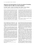
Fig. 1. F420H2:heterodisulfide oxidoreductase activity in Methano- lysates. Cuvettes contained 600 lL of Buffer A, sarcina mazei cell 10 lM F420H2, 250 lg of cell lysate and 33 lM heterodisulfide under a N2 atmosphere. d, Wild-type cell lysate; m, DfpoA-O cell lysate; j, DfpoF cell lysate.
F420H2 þ CoM-S-S-CoB ! F420 þ HS-CoM ð1Þ þHS-CoB;
where HS-CoB is N-7-mercaptoheptanoyl-l-threonine 2-mercaptoethanesulfonate phosphate, HS-CoM is and CoM-S-S-CoB is a heterodisulfide of HS-CoM and HS-CoB. F420H2:heterodisulfide oxidoreductase activity was exclusively found in the membrane frac- tion of the wild-type Ms. mazei, with a specific activ- ity of about 130 mUÆmg)1 of membrane protein; in the F420H2:heterodisulfide oxidoreductase contrast, activity was < 1 mUÆmg)1 of membrane protein in the membrane fractions of the DfpoF and DfpoA-O mutant strains (data not shown). These results are in agreement with the hypothesis that Fpo is the only membrane-integral enzyme that is able to channel electrons from F420H2 directly into the respiratory chain [1]. However, F420H2 is a stringent intermediate in methanogenesis from methylated substrates, such as trimethylamine, where part of the methyl groups are oxidized to CO2 and electrons are transferred to F420 in the course of the methylene-H4SPT reductase (Eqn 2) and dehydrogenase (Eqn 3) reactions [5,6], as fol- lows:
Methyl-H4SPT þ F420 ! methylene ð2Þ -H4SPT þ F420H2
Methylene-H4SPT þ F420 ! methenyl ð3Þ -H4SPT þ F420H2;
where H4SPT is tetrahydrosarcinapterin.
Table 2. Activities of the Frh in cell lysates.
Cell lysate
Electron donor
Electron acceptor
Reduction of electron acceptor (nmolÆmin)1 Æmg protein)1)a
0.65
F420H2 H+
This finding raised the question of whether Frh is also involved in cofactor regeneration in Ms. mazei. As evident form Table 2, electrons from F420H2 were transferred to H+ in the cell lysates and escaped as H2 when no external electron acceptor (heterodisulfide) was added to the cell extracts. The activities were rather low (about 0.5 mUÆmg)1 of cellular protein) but were approximately the same in the parental strain and in the deletion mutants. In contrast, the reverse reaction, with H2 as the electron donor, was catalyzed by Frh with 35 mUÆmg)1 of cellular protein and there- fore was more than 50-fold faster (Table 2). Neverthe- less, the Fpo mutants are obviously able to take advantage of the H2-evolving activity of the Frh when F420H2 accumulates in these mutants. However, it is evident that the low hydrogen-producing activity of the Frh cannot compensate for the activity of the Fpo the DfpoF and and leads to impaired growth of
0.55 0.45
Accordingly, F420H2 has to be reoxidized in the wild-type and the mutant strains. As there is no mem- brane-bound Fpo activity, the two Dfpo mutant strains obviously depend on alternative components to cata- lyse this reaction. Indeed, the cell lysate of the two mutant strains exhibited F420H2-dependent CoM-S-S- CoB reductase activity of (cid:2) 4–5 mUÆmg)1 of protein (Fig. 1), whereas in the cell lysate of the parental strain the activity was (cid:2) 50 mUÆmg)1 of protein. These find- ings clearly indicate that the predominant part of F420H2 oxidation is performed by the membrane- bound Fpo complex. Kulkani et al.
35 360
Methanosarcina mazei b Ms. mazei DfpoA-O F420H2 H+ F420H2 H+ Ms. mazei DfpoF Ms. mazei b F420 H2 Methanosarcina F420 H2 barkeri b
[4] showed that in Ms. barkeri, the soluble Frh is the key enzyme in the reoxidation of F420H2: ð4Þ F420H2 ! F420 þ H2
(DG0
ð5Þ
0 = +11.6 kJÆmol)1) H2 þ CoM-S-S-CoB ! HS-CoM þ HS-CoB 0 = )49.2 kJÆmol)1)
a Tests were conducted, as indicated in the Materials and methods, with 10 lM F420H2 or 100% H2 in the head space as electron donors, and with 10)7 M H+ (pH 7) or 10 lM F420 as electron acceptors. b Wild type.
FEBS Journal 278 (2011) 1277–1287 ª 2011 The Authors Journal compilation ª 2011 FEBS
1279
(DG0
C. Welte and U. Deppenmeier
Role of F420 dehydrogenase in Ms. mazei
amines, where Fdred provides one-third of the reducing equivalents needed for heterodisulfide reduction. Although Ech hydrogenase is evidently missing, Fdred can still be oxidized by an unknown mechanism. To shed light on this phenomenon, the Ms. mazei mutants and wild-type organisms were analyzed for enzymatic activities producing reduced F420 from Fdred: DfpoA-O mutants. This situation might be different in Ms. barkeri because the cell extract of this organism had an Frh activity of about 360 mUÆmg)1 of protein, which was 10-fold higher than the activity found in the cell extracts of Ms. mazei (Table 2). This finding is in line with the observation that hydrogen is a preferred intermediate in the energy-conserving electron trans- port chain of Ms. barkeri [4]. ð8Þ 2Fdred þ F420 þ 2Hþ ! 2Fd þ F420H2
0 = )11.6 kJÆmol)1)
(DG0
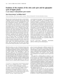
The Fd:F420 oxidoreductase reaction was not cataly- sed by membrane fractions from either the wild-type organism or the deletion mutants. In contrast, direct measurement of Fd:F420 oxidoreductase activity in cytoplasmic fractions revealed that the Fdred-dependent F420 reduction occurred in the wild-type organism and in the Dech and the DfpoA-O mutants (Fig. 2). Inter- estingly, this activity was essentially absent in the cyto- plasmic fraction of the DfpoF mutant.
A second possibility for F420H2 oxidation in fpo mutants is the utilization of NAD(P) as an elec- catalysed by an F420:NAD(P) tron acceptor, as oxidoreductase, which has been characterized in Methanococcus vannielii [7], Archaeoglobus fulgidus [8], Methanobacterium thermoautotrophicum [9] and Metha- nosphaera stadtmanae [10]. Ms. mazei is also able to produce such an enzyme, which is encoded by the gene mm0977 (unpublished results). In the course of the reaction, reduced nicotinamide adenine dinucleotide would be formed. However, neither NADPH nor NADH function as electron donors for the membrane- bound electron transport systems in Ms. mazei. There- fore, the F420:NAD(P) oxidoreductase cannot partici- pate in energy metabolism. In summary, there is a wealth of evidence that the Fpo is of major importance for F420H2-dependent heterodisulfide reduction in Ms. mazei. The possible electron transfer via H2 in the DfpoF and DfpoA-O mutants is probably only a rescue pathway that allows relatively slow growth of the cells with increased doubling times.
Electron transport in Dech mutants from A. fulgidus
This observation prompted investigation into the function of FpoF in more detail. The corresponding gene, mm0627, was expressed in E. coli and the protein was purified by affinity chromatography to apparent homogeneity (Fig. S1). UV–Vis spectra revealed the (FeS) clusters presence of flavins and iron–sulfur (Fig. S2). The determination of iron and sulfur yielded 8.7 ± 0.1 mol of nonheme iron and 5.1 ± 0.2 mol of acid-labile sulfur per mol of protein. These findings indicate the presence of two FeS clusters, as predicted from the amino acid sequence, and are in accordance with the FeS cluster content of the homologous pro- tein, FqoF, [12]. Finally, HPLC analysis revealed the presence of 1.0 ± 0.1 mol FAD per mol protein. The purified FpoF protein from
Another question in methanogenic bioenergetics con- cerns the fate of Fdred that is produced in aceticlastic and methylotrophic methanogenesis in the course of the acetyl-CoA synthase ⁄ CO dehydrogenase and form- ylmethanofuran dehydrogenase reactions, respectively.
Acetyl-CoA þ H4SPT þ 2Fd ! CH3-H4SPT þ CO2 þ 2Fdred þ HS-CoA
) M µ (
ð6Þ
2 H 0 2 4 F
ð7Þ Formyl-MF þ 2Fd ! MF þ CO2 þ 2Fdred
where MF is methanofuran and HS-CoA is coenzyme A.
Time (s)
Fig. 2. Fd:F420 oxidoreductase activity in Methanosarcina mazei cytoplasm. Assays contained 500 lg of cytoplasmic protein, 15 lM F420, 50 lg of CO dehydrogenase and 5 lg of clostridial Fd in 600 lL of Buffer A under a 5% CO ⁄ 95% N2 atmosphere. d, Wild- type cytoplasm; m, DfpoA-O cytoplasm; D, Dech cytoplasm; j, DfpoF cytoplasm.
FEBS Journal 278 (2011) 1277–1287 ª 2011 The Authors Journal compilation ª 2011 FEBS
1280
In many Methanosarcina spp., Fdred is oxidized by the membrane-bound, proton-translocating and H2- forming Ech hydrogenase [11]. The H2 thus produced is scavenged by the membrane-bound hydrogenase (Vho). Surprisingly, both Ms. mazei and Ms. barkeri Dech mutants are still capable of growth on methylated
C. Welte and U. Deppenmeier
Role of F420 dehydrogenase in Ms. mazei
M
C
4 µL
2 µL
8 µL
4 µL
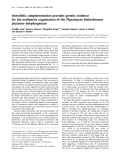
Fig. 4. Immunoblot used for FpoF quantification. Proteins blotted onto nitrocellulose membrane were detected with rabbit anti-FpoF and horseradish peroxidise-conjugated goat anti-(rabbit IgG). Lanes 1–5, 10–100 ng of FpoF; lane 6, molecular mass standard; lanes 7 and 8, solubilized membrane preparation from Methanosarcina mazei, lanes 9 and 10, membrane-free cytoplasm from Ms. mazei.
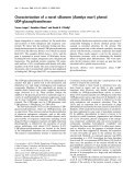
Fig. 3. Kinetic parameters of the purified Fd:F420 oxidoreductase, FpoF. Dependence of activity on the Fd concentration. Assays con- tained 50 lg of CO dehydrogenase and 10 lM F420 in 600 lL of buffer A under a 5% CO ⁄ 95% N2 atmosphere, as well as variable amounts of Fd.
indicating that
analysing the reduction of CoM-S-S-CoB, as catalysed by the membrane-bound heterodisulfide reductase, to exclude the possibility that FpoF connected to the Fpo complex interfered with the quantification of soluble FpoF. The specific activity of heterodisulfide reductase was < 1 mUÆmg)1 of protein, the cytoplasmic fraction was free of membrane particles. Hence, it is obvious that a large amount (about 75%) of the total FpoF protein is membrane-bound and that a smaller, yet significant, amount of this protein (about 25%) is also present in the cytoplasmic fraction.
Ms. mazei was able to oxidize clostridial Fdred and reduce F420. The kinetic parameters of the reaction exhibited a maximal velocity (Vmax) of 225 mUÆmg)1 of protein and KM values of 2 and 0.5 lm for F420 and Fdred, respectively (Fig. 3). The relatively low activity of purified FpoF was probably partly because a clos- tridial Fd was used as an electron donor, which might be responsible for an impaired transfer of electrons onto FpoF. Further studies will indicate which of the Methanosarcina Fd proteins function as natural elec- tron carriers.
Localization and dual function of FpoF
These findings led to the hypothesis that FpoF might have a dual function. First, when it is connected to the membrane integral Fpo complex, it functions as an electron input module of the Fpo. Second as a solu- ble single subunit it is involved in the reoxidation of Fdred in the cytoplasm, whereby reduced F420 is pro- duced that is then reoxidized by the complete Fpo complex. In summary, these experiments led to the conclusion that soluble FpoF can function as an Fd:F420 oxidoreductase when Fdred accumulates in the cytoplasm, as predicted for the Dech mutant.
Discussion
Reduced coenzyme F420 as electron donor of the respiratory chain
hydrogenotrophic
to the total cellular protein content,
FEBS Journal 278 (2011) 1277–1287 ª 2011 The Authors Journal compilation ª 2011 FEBS
1281
F420H2 is the key electron donor in methanogens and is formed by the reduction of F420 in methylotrophic and methanogenesis. When Ms. mazei grows on H2 ⁄ CO2, H2 can take two routes into the metabolism: it is either oxidized by a mem- brane-bound, proton-translocating H2:heterodisulfide oxidoreductase system (Fig. 5A), or it is oxidized by a soluble, nonrespiratory Frh that reduces F420 [13–18]. The resulting F420H2 is mainly used for the reduction in the methanogenic pathway where the of CO2 heterodisulfide CoM-S-S-CoB (as a terminal electron acceptor) is formed, which is reduced by the energy- As a prerequisite for the in vivo action of FpoF as an Fd:F420 oxidoreductase, the protein has to be present in both the cytoplasm and the membrane-bound Fpo complex. To examine this, antibodies directed against FpoF were produced using purified FpoF as the antigen, and used in subsequent immunoblotting experiments. Membrane-free cytoplasm and washed membrane fractions were analyzed using SDS ⁄ PAGE and immunoblotting. Known concentrations of FpoF were also applied and were used to quantify FpoF based on relative band intensities (Fig. 4). When the bands for the cytoplasm and the membrane fraction were compared with the calibration curve, about 76 ngÆlL)1 of membrane preparation could be identi- fied as FpoF and about 4 ng of FpoF could be detected per lL of cytoplasm. Taking into account the these preparations with protein concentrations of respect the amount of soluble FpoF was about 0.9 lgÆmg)1 of protein and 3 lgÆmg)1 of protein was present in the membrane fraction. The cytoplasmic fraction was also tested for contamination with membrane proteins by
C. Welte and U. Deppenmeier
Role of F420 dehydrogenase in Ms. mazei
the heterodisulfide (DG0
A
B
0 = exergonic reduction of )42.1 kJÆmol)1). Evidence in favour of this hypothesis came from the finding that the rate of F420H2-depen- dent heterodisulfide reduction in cell lysates of the DfpoA-O mutant or the DfpoF mutant was in the range of 5 mUÆmg)1 of protein and hence 10-fold higher than the rate of H2 production from F420H2 in the absence of CoM-S-S-CoB. The H2 produced could then be used by the membrane-bound H2:heterodisul- fide oxidoreductase and would yield the same amount of translocated protons compared with the F420H2:hete- rodisulfide oxidoreductase system (Fig. 5A).
In contrast to the fpo mutants, cell lysates of the wild-type organism exhibited an activity of about 50 mUÆmg)1 of protein with the substrate combination F420H2 plus heterodisulfide. This clearly demonstrates that, under methylotrophic growth conditions, 90% of the cellular F420H2 oxidation in Ms. mazei is per- formed by the Fpo connected to heterodisulfide reduc- tase via methanophenazine. Only 10% of the cellular F420H2 oxidation is accomplished by the cytoplasmic leading to H2 efflux that is scavenged by the Frh, membrane-bound H2:heterodisulfide oxidoreductase (Fig. 5A,B).
transport
in Ms. mazei. Vho ⁄ Vht,
F420
Fig. 5. Model of the branched electron transport pathway in Met- hanosarcina mazei. (A) Membrane-bound and (B) cytoplasmic elec- tron non-reducing hydrogenase; Hdr, heterodisulfide reductase; Frh, F420 (reducing) hydrogenase; Fpo, F420H2 dehydrogenase; FpoF, F-subunit of Fpo; MP, methanophenazine; CoM, Coenzyme M; H4SPT, tetrahydros- arcinapterin; A1AO, A1AO ATP synthase.
activity of a much higher
et al. [20]
In fact,
in Ms. mazei
FEBS Journal 278 (2011) 1277–1287 ª 2011 The Authors Journal compilation ª 2011 FEBS
1282
conserving H2:heterodisulfide oxidoreductase. In meth- ylotrophic methanogenesis, F420H2 is formed during methyl group oxidation to CO2 (Fig. 5B). In principle, the CO2-reduction pathway of the hydrogenotrophic methanogens acts in reverse, and F420H2 can be used in the respiratory chain (Fig. 5B). In Ms. mazei, the Fpo was characterized in detail as part of the mem- brane-bound energy-conserving F420H2:heterodisulfide oxidoreductase system [2,3]. The soluble Frh is also present in this organism under methylotrophic growth conditions [19]. The question arose of whether this enzyme can act in the reverse direction compared with hydrogenotrophic methanogenesis, namely producing H2 through the oxidation of F420H2 (Fig. 5B). As shown before, the Frh activity in the cell extracts of the parental strain and of the mutants was low, proba- bly because the overall reaction is thermodynamically unfavourable under standard conditions. The activity might be higher when the reaction is coupled to the As already indicated, the F420H2-oxidizing ⁄ H2-evolv- ing activity of cell lysates was only about 0.5 mUÆmg)1 of cellular protein. However, the reverse reaction (i.e. oxidation of H2 coupled to the reduction of F420) is catalyzed at about 35 mUÆmg)1 of cellular protein and is probably physio- logical when hydrogenotrophic methanogenesis is In Ms. barkeri, H2:F420 oxidoreductase performed. activity was observed at a much higher activity in cell lysates. We found an activity of about 360 mUÆmg)1 of protein and Michel even reported 870 mUÆmg)1 of protein in methanol-grown cells. This is a 10- to 25-fold higher activity than observed in Ms. mazei, and an indicator for a stronger activity of the reverse reaction, namely F420H2 oxidation with H2 production. It is tempting to speculate that the role of Frh in Ms. barkeri under methylotrophic growth con- ditions is more important than the role of the same enzyme in Ms. mazei. this was actually observed by Kulkarni et al. [4], who reported that knockouts of fpoA-O or fpoF in in Ms. barkeri do not significantly alter growth parameters, whereas the same strongly decreased the knockouts doubling time. In Methanosarcina acetivorans, a close relative of both Ms. mazei and Ms. barkeri, the appar- ent lack of hydrogenase activity [21–23] makes the route of F420H2 oxidation by Frh impossible. In addi- tion, only very low hydrogenase activities were observed in Methanosarcina thermophila [24] and in
C. Welte and U. Deppenmeier
Role of F420 dehydrogenase in Ms. mazei
obligate methylotrophic methanogens such as Metha- nolobus tindarius [25] and Methanococcoides burtonii [26,27]. Taking these findings together, we conclude that almost all methylotrophic methanogens rely on the Fpo to feed reducing equivalents into the respira- tory chain (Fig. 5A). Obviously, the only known exception is Ms. barkeri, where reducing equivalents from methyl group oxidation seem to be preferentially passed to molecular H2 by the cytoplasmic F420-reduc- ing hydrogenase. Hence, Ms. barkeri is not an ideal model for the principal electron transport chain of methylotrophic methanogens and a re-analysis of in Methanosarcina energy-conservation mechanisms species, as suggested by Kulkarni et al. is not [4], warranted.
Dual function of the FpoF subunit of the Fpo complex
is highly similar The existence of an Fd:F420 oxidoreductase, identi- fied as soluble FpoF, surely contributes to the regener- ation of oxidized Fd and to the survival of the Ms. mazei Dech mutant. However, this cytoplasmic enzyme does not explain the relatively high reduction rate of CoM-S-S-CoB at the expense of Fdred observed in the membrane fraction of this mutant. Therefore, it is tempting to speculate that besides FpoF there is another, still-unknown protein that is able to channel electrons from Fdred into the respiratory chain. Fur- thermore, it is important to note that the efficiency of energy conservation using Fdred is higher compared with F420H2 because the membrane-bound Fd:CoM-S- S-CoB oxidoreductase system translocates more the cytoplasmic membrane than the protons over F420H2:CoM-S-S-CoB oxidoreductase system [11]. Hence, the cells have to avoid a major transfer of elec- trons from Fdred to F420, which is accomplished by the low activity of the Fd:F420 oxidoreductase. Instead, the enzyme may act as a valve to establish the equilibrium of different reducing-equivalent concentrations, as well as in the transition of methanogenic pathways (e.g. from aceticlastic growth to methylotrophic growth).
Materials and methods
Strains, culture conditions and growth measurement
Methanosarcina mazei Go¨ 1 (DSM 7222) was used as the parent strain for mutant generation and as the wild-type strain. Furthermore, an Ms. mazei mutant lacking Ech hydrogenase (Ms. mazei Dech) [28], as well as DfpoF and DfpoA-O mutant strains (the generation of these strains is described below) were used. In addition, Ms. barkeri (DSM 800T) was used for cell-lysate experiments. All strains were grown in DSM medium 120 containing trimethylamine (50 mm final concentration) or acetate (100 mm final con- centration) as the substrate. To monitor growth, 50-mL cultures were grown and samples were taken at different time-points. These samples were reduced with sodium dithi- onite and the D at 600 nm was determined using a Helios Epsilon spectrophotometer (Thermo Scientific, Schwa¨ bisch- Gmu¨ nd, Germany). All growth experiments were performed with at least four separate cultures.
The cluster encoding the Fpo of Ms. mazei and related Methanosarcina strains is composed of 12 genes (fpoA– O) [3]. Analysis of the Fpo subunits showed that the enzyme to proton-translocating NADH dehydrogenases (NDH-1). The gene products FpoAHJKLMN are hydrophobic and homologous to subunits that form the membrane integral module of NDH-1. FpoBCDI have their counterparts in the amphipathic membrane-associated module of NDH-1. Homologues to the hydrophilic NADH-oxidizing sub- unit of NDH-1 are not present in Ms. mazei. Instead, the gene product FpoF is responsible for F420H2 oxidation and functions as the electron-input device [3]. Interestingly, the gene fpoF is not part of the operon and is located at a different site on the chro- mosome.
Cultures of wild-type and mutant Ms. mazei, grown to mid-exponential phase, were harvested, washed once in stabilizing buffer (2 mm KH2PO4 ⁄ K2HPO4, 2 mm MgSO4, 20 mm NaCl, 200 mm sucrose, pH 6.8) and resuspended in the same buffer to yield a final protein content of
Measurements of resting cell suspensions
FEBS Journal 278 (2011) 1277–1287 ª 2011 The Authors Journal compilation ª 2011 FEBS
1283
Previously it was only known that the complex of FpoA–O and FpoF has a role in F420H2 oxidation, but is not involved in the reaction with Fd. This fact still holds true for the entire complex, but the single subunit FpoF may also interact with Fdred (Fig. 5B). A slow reduction of F420 with Fdred as the substrate was found to occur in the cytoplasm of the parental strain and of the DfpoA–O and the Dech mutants. However, this activity was essentially absent in the DfpoF mutant. Finally, we showed that the purified FpoF functions as an Fd:F420 oxidoreductase. The results of the immunoblotting experiments indicated that FpoF is present at relatively high concentrations in the cytoplasm. This fact cannot be explained by trafficking of premature FpoF intended for binding to the FpoA–O complex but points to a second function of the soluble form of FpoF that is characterized by a slow electron transfer from Fdred onto F420.
C. Welte and U. Deppenmeier
Role of F420 dehydrogenase in Ms. mazei
0.5–1 mgÆmL)1. The cells were starved at 37 (cid:2)C for 30 min before methane formation was induced by the addition of 5 mm trimethylamine. If desired, the 100% N2 in the head- space was replaced with 100% H2. At various reaction time-points, 50 lL of the headspace was injected into a gas chromatograph (GC-14A; Shimadzu, Kyoto, Japan) with N2 as the carrier gas. Methane was analyzed using a flame ionization detector and quantified by comparison with a standard curve.
Preparation of proteins, antibodies, membranes and cofactors
primers
(employing
and
induction medium [35]
(32 gÆL)1 of
for
the lysate (25 000 g, 15 min, 4 (cid:2)C),
The CO dehydrogenase from Moorella thermoacetica was purified as described previously [31], with the modifications as also described previously [28]. Purification of Fd from Clos- tridium pasteurianum was performed as outlined by Morten- son [32] with replacement of the last two steps (crystallization and dialysis) by ultrafiltration. Heterodisulfide was synthe- sized as specified previously [33], and cofactor F420 was puri- fied and reduced as described by Abken et al. [34]. Cell lysate was obtained by anaerobically harvesting trimethylamine- grown cells and resuspending in Buffer A (40 mm K2HPO4 ⁄ KH2PO4, pH 7.0, 5 mm dithioerythritol, 1 lgÆmL)1 of resazurin) containing desoxyribonuclease to yield a final protein content of about 5 mgÆmL)1. Ms. mazei cells are immediately lysed in Buffer A by osmotic shock; Ms. barkeri cells were broken by treatment in a French pressure cell press at 1000 psi. Subsequent preparation of membranes of Ms. mazei was carried out as described by Welte et al. [28]. The cytoplasmic supernatant was ultracentrifuged twice (120 000 g, 45 min) and only the top 60% of the ultracentrifu- gation supernatant was defined as ‘membrane-free cytoplasm’. For antibody production and enzyme assays, FpoF was heterologously produced in E. coli DH5a. The fpoF gene (mm0627) was cloned into pASK-IBA3 (containing a C-ter- minal StrepII-Tag; IBA, Go¨ ttingen, Germany). The fpoF PCR fragment was generated with the primers 5¢-ATGG TACGTCTCAAATGCCACCAAAGATTGCAGAAGTCA TT-3¢ 5¢-ATGGTACGTCTCAGCGCTGACTGTT TCACTGCGGATTCCG-3¢. It was digested with BsmBI and cloned into the BsaI sites of pASK-IBA3. The resulting construct was checked by sequencing (StarSEQ, Mainz, Germany). Expression was performed in 200 mL of maxi- trypton and mal 20 gÆL)1 of yeast extract) containing M9 salts, 100 lm CaCl2, 1 mm MgSO4 and 1 lm FeNH4 citrate. The cells were grown until an D of (cid:2) 1 was reached, then protein production was induced with 200 ngÆmL)1 of anhydrotetra- cyclin. Subsequently, 30 lm FeNH4 citrate and 40 lgÆmL)1 of riboflavin were added and the culture was flushed with N2 and sealed with a rubber stopper to achieve anoxic con- ditions. After a 4 h induction period at 30 (cid:2)C with gentle shaking, the culture was harvested anaerobically (10 000 g, 15 min). To facilitate lysis of the cells, the pellet was frozen and thawed, then resuspended in Buffer W (150 mm Tris, pH 8.0, 5 mm dithioerythritol, 1 lgÆmL)1 of resazurin) con- taining a small amount of lysozyme, desoxyribonuclease and 1 mL of B-PER protein extraction solution (Thermo Fisher, Schwerte, Germany) per mL of Buffer W. After clarification of the recombinant protein was purified anaerobically in an anaer- obic chamber (3% H2, 97% N2; Coy Laboratories, Grass Lake, MI, USA) using Strep-Tactin sepharose, as described by the manufacturer (IBA).
Methanosarcina mazei DfpoF and DfpoA-O were generated using homologous recombination, as described by Metcalf et al. [29]. For the creation of knockout vectors, fragments of the upstream and downstream regions of the respective genes or gene clusters were amplified by PCR and cloned into the two multiple cloning sites of the pJK3 vector [29]. For the DfpoF knockout, a 1.2-kb fragment upstream of mm0627 (fpoF) was cloned into pJK3 using XhoI and EcoRV (employing the primers 5¢-CCGGCTCGAGTG CAATTAACATCTATTGTA-3¢ and 5¢-CCTTGATATCT CAGTTACCTCCACTGCCT-3¢), and a 1.2-kb fragment downstream of mm0627 was cloned into pJK3 using SpeI and NotI 5¢-GTAACTAG the TCAGTCTGAAGTCCGAAACTT-3¢ and 5¢-AGCGCGG CCGCGGAAAGTGGTCT ACCTTA-3¢). The knockout vector was linearized with XhoI and transformed into Ms. mazei, as described previously [30]. For the DfpoA-O knockout, a 1.2-kb fragment upstream of mm2491 (fpoA) was cloned into pJK3 using XhoI and HindIII (employing the primers 5¢-CCTTCTCGAGGCCCTCCAAGTCCTG CACCT-3¢ and 5¢-CATGAAGCTTAGTGCAGCA AT CTGAAATTGC-3¢), and a 1.2-kb fragment downstream of mm2479 (fpoO) was cloned using SacI and BcuI (employing the primers 5¢-AACGCTGC AGGAACACGTACACCC GCATTA-3¢ and 5¢-TACTACTAGTCCTCAGTTGGACG TTTACTC-3¢). The DfpoA-O knockout vector was linear- ized with BcuI and transformed into Ms. mazei. After clonal separation on agar plates, the mutant cultures were fpoD confirmed by PCR. Gene-specific primers (mm2488) and for fpoF (mm0627) revealed the presence or absence of the respective genes in the mutant strains. In parallel, a control PCR was performed with wild-type DNA or cell material. Furthermore, primers specific for the puromycin resistance cassette (pac) were used to ver- ify the presence of the pac cassette in the mutant ge- nomes. The absence of the FpoF protein in the DfpoF mutant was also verified by western blotting and anti- body detection with antibodies directed against FpoF. More information about these genes and proteins can be found in the database KEGG (http://www.genome.jp/ the kegg/) using the abovementioned locus number of genes from Ms. mazei (MM_0627, MM_2488, MM_2479 and MM_2491).
FEBS Journal 278 (2011) 1277–1287 ª 2011 The Authors Journal compilation ª 2011 FEBS
1284
Generation of mutant strains
C. Welte and U. Deppenmeier
Role of F420 dehydrogenase in Ms. mazei
Antibodies were produced by Seqlab (Go¨ ttingen, Ger- many) with the 3-month protocol using rabbits for immuni- zation. Horseradish peroxidase-conjugated goat anti-(rabbit IgG) was purchased from Rockland Inc. (Gilbertsville, PA, USA).
Western blot
Nonheme iron was quantified as described by Landers and Sak [36]. Acid-labile sulfide was quantified photometrically at 670 nm by measuring the formation of methylene blue the addition of N,N-dimethyl-p-phenylenediamine, after with Na2S as standard [37]. Flavin identity and content were determined by HPLC analysis (Knauer Smartline, Ber- lin, Germany) with a reverse-phase C-18 column (Varian Microsorb-MV, 250 mm · 4.6 mm) at a flow rate of 0.75 mLÆmin)1 with a lineacr gradient of 0–100% methanol in ammonium acetate buffer (50 mm, pH 6.0). Flavins were detected at 436 nm [12] with a retention time of 12.6 min for FAD and a retention time of 14.9 min for FMN. Prior to loading onto the HPLC column, protein samples were treated with 5% trichloroacetic acid for 15 min (Adams and Jia 2006), then precipitated protein was collected by centrifugation (8000 g, 2 min) and protein-free supernatant was applied to the HPLC column. Peak areas were com- pared with a standard curve created using 0–20 lm FAD.
Cofactor quantification
All enzyme assays were performed in rubber-stoppered cuvettes or vials filled with Buffer A with 100% N2 in the headspace, unless indicated otherwise.
Before SDS ⁄ PAGE, membrane proteins were solubilized in 1% decylmaltoside for 3 h or overnight, then mixed with SDS ⁄ PAGE loading dye [38] and applied directly to the gel. Cytoplasmic proteins, as well as FpoF, were heated in SDS ⁄ PAGE loading dye for 5 min at 95 (cid:2)C before loading onto the gel. To the gel, 10–100 lg of membrane or cytoplas- mic protein fractions and 10–100 ng of purified FpoF were applied. All proteins were separated by SDS/PAGE (12.5% gel) according to Laemmli [38] and blotted onto a nitrocellu- lose membrane using a semi-dry blotting device (Biozym, Hessisch Oldendorf, Germany). The membrane was blocked in NaCl ⁄ Pi (140 mm NaCl, 2.7 mm KCl, 4.3 mm Na2HPO4, 1.5 mm KH2PO4, pH 7.3) containing 5% milk powder for 1 h at room temperature. Then, the membrane was washed (3 · 5 min with NaCl ⁄ Pi) and the primary antibody directed against FpoF (rabbit-aFpoF; obtained on the second bleed) was applied at a 1 : 1000 dilution in NaCl ⁄ Pi. This was fol- lowed by a washing step with PBST (NaCl ⁄ Pi containing 0.05% Tween 20; three, 10-min washes) and incubation with the secondary antibody [horseradish peroxidase-conjugated goat anti-(rabbit IgG)] at a 1 : 5000 dilution. The membrane was washed again with PBST (3 · 10 min), and detection was performed in 20 mL of NaCl ⁄ Pi containing 200 lL of 4-chloro-1-naphthol (3% w ⁄ v) and 20 lL of H2O2 (30%). Quantification was performed using a Canon CanoScan 4400F flatbed scanner (Canon, Krefeld, Germany) and Adobe Photoshop with the method outlined at http://luke miller.org/journal/2007/08/quantifying-western-blots-without. html. Briefly, membranes were scanned in black and white, the colours were inverted and the relative intensity (lumines- cence · occupied pixel) was determined using the ‘histogram’ option. A calibration curve was constructed using different amounts of FpoF, and the amount of FpoF was estimated in membrane and cytoplasmic fractions.
Enzyme assays
Acknowledgements
F420H2:heterodisulfide oxidoreductase activity was deter- mined in 600 lL of buffer A using 10 lm F420H2, 20 lm lysate, membrane CoM-S-S-CoB and 100–200 lg of cell fraction or cytoplasmic fraction. The increase in absorbance at 420 nm was recorded (Jasco, Gross-Umstadt, Germany) using a V-550 UV ⁄ Vis spectrophotometer (Gross-Umstadt, Germany), and a molar extinction coefficient of F420 of 40 mm)1Æcm)1 was used to calculate enzyme activity. For the measurement of Frh, heterodisulfide was omitted and the amount of cell lysate was increased to 500 lg. For the reverse reaction, 100% H2 in the headspace, 100–200 lg of cell lysate and 10 lm F420 were used.
the Deutsche Forschungsgemeinschaft We thank Elisabeth Schwab for technical assistance and Paul Schweiger for critical reading of the manu- script. Many thanks also go to Gunes Bender and Steve Ragsdale, Department of Biological Chemistry, University of Michigan Medical School for providing the CO dehydrogenase ⁄ acetyl-CoA synthase from Moorella thermoacetica. This work was supported by (grant De488 ⁄ 9-1).
References
1 Deppenmeier U & Mu¨ ller V (2008) Life close to the
thermodynamic limit: how methanogenic archaea con- serve energy. Results Probl Cell Differ 45, 123–152.
FpoF activity was determined by observing the change in absorbance at 420 nm caused by F420 reduction. The forward reaction (Fdred fi F420) was measured using 600 lL of Buf- fer A under a 5% CO ⁄ 95% air atmosphere, 5 lg of Fd, 50 lg of CO dehydrogenase and 15 lm F420. The initial substrate CO passes electrons to the M. thermoacetica CO dehydrogenase ⁄ acetyl-CoA synthase, which reduces C. paste- urianum Fd. Fdred is then used by FpoF to reduce F420. For KM and Vmax calculations, variable amounts of Fd (0.5–20 lg) and F420 (1.2–10 lm) were used, whereas the other compo- nents were used at the concentrations described above.
FEBS Journal 278 (2011) 1277–1287 ª 2011 The Authors Journal compilation ª 2011 FEBS
1285
C. Welte and U. Deppenmeier
Role of F420 dehydrogenase in Ms. mazei
14 Brodersen J, Ba¨ umer S, Abken HJ, Gottschalk G &
2 Ba¨ umer S, Murakami E, Brodersen J, Gottschalk G,
Deppenmeier U (1999) Inhibition of membrane-bound electron transport of the methanogenic archaeon Met- hanosarcina mazei Go¨ 1 by diphenyleneiodonium. Eur J Biochem 259, 218–224.
15 Alex LA, Reeve JN, Orme-Johnson WH & Walsh CT
Ragsdale SW & Deppenmeier U (1998) The F420H2:heterodisulfide oxidoreductase system from Methanosarcina species. 2-Hydroxyphenazine mediates electron transfer from F420H2 dehydrogenase to hetero- disulfide reductase. FEBS Lett 428, 295–298.
3 Ba¨ umer S, Ide T, Jacobi C, Johann A, Gottschalk G & Deppenmeier U (2000) The F420H2 dehydrogenase from Methanosarcina mazei is a redox-driven proton pump closely related to NADH dehydrogenases. J Biol Chem 275, 17968–17973.
(1990) Cloning, sequence determination, and expression of the genes encoding the subunits of the Nickel-con- taining 8-hydroxy-5-deazaflavin reducing hydrogenase from Methanobacterium thermoautotrophicum DH. Biochemistry 29, 7237–7244.
4 Kulkarni G, Kridelbaugh DM, Guss AM & Metcalf
WW (2009) Hydrogen is a preferred intermediate in the energy-conserving electron transport chain of Methano- sarcina barkeri. Proc Natl Acad Sci USA 106, 15915– 15920.
16 Fox JA, Livingston DJ, Orme-Johnson WH & Walsh CT (1987) 8-hydroxy-5-deazaflavin-reducing hydroge- nase from Methanobacterium thermoautotrophicum. 1. Purification and characterization. Biochemistry 26, 4219–4227.
5 Tebrommelstroet BWJ, Geerts WJ, Keltjens JT,
17 Livingston DJ, Fox JA, Orme-Johnson WH & Walsh CT (1987) 8-hydroxy-5-deazaflavin-reducing hydroge- nase from Methanobacterium thermoautotrophicum .2. Kinetic and hydrogen-transfer studies. Biochemistry 26, 4228–4237.
Vanderdrift C & Vogels GD (1991) Purification and properties of 5,10-methylenetetrahydromethanopterin dehydrogenase and 5,10-methylenetetrahydromethanop- terin reductase, 2 coenzyme-F420-dependent enzymes, from Methanosarcina barkeri. Biochim Biophys Acta 1079, 293–302.
6 Ma KS & Thauer RK (1990) N5, N10-methylenetetra- hydromethanopterin reductase from Methanosarci- na barkeri. FEMS Microbiol Lett 70, 119–124.
18 Jacobson FS, Daniels L, Fox JA, Walsh CT & Orme- Johnson WH (1982) Purification and properties of an 8-hydroxy-5-deazaflavin reducing hydrogenase from Methanobacterium thermoautotrophicum. J Biol Chem 257, 3385–3388.
7 Yamazaki S & Tsai L (1980) Purification and properties of 8-hydroxy-5-deazaflavin-dependent NADP+ reduc- tase from Methanococcus vannielii. J Biol Chem 255, 6462–6465.
19 Hovey R, Lentes S, Ehrenreich A, Salmon K, Saba K, Gottschalk G, Gunsalus RP & Deppenmeier U (2005) DNA microarray analysis of Methanosarcina mazei Go¨ 1 reveals adaptation to different methanogenic substrates. Mol Genet Genomics 273, 225–239.
20 Michel R, Massanz C, Kostka S, Richter M & Fiebig
8 Kunow J, Schwo¨ rer B, Stetter KO & Thauer RK (1993) A F420-dependent NADP reductase in the extremely thermophilic sulfate-reducing Archaeoglobus fulgidus. Arch Microbiol 160, 199–205.
9 Berk H & Thauer RK (1998) F420H2 : NADP
K (1995) Biochemical characterization of the 8- hydroxy-5-deazaflavin-reactive hydrogenase from Methanosarcina barkeri Fusaro. Eur J Biochem 233, 727–735.
oxidoreductase from Methanobacterium thermoauto- trophicum: identification of the encoding gene via functional overexpression in Escherichia coli. FEBS Lett 438, 124–126.
21 Guss AM, Mukhopadhyay B, Zhang JK & Metcalf WW (2005) Genetic analysis of mch mutants in two Methanosarcina species demonstrates multiple roles for the methanopterin-dependent C1 oxidation ⁄ reduction pathway and differences in H2 metabolism between closely related species. Mol Microbiol 55, 1671–1680.
10 Elias DA, Juck DF, Berry KA & Sparling R (2000) Purification of the NADP+: F420 oxidoreductase of Methanosphaera stadtmanae. Can J Microbiol 46, 998– 1003.
22 Guss AM, Kulkarni G & Metcalf WW (2009)
Differences in hydrogenase gene expression between Methanosarcina acetivorans and Methanosarcina barkeri. J Bacteriol 191, 2826–2833.
11 Welte C, Kra¨ tzer C & Deppenmeier U (2010) Involve- ment of Ech hydrogenase in energy conservation of Methanosarcina mazei. FEBS J 277, 3396–3403. 12 Bru¨ ggemann H, Falinski F & Deppenmeier U (2000) Structure of the F420H2:quinone oxidoreductase of Archaeoglobus fulgidus. Identification and overproduc- tion of the F420H2-oxidizing subunit. Eur J Biochem 267, 5810–5814.
23 Rohlin L & Gunsalus RP (2010) Carbon-dependent control of electron transfer and central carbon pathway genes for methane biosynthesis in the archaean Methanosarcina acetivorans strain C2A. BMC Microbiol 10, 62.
24 Zinder SH & Mah RA (1979) Isolation and character- ization of a thermophilic strain of Methanosarcina unable to use H2-CO2 for methanogenesis. Appl Environ Microbiol 38, 996–1008.
13 Muth E, Mo¨ rschel E & Klein A (1987) Purification and characterization of an 8-hydroxy-5-deazaflavin-reducing hydrogenase from the archaebacterium Methanococ- cus voltae. Eur J Biochem 169, 571–577.
FEBS Journal 278 (2011) 1277–1287 ª 2011 The Authors Journal compilation ª 2011 FEBS
1286
C. Welte and U. Deppenmeier
Role of F420 dehydrogenase in Ms. mazei
tions with highly purified methyl-CoM reductase (com- ponent C) from Methanobacterium thermoautotrophicum (strain Marburg). Eur J Biochem 172, 669–677.
25 Deppenmeier U (1991) Identifizierung und Charakteri- sierung membrangebundener protonentranslozierender Redox-Systeme in methanogenen Bakterien. PhD thesis, University of Go¨ ttingen.
26 Allen MA, Lauro FM, Williams TJ, Burg D, Siddiqui
34 Abken HJ, Tietze M, Brodersen J, Ba¨ umer S, Beifuss U & Deppenmeier U (1998) Isolation and characterization of methanophenazine and function of phenazines in membrane-bound electron transport of Methanosarci- na mazei Go¨ 1. J Bacteriol 180, 2027–2032.
KS, De Francisci D, Chong KWY, Pilak O, Chew HH, De Maere MZ et al. (2009) The genome sequence of the psychrophilic archaeon Methanococcoides burtonii: the role of genome evolution in cold adaptation. ISME J 3, 1012–1035.
35 Mott JE, Grant RA, Ho YS & Platt T (1985) Maximiz- ing gene expression from plasmid vectors containing the Lambda-Pl promoter – strategies for overproducing transcription termination factor Rho. Proc Natl Acad Sci USA 82, 88–92.
36 Landers JW & Zak B (1958) Determination of serum
27 Franzmann PD, Springer N, Ludwig W, Demacario EC & Rohde M (1992) A methanogenic archaeon from Ace Lake, Antarctica – Methanococcoides burtonii sp. nov. Syst Appl Microbiol 15, 573–581.
28 Welte C, Kallnik V, Grapp M, Bender G, Ragsdale S
copper and iron in single small sample. Tech Bull Regist Med Technol 28, 98–100.
& Deppenmeier U (2010) Function of Ech hydrogenase in ferredoxin-dependent, membrane-bound electron transport in Methanosarcina mazei. J Bacteriol 192, 674–678.
29 Metcalf WW, Zhang JK, Apolinario E, Sowers KR &
37 Beinert H (1983) Semi-micro methods for analysis of labile sulfide and of labile sulphide plus sulfane sulfur in unusually stable iron–sulfur proteins. Anal Biochem 131, 373–378.
38 Laemmli UK (1970) Cleavage of structural proteins
during assembly of head of bacteriophage T4. Nature 227, 680–685.
Wolfe RS (1997) A genetic system for archaea of the genus Methanosarcina: liposome-mediated transforma- tion and construction of shuttle vectors. Proc Natl Acad Sci USA 94, 2626–2631.
Supporting information
30 Ehlers C, Weidenbach K, Veit K, Deppenmeier U, Met- calf WW & Schmitz RA (2005) Development of genetic methods and construction of a chromosomal glnK1 mutant in Methanosarcina mazei strain Go¨ 1. Mol Genet Genomics 273, 290–298.
31 Ragsdale SW, Ljungdahl LG & Der Vartanian DV (1983) Isolation of carbon monoxide dehydrogenase from Acetobacterium woodii and comparison of its properties with those of the Clostridium thermoaceticum enzyme. J Bacteriol 155, 1224–1237.
32 Mortenson LE (1964) Purification and analysis of ferre-
doxin from Clostridium pasteurianum. Biochim Biophys Acta 81, 71–77.
33 Ellermann J, Hedderich R, Bocher R & Thauer RK
The following supplementary material is available: Fig. S1. SDS ⁄ PAGE of recombinant FpoF. Fig. S2. UV ⁄ Visible spectra of recombinant FpoF. This supplementary material can be found in the online version of this article.
(1988) The final step in methane formation. Investiga-
FEBS Journal 278 (2011) 1277–1287 ª 2011 The Authors Journal compilation ª 2011 FEBS
1287
Please note: As a service to our authors and readers, this journal provides supporting information supplied by the authors. Such materials are peer-reviewed and may be re-organized for online delivery, but are not copy-edited or typeset. Technical support issues arising from supporting information (other than missing files) should be addressed to the authors.