
RESEARCH Open Access
Promising cytotoxic activity profile of fermented
wheat germ extract (Avemar
®
) in human cancer
cell lines
Thomas Mueller, Karin Jordan and Wieland Voigt
*
Abstract
Fermented wheat germ extract (FWGE) is currently used as nutrition supplement for cancer patients. Limited recent
data suggest antiproliferative, antimetastatic and immunological effects which were at least in part exerted by two
quinones, 2-methoxy benzoquinone and 2,6-dimethoxybenzquinone as ingredients of FWGE. These activity data
prompted us to further evaluate the in vitro antiproliferative activity of FWGE alone or in combination with the
commonly used cytotoxic drugs 5-FU, oxaliplatin or irinotecan in a broad spectrum of human tumor cell lines. We
used the sulforhodamine B assay to determine dose response relationships and IC
50
-values were calculated using
the Hill equation. Drug interaction of simultaneous and sequential drug exposure was estimated using the model
of Drewinko and potential clinical activity was assessed by the model of relative antitumor activity (RAA). Apoptosis
was detected by DNA gel electrophoresis.
FWGE induced apoptosis and exerted significant antitumor activity in a broad spectrum of 32 human cancer cell
lines. The highest activity was found in neuroblastoma cell lines with an average IC
50
of 0.042 mg/ml. Furthermore,
IC
50
-range was very narrow ranging from 0.3 mg/ml to 0.54 mg/ml in 8 colon cancer cell lines. At combination
experiments in colon cancer cell lines when FWGE was simultaneously applied with either 5-FU, oxaliplatin or
irinotecan we observed additive to synergistic drug interaction, particularly for 5-FU. At sequential drug exposure
with 5-FU and FWGE the observed synergism was abolished.
Taken together, FWGE exerts significant antitumor activity in our tumor model. Simultaneous drug exposure with
FWGE and 5-FU, oxaliplatin or irinotecan yielded in additive to synergistic drug interaction. However, sequential
drug exposure of 5-FU and FWGE in colon cancer cell lines appeared to be schedule-dependent (5-FU may
precede FWGE).
Further evaluation of FWGE as a candidate for clinical combination drug regimens appeared to be warranted.
Introduction
The exact chemical composition of FWGE, which is
currently used as nutriment for cancer patients is not
completely known [1]. It contains two quinones, 2-
methoxy benzoquinone and 2,6-dimethoxybenzquinone
that likely play a significant role in exerting several of its
biological properties [2]. Preclinical in vitro and in vivo
data suggested antiproliferative, antimetastatic and
immunological effects of FWGE [1-7]. In cell lines stu-
dies, FWGE induced programmed cell death via the cas-
pase - PARP-pathway [7,8]. But the exact mechanism by
which this multi-molecule composition triggers cell
death is still obscure. In previous studies several groups
could demonstrate that FWGE interferes with enzymes
of the anaerobic glycolisis and pentose cycle [2,9,10].
Known targets are the transketolase, glucose-6-phos-
phate dehydrogenase, lactate dehydrogenase and hexoki-
nase which are necessary for the allocation of precursors
for DNA-synthesis [9]. Also involved in DNA-synthesis
is ribonucleotide reductase [6]. This enzyme is upregu-
lated in various types of cancer and is an attractive tar-
get in cancer chemotherapy. Several established
anticancer drugs like fludarabine, cytarabine and gemci-
tabine exert at least in part their cytotoxic activity by
inhibiting ribonucleotide reductase [11]. An inhibitory
activity on ribonucleotide reductase could also be
* Correspondence: wieland.voigt@medizin.uni-halle.de
University of Halle, Department Internal Medicine, Oncology/Hematology
and Hemostaseology, Ernst-Grube Str. 40, 06120 Halle/Saale, Germany
Mueller et al.Journal of Experimental & Clinical Cancer Research 2011, 30:42
http://www.jeccr.com/content/30/1/42
© 2011 Mueller et al; licensee BioMed Central Ltd. This is an Open Access article distributed under the terms of the Creative Commons
Attribution License (http://creativecommons.org/licenses/by/2.0), which permits unrestricted use, distribution, and reproduction in
any medium, provided the original work is properly cited.

demonstrated for FWGE, allowing FWGE to interfere
with nucleic acid-synthesis by several pathways [1,8,11].
Beside the single agent cytotoxic activity of FWGE
against human tumor cell lines and human tumor xeno-
grafts some data suggest synergistic drug interaction
between 5-FU or DTIC in a limited number of cell lines
[2,6].
In addition to the preclinical data there are already a
few clinical studies published which suggest some ben-
eficial effect of FWGE in human cancer therapy. The
most impressive data were generated in a randomized
Phase II trial by Demidov et al. who observed a signifi-
cant gain in progression free survival and overall survi-
val for the combination of DTIC and FWGE as
compared to DTIC alone in melanoma patients [12]. A
study conducted by Jakab et al. in patients with color-
ectal cancer found an enhanced survival and reduced
metastasis formation for the combination of che-
motherapy and FWGE as compared to chemotherapy
alone group. In a multivariate analysis of this study
only tumor stage and FWGE treatment were the only
significant predictors of survival [13]. However, this
data have to be interpreted with caution since the
study had a non randomized design and the patient
groups were not balanced [1,13]. Of similar impor-
tance, several studies including the ones cited above
suggested an improvement of quality of life due to co
treatment with FWGE [14].
Overall, the limited preclinical and clinical data avail-
able suggest some promising activity profile of FWGE as
a nutriment for cancer patients but also a potential
anticancer agent.
In this broad in vitro study we aimed to analyze the
single agent activity of FWGE as well as its interaction
with the commonly used drugs 5-FU, oxaliplatin and iri-
notecan in a large panel of human cancer cell lines from
different tumor entities. These data are of potential
value to direct the further development FWGE in differ-
ent cancer types and to help to select potential drug
partners for the future development of combinations of
chemotherapy regimens with FWGE.
Materials and methods
Drugs and chemicals
FWGE was a generous gift from Biropharma Ltd, Kunfe-
herto, Hungary. FWGE was stored as dried powder at 4°
C until use. For experimentation, FWGE was freshly
prepared in sterile water to a final concentration of 100
mg/ml. After solution FWGE was centrifuged with 150
g to remove the insoluble material. 5-FU, Irinotecan,
Oxaliplatin and Sulforhodamine B were purchased from
Sigma Chemical Company, Germany. RPMI 1640 and
Penicillin/Streptomycin were obtained from PAA,
Pasching, Austria. FBS was purchased Biochrom AG,
Berlin, Germany.
Cell lines and culture
The following human cancer cell lines were used for
experimentation: testicular cancer (H12.1, 2102EP,
1411HP, 1777NRpmet), colon cancer (HCT-8, HCT-15,
HCT-116, HT-29, DLD-1, SW480, COLO205,
COLO320DM), NSCLC (A549, A427, H322, H358),
head and neck cancer (FADU, A253), cervical epider-
moid carcinoma (A431), mammary adenocarcinoma
(MCF-7, BT474), ovarian adenocarcinoma (A2780), gas-
tric cancer (M2), anaplastic thyroid cancer (8505C,
SW1736), papillary thyroid cancer (BCPAP), follicular
thyroid cancer (FTC133), melanoma (518A2), hepatoma
(HepG2), glioblastoma (U87MG), neuroblastoma
(SHSY5Y, SIMA). All cell lines were grown as mono-
layers of up to 80% confluence in RPMI 1640 supple-
mented with 10% FBS and 1% Penicillin/Streptomycin at
37°C, 5% CO
2
and humidified air.
Growth inhibition experiments
To assess antiproliferative effects, the total protein sul-
forhodamine B (SRB) assay was used as described pre-
viously [15]. In brief, cells were seeded in 96 well plates
at a cell line specific density to ensure exponential
growth throughout the whole period of the assay. These
cell numbers were determined previously by cell growth
kinetics. After 24 h, exponentially growing cells were
exposed to serial dilutions of each drug alone or drug
combinations for the indicated times continuously. To
investigate the influence of drug schedules drug A was
added 24 h after cell seeding followed by drug B another
24 h later or vice versa. Corresponding control plates
with single agents were treated in parallel.
After 120 h total assay time, media was removed and
cells were fixed with 10% TCA and processed according
to the published SRB assay protocol [15]. Absorbency
was measured at 570 nm using a 96-well plate reader
(Rainbow, SLT, Germany).
DNA gel electrophoresis
To detect apoptosis by DNA gel electrophoresis the
floating cells after drug treatment with an IC
90
of
FWGE for 48 h were used. After washing cells twice
with PBS they were lysed in lysis-buffer (100 mM TRIS-
HCL (pH8.0), 20 mM EDTA, 0,8% SDS). Subsequent to
treatment with RNaseA for 2 h at 37°C and proteinase
K (Roche Molecular Biochemicals) overnight at 50°C,
lysastes were mixed with DNA loading buffer. To sepa-
rate DNA fragments, probes were run on a 1.5% agarose
gel followed by ethidium bromide staining and rinsing
with destilled water. DNA ladders were visualized under
Mueller et al.Journal of Experimental & Clinical Cancer Research 2011, 30:42
http://www.jeccr.com/content/30/1/42
Page 2 of 7

UV light and documented on a BioDocAnalyse instru-
ment (Biometra).
Data analysis
Dose response curves were generated by Sigma Plot
(Jandel Scientific, San Rafael, CA) and IC
50
values were
calculated based on the Hill equation. Drug interaction
was assessed using the model of Drewinko [16]. In
brief, a hypothetical curve was calculated by multiply-
ing the ratio of treated and untreated control with the
dose response data points of the single drug curve.
Synergy could be assumed if the hypothetical curve
runs above the combination curve and antagonism is
indicated if the hypothetical curve runs below the
combination curve. In case of additivity both curve
were superimposed.
Statistical significance was probed with the two tailed,
unpaired student’s t-test. Significance was assumed at a
p-value < 0.05.
Potential clinical activity was estimated by relative
antitumor activity (RAA), which was defined as the ratio
of peak plasma level and in vitro IC
50
value [17]. A
RAA > 1 indicates potential clinical activity.
Results
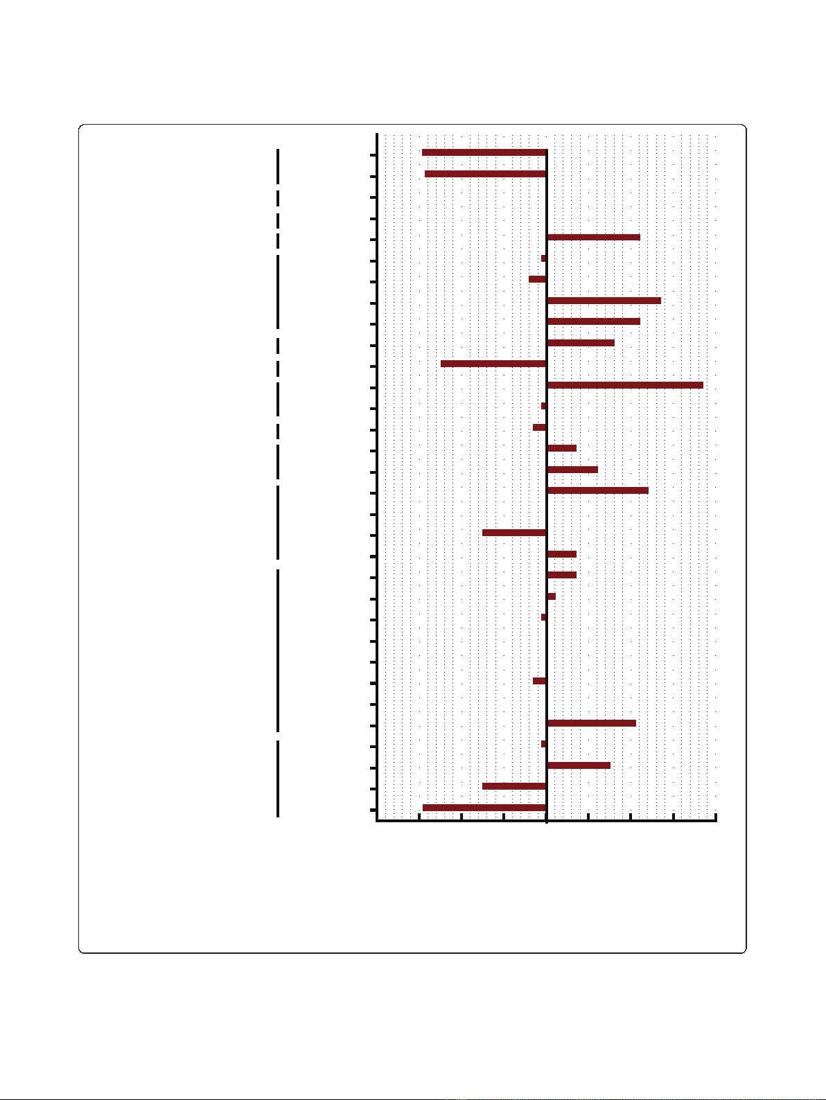
Single agent antiproliferative activity of FWGE in human
cancer cell lines
The antiproliferative activity of a 96 hour continuous
exposure to FWGE was evaluated in a large panel of
human tumor cell lines using the SRB-assay. IC
50
-values
were calculated using the Hill equation and the obtained
data from at least three independent experiments were
summarized as a mean graph (Figure 1). IC
50
of FWGE
ranged from 0.038 mg/ml to 0.7 mg/ml with a median
IC
50
of 0.33 mg/ml.
Notably, the estimated peak plasma concentration
after the oral intake of a standard dose of 9 g/day
FWGE in patients is 0.5-1 mg/ml [7]. Considering this
peak plasma concentration and the observed IC
50
in our
cell line screen, the calculated RAA is at least 1 or
higher which could indicate potential clinical activity.
The highest activity of FWGE was found in neuroblas-
toma cell lines with an average IC
50
of 0.042 mg/ml
(RAA ≈12-24). Of note, the 8 colon cancer cell lines
included in this screen had a very narrow IC
50
range
varying from 0.3 mg/ml to 0.54 mg/ml yielding in a
RAA of 1.7-3.3 (Figure 1).
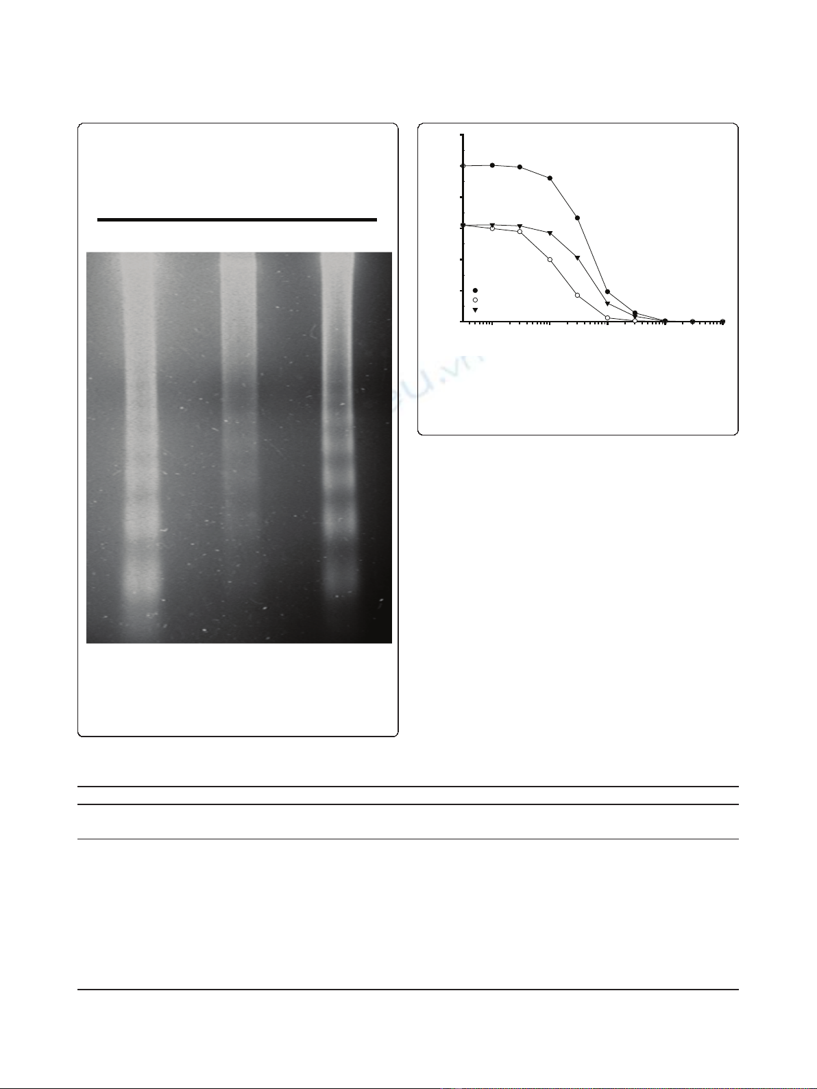
Detection of the mode of cell death induced by FWGE in
a panel of cell lines
In order to distinguish the mode of cell death induced by
FWGE we treated a representative panel of human cancer
cell lines with an IC
90
of FWGE for 48 h. Subsequent to
treatment, floating cells were harvested and an DNA gel
electrophoresis was performed. Clearly, in all treated cell
lines the typical 180 bp DNA laddering structure indica-
tive for specific DNA degradation during the process of
apoptosis could be detected (Figure 2).
Combination of FWGE with 5-FU, Oxaliplatin and
Irinotecan in human colon cancer cell lines
The combined drug effect of a parallel exposure to
FWGE and either 5-FU, irinotecan or oxaliplatin was
assessed in a panel of 8 colon cancer cell lines. The
mode of drug interaction was analyzed by the method
of Drewinko and the data summarized in table 1. Over-
all, mainly significant synergy was observed for the com-
binations of FWGE and 5-FU (6 out of 8 cell lines) and
to a lesser extend for irinotecan and oxaliplatin (2 out
of 8 cell lines). Drug interaction for the remaining cell
lines was additive. Importantly, no significant antagon-
ism was found for simultaneous drug exposure. A repre-
sentative plot for synergistic drug interaction is
presented in Figure 3.
Sequential drug application of FWGE and 5-FU in the
human colon cancer cell lines HT29 and HCT-8
To evaluate the influence of drug scheduling, exponen-
tially growing cells were exposed to an IC
30
of FWGE
24 h after seeding which was followed by serial dilu-
tions of 5-FU after further 24 hours or vice versa. Cells
were fixated after 120 h total assay time and processed
according to the SRB protocol. IC
50
values were calcu-
lated based on the Hill equation using Sigma plot and
the data were summarized in table 2. In both cell lines,
if 5-FU was followed by FWGE, we observed an addi-
tive drug interaction. On the other hand, if FWGE pre-
cedes 5-FU for 24 hours, we observed a trend to
antagonism in both cell lines. However, this antagon-
ism did not reach statistical significance. Taken
together, these findings suggest that the interactions
between 5-FU and FWGE are schedule-dependent.
Schedules in which FWGE precedes 5-FU should be
avoided.
Discussion
FWGE belongs to the group of nutraceuticals that are
approved as dietary food for special medical purposes
for cancer patients. It is well tolerated at the recom-
mended doses and possesses a broad therapeutic win-
dow [2]. Beside its use as nutrition supplement to
ameliorate cancer symptoms in patients there is incre-
mental evidence that FWGE might exert some antican-
cer properties as well [1-3]. However, up to now this
antitumor effect is only sparsely investigated.
Thus, we screened the preclinical cytotoxic activity of
FWGE as a single agent or in combination with the
commonly used cytostatics 5-FU, oxaliplatin or
Mueller et al.Journal of Experimental & Clinical Cancer Research 2011, 30:42
http://www.jeccr.com/content/30/1/42
Page 3 of 7
IC
50
(mg/ml); n = 3-4
0,03 0,13 0,23 0,33 0,43 0,53 0,63 0,7
3
H12.1
2102EP
1411HP
1777N
HCT-8
HCT15
HCT116
HT29
DLD-1
SW480
COLO205
COLO320
A549
A427
H322
H358
FADU
A253
A431
MCF-7
BT474
A2780
M2
8505C
SW1736
BCPAP
FTC133
518A2
HepG2
U87MG
SHSY5Y
SIMA
NSCLC
Colon cancer
Testicular cancer
Neuroblastoma
Thyroid cancer
Head and neck cancer
Glioblastoma
Hepatoma
518A2
Gastric cancer
Ovarian cancer
Breast cancer
cervix cancer
Figure 1 Illustration of IC
50
of FWGE as a mean graph.IC
50
of at least 3 independent experiments per cell line were averaged and
summarized as a mean graph for better comparison of the different activity. The average IC
50
is 0.33 mg/ml. The highest activity of FWGE was
found on neuroblastoma and ovarian cancer cell lines. It’s interesting to note that the IC
50
-values of the 8 human CRC cell lines included in this
screen range close to the average IC
50
.
Mueller et al.Journal of Experimental & Clinical Cancer Research 2011, 30:42
http://www.jeccr.com/content/30/1/42
Page 4 of 7
irinotecan in a large panel of human tumor cell lines to
evaluate its potential antitumor properties.
Human tumor cell lines or human tumor xenografts
commonly serve as models for preclinical drug screen-
ing. Still, care has to be taken in the interpretation of
results since their positive predictive value is limited to
approximately 60-70% [18,19]. The predictive value of
preclinical cytotoxicity data could by strengthened by
the model of relative antitumor activity. It allows to esti-
mate the potential activity of a drug in a certain tumor
type by taking the preclinical IC
50
value and clinically
achievable peak plasma concentrations into account
[20]. Only if the preclinical IC
50
value is clearly below
the plasma concentration that can be achieved in a
patient one can assume potential clinical activity.
In the present study we observed a significant antipro-
liferative activity of FWGE as assessed by IC
50
H12.1
2102E
P
HCT-8
Figure 2 Induction of apoptosis by FWGE. A representative panel
of human tumor cell lines was treated with an IC
90
of FWGE for 48
h and floating cells were harvested by centrifugation for DNA
extraction. DNA was seperated by DNA gel electrophoresis and
stained with ethidium bromide subsequently. Typical DNA laddering
indicative for apoptosis was visualized by UV light illumination.
Table 1 Summary of drug combinations
IC50 (μM)
Cell line Oxaliplatin ± FWGE p-value 5-FU ± FWGE p-value CPT-11 ± FWGE p-value
-+ - + -+
HCT-8 0,43 ± 0,03 0,45 ± 0,03 0,52 2,65 ± 0,35 1,2 ± 0,6 0,023* 2,0 ± 0,46 1,8 ± 0,32 0,63
HCT-15 0,95 ± 0,19 0,57 ± 0,25 0,05 4,45 ± 0,72 1,45 ± 0,61 0,0001* 4,5 ± 0,3 3,4 ± 0,31 0,001*
HCT116 0,39 ± 0,06 0,19 ± 0,09 0,01* 4,6 ± 0,38 2,9 ± 0,9 0,01* 1,2 ± 0,1 0,96 ± 0,11 0,01*
HT29 0,32 ± 0,09 0,35 ± 0,05 0,53 0,99 ± 0,31 1,3 ± 0,6 0,39 3,5 ± 0,3 4,1 ± 0,23 0,05
DLD-1 2,47 ± 0,17 2,2 ± 0,8 0,61 3,2 ± 0,21 1,6 ± 0,7 0,02* 6,6 ± 0,6 6,1 ± 0,85 0,43
Colo205 0,45 ± 0,05 0,24 ± 0,05 0,001* 0,54 ± 0,12 0,44 ± 0,1 0,26 1,2 ± 0,19 1,1 ± 0,19 0,24
Colo320 1,1 ± 0,34 0,84 ± 0,13 0,33 1,35 ± 0,133 0,57 ± 0,03 0,001* 8,5 ± 3,4 8,7 ± 3,1 0,92
SW48 0,13 ± 0,02 0,1 ± 0,02 0,09 3,4 ± 0,2 2,2 ± 0,2 0,002* 2,4 ± 0,35 2,1 ± 0,29 0,18
SW480 0,57 ± 0,11 0,37 ± 0,12 0,06 2,7 ± 0,17 2,9 ± 1,5 0,83 6,4 ± 1,2 6,9 ± 2,3 0,72
n≥3, asterisk indicates significant synergistic drug interaction
c
(
μM
)
0,1 1 10 100 100
0
% control
0
20
40
60
80
100
120
5-FU
5-FU + 0.4 mg/ml FWGE
hypothetical curve
Figure 3 Synergy between FWGE and 5-FU in human colon
cancer cell line HCT15. Plots represent the average of 3
independent experiments. The hypothetical curve was calculated as
described by Drewinko et al. [16]. Synergy is indicated by the
hypothetical curve which runs above the combination curve.
Mueller et al.Journal of Experimental & Clinical Cancer Research 2011, 30:42
http://www.jeccr.com/content/30/1/42
Page 5 of 7


![Vaccine và ứng dụng: Bài tiểu luận [chuẩn SEO]](https://cdn.tailieu.vn/images/document/thumbnail/2016/20160519/3008140018/135x160/652005293.jpg)


%20--%3e%3cdefs%3e%3cstyle%3e%20.st0%20{%20fill:%20%23fff;%20}%20.st1%20{%20fill:%20%237800fa;%20}%20%3c/style%3e%3c/defs%3e%3cpath%20class='st1'%20d='M117.78,12.18H43.11c2.9,3.47,4.65,7.94,4.65,12.82,0,5.6-2.3,10.66-6.01,14.29h76.02l7.22-13.56-7.22-13.56Z'/%3e%3cg%3e%3cpath%20class='st0'%20d='M53.58,26.17h-.59v-1.46h.59v-4.96h2.83c1.78,0,2.67.94,2.67,2.82v5.76c0,1.87-.89,2.81-2.67,2.81h-2.83v-4.96ZM55.36,21.37v3.34h1.1v1.46h-1.1v3.34h1.01c.61,0,.91-.37.91-1.1v-5.93c0-.74-.3-1.1-.91-1.1h-1.01Z'/%3e%3cpath%20class='st0'%20d='M65.99,31.14h-1.8l-.31-2.07h-2.19l-.31,2.07h-1.64l1.82-11.39h2.62l1.82,11.39ZM65.28,18.04c-.25.46-.51.77-.75.94-.21.15-.47.22-.79.22-.26,0-.57-.07-.92-.22l-.38-.15c-.14-.05-.26-.07-.37-.07-.3,0-.53.18-.71.54l-.91-.68c.25-.46.51-.77.75-.94.21-.14.48-.21.79-.21.26,0,.57.07.92.21l.38.15c.14.05.26.07.37.07.3,0,.53-.18.71-.54l.91.68ZM61.91,27.52h1.73l-.87-5.76-.87,5.76Z'/%3e%3cpath%20class='st0'%20d='M74.53,26.89v1.52c0,1.91-.89,2.86-2.67,2.86s-2.67-.95-2.67-2.86v-5.93c0-1.91.89-2.86,2.67-2.86s2.67.95,2.67,2.86v1.11h-1.69v-1.22c0-.75-.31-1.12-.93-1.12s-.93.37-.93,1.12v6.15c0,.74.31,1.11.93,1.11s.93-.37.93-1.11v-1.63h1.69Z'/%3e%3cpath%20class='st0'%20d='M81.4,31.14h-1.8l-.31-2.07h-2.19l-.31,2.07h-1.64l1.82-11.39h2.62l1.82,11.39ZM75.9,19.2l1.52-1.91h1.71l1.51,1.91h-1.61l-.76-.95-.75.95h-1.61ZM77.32,27.52h1.73l-.87-5.76-.87,5.76ZM83.1,15.99l-1.76,1.91h-1.26l1.17-1.91h1.86Z'/%3e%3cpath%20class='st0'%20d='M84.86,19.75c1.78,0,2.67.94,2.67,2.82v1.48c0,1.87-.89,2.81-2.67,2.81h-.85v4.28h-1.79v-11.39h2.64ZM84.01,21.37v3.86h.85c.58,0,.87-.36.87-1.08v-1.71c0-.71-.29-1.07-.87-1.07h-.85Z'/%3e%3cpath%20class='st0'%20d='M93.51,19.75c1.78,0,2.67.94,2.67,2.82v1.48c0,1.87-.89,2.81-2.67,2.81h-.85v4.28h-1.79v-11.39h2.64ZM92.66,21.37v3.86h.85c.58,0,.87-.36.87-1.08v-1.71c0-.71-.29-1.07-.87-1.07h-.85Z'/%3e%3cpath%20class='st0'%20d='M98.8,31.14h-1.79v-11.39h1.79v4.88h2.03v-4.88h1.83v11.39h-1.83v-4.88h-2.03v4.88Z'/%3e%3cpath%20class='st0'%20d='M105.36,24.55h2.46v1.62h-2.46v3.34h3.09v1.63h-4.88v-11.39h4.88v1.63h-3.09v3.18ZM108.17,17.29l-1.76,1.91h-1.26l1.17-1.91h1.86Z'/%3e%3cpath%20class='st0'%20d='M112.2,19.75c1.78,0,2.67.94,2.67,2.82v1.48c0,1.87-.89,2.81-2.67,2.81h-.85v4.28h-1.79v-11.39h2.64ZM111.35,21.37v3.86h.85c.58,0,.87-.36.87-1.08v-1.71c0-.71-.29-1.07-.87-1.07h-.85Z'/%3e%3c/g%3e%3ccircle%20class='st1'%20cx='25'%20cy='25'%20r='20'/%3e%3cpath%20class='st0'%20d='M32.78,19.27c2.92,0,4.43,2.55,5.28,5.33l.71,2.17c.14.38-.33.75-.71.75h-5.61c.19-.33.24-.71.09-1.08l-.75-2.45c-.43-1.32-.99-2.64-1.79-3.77.75-.57,1.65-.94,2.78-.94h0ZM25,18.38c3.25,0,4.9,2.78,5.89,5.89l.76,2.45c.14.42-.33.8-.8.8h-11.69c-.42,0-.94-.38-.8-.8l.75-2.45c.99-3.11,2.64-5.89,5.89-5.89h0ZM25,11.35c1.74,0,3.11,1.37,3.11,3.11s-1.37,3.11-3.11,3.11-3.11-1.41-3.11-3.11,1.41-3.11,3.11-3.11h0ZM17.27,19.27c1.08,0,1.98.38,2.73.94-.8,1.13-1.37,2.45-1.74,3.77l-.8,2.45c-.14.38-.05.75.09,1.08h-5.56c-.42,0-.9-.38-.75-.75l.71-2.17c.9-2.78,2.41-5.33,5.33-5.33h0ZM17.27,12.91c1.51,0,2.78,1.27,2.78,2.83s-1.27,2.83-2.78,2.83-2.83-1.27-2.83-2.83,1.27-2.83,2.83-2.83h0ZM32.78,12.91c1.56,0,2.78,1.27,2.78,2.83s-1.23,2.83-2.78,2.83-2.83-1.27-2.83-2.83,1.27-2.83,2.83-2.83h0ZM27.07,28.56v.09c0,.57-.24,1.08-.61,1.46h0v.05c-.38.33-.9.57-1.46.57s-1.08-.24-1.46-.61h0c-.38-.38-.61-.9-.61-1.46v-.09h1.41v.09c0,.19.05.38.19.47v.05c.09.09.28.19.47.19s.38-.09.47-.19v-.05c.14-.09.24-.28.24-.47t-.05-.09h1.41ZM30.99,28.56v.09c0,1.65-.66,3.16-1.74,4.24-1.08,1.08-2.59,1.79-4.24,1.79s-3.16-.71-4.24-1.79l-.05-.05c-1.04-1.08-1.7-2.55-1.7-4.2v-.09h1.41v.09c0,1.27.47,2.4,1.27,3.25h.05c.85.85,1.98,1.37,3.25,1.37s2.4-.52,3.25-1.37c.85-.8,1.37-1.98,1.37-3.25v-.09h1.37ZM34.99,28.56v.09c0,2.78-1.13,5.28-2.92,7.07-1.79,1.79-4.29,2.92-7.07,2.92s-5.23-1.13-7.07-2.92c-1.79-1.79-2.92-4.29-2.92-7.07v-.09h1.41v.09c0,2.4.94,4.53,2.5,6.08,1.56,1.56,3.72,2.5,6.08,2.5s4.52-.94,6.08-2.5c1.56-1.56,2.5-3.68,2.5-6.08v-.09h1.41Z'/%3e%3c/svg%3e)