Essential role of the C-terminus in Melanocarpus albomyces laccase for enzyme production, catalytic properties and structure Martina Andberg1, Nina Hakulinen2, Sanna Auer1, Markku Saloheimo1, Anu Koivula1, Juha Rouvinen2 and Kristiina Kruus1
1 VTT Technical Research Center of Finland, Finland 2 Department of Chemistry, University of Joensuu, Finland
Keywords ascomycete; C-terminal plug; multicopper oxidase; mutants; site-directed mutagenesis
Correspondence M. Andberg, VTT Technical Research Center of Finland, P.O. Box 1000, FIN-02044 VTT, Finland Fax: +358 20 722 7071 Tel: +358 20 722 5124 E-mail: martina.andberg@vtt.fi Website: http://www.vtt.fi/research/bic/ ?lang=en
Database The atomic coordinates and structure factors have been submitted to the Protein Data Bank under the accession number 3DKH
(Received 2 July 2009, revised 17 August 2009, accepted 28 August 2009)
doi:10.1111/j.1742-4658.2009.07336.x
The C-terminus of the fungal laccase from Melanocarpus albomyces (MaL) is processed during secretion at a processing site conserved among the ascomycete laccases. The three-dimensional structure of MaL has been solved as one of the first complete laccase structures. According to the crystal structure of MaL, the four C-terminal amino acids of the mature protein penetrate into a tunnel leading towards the trinuclear site. The C-terminal carboxylate group forms a hydrogen bond with a side chain of His140, which also coordinates to the type 3 copper. In order to analyze the role of the processed C-terminus, site-directed mutagenesis of the MaL cDNA was performed, and the mutated proteins were expressed in Tricho- derma reesei and Saccharomyces cerevisiae. Changes in the C-terminus of MaL caused major defects in protein production in both expression hosts. The deletion of the last four amino acids dramatically affected the activity of the enzyme, as the deletion mutant delDSGL559 was practically inactive. the purified L559A mutant expressed in Detailed characterization of S. cerevisiae showed the importance of the C-terminal plug for laccase activity, stability, and kinetics. Moreover, the crystal structure of the L559A mutant expressed in S. cerevisiae showed that the C-terminal muta- tion had clearly affected the trinuclear site geometry. The results in this study clearly confirm the critical role of the last amino acids in the C-terminus of MaL.
Introduction
are
copper-containing metalloenzymes
Laccases (EC 1.10.3.2; p-dihenol dioxygen oxidoreduc- that tases) oxidize various phenolic compounds, anilines and even some nonaromatic compounds by a one-electron removal mechanism, which usually generates radicals.
Oxidation of reducing substrates occurs concomitantly with the reduction of molecular oxygen to water. Lac- cases are ubiquitous enzymes found in various micro- organisms, insects, and plants. They share structural similarities with other blue multicopper oxidases,
FEBS Journal 276 (2009) 6285–6300 ª 2009 The Authors Journal compilation ª 2009 FEBS
6285
Abbreviations 2,6-DMP, 2,6-dimethoxyphenol; ABTS, 2,2¢-azinobis(3-ethylbenzo-6-thiazolinesulfonic acid); BsL, Bacillus subtilis laccase; MaL, Melanocarpus albomyces laccase; rMaL, recombinant MaL expressed in T. reesei; Sc(delDSGL559), Melanocarpus albomyces laccase delDSGL559 mutant expressed in Saccharomyces cerevisiae; Sc(L559A), Melanocarpus albomyces laccase L559A mutant expressed in Saccharomyces cerevisiae; ScMaL, Melanocarpus albomyces laccase expressed in Saccharomyces cerevisiae; Tr(delDSGL559), Melanocarpus albomyces laccase delDSGL559 mutant expressed in Trichoderma reesei; Tr(L559G), Melanocarpus albomyces laccase L559G mutant expressed in Trichoderma reesei.
M. Andberg et al. Function of C-terminus in M. albomyces laccase
including ascorbate oxidase, ceruloplasmin, CueO, and Fet3p. For catalytic activity, all four copper atoms are needed: one type 1 (T1) copper forming a mononuclear site, and one type 2 (T2) copper and two type 3 (T3 and T3¢) coppers forming a trinuclear site.
known laccases, this cavity is open, and it is thought to provide access to the fresh oxygen molecules needed in the catalytic cycle. The C-terminus of MaL blocks this route, as the packing of the C-terminus against the tunnel is extensive, and there is no space for dioxygen or any other molecules to enter. Further- more, the C-terminal carboxylate group in MaL is hydrogen-bonded to the side chain of His140, which is one of the His residues coordinating the T2 copper in the trinuclear center.
C-terminal sequencing of the mature MaL showed that the C-terminus is post-translationally processed after Leu559, leading to removal of the last 14 amino
Melanocarpus albomyces is a thermophilic fungus expressing a laccase with substantial thermal stability and a pH optimum with phenolic substrates in the neutral pH region [1]. These unusual properties, as compared with most reported fungal laccases, makes M. albomyces laccase (MaL) an interesting enzyme for many applications. The three-dimensional structure of MaL has been solved as one of the first complete laccase structures (Protein Data Bank codes: 1GW0, laccase from M. albomyces; 2IH8, a low-dose crystal structure of a recombinant MaL; 2IH9, a high-dose crystal structure of a recombinant MaL; and 2Q9O, near-atomic resolution structure of recombinant MaL) [2–4]. The enzyme is composed of three cupredoxin domains, A, B, and C (or 1, 2, and 3), which all have a similar Greek key b-barrel structure. The mononu- clear site is located in domain C, whereas the trinucle- ar site is between domains A and C. Electrons are withdrawn from the mononuclear site and further transferred about 13 A˚ along a conserved Cys–His pathway into the trinuclear site, where dioxygen is reduced to water. The MaL structure was the first solved three-dimensional structure showing dioxygen binding [2]. Since then, dioxygen binding has also been found for Bacillus subtilis laccase (BsL) [5] and cerulo- plasmin [6]. Other solved laccase crystal structures have shown only one oxygen atom between the two T3 coppers [7–10]. The crystal structure of MaL also has a chloride ion attached to the T2 copper, whereas other crystal structures of multicopper oxidases have ion ⁄ water in this position. The role of the hydroxyl chloride ion is unknown. A number of anions, i.e. ), and F), are known to act as effective lac- CN), N3 case inhibitors [11,12]. However, chloride ion does not act as an inhibitor for MaL, as shown by Kiiskinen et al. [1]. Instead, azide is a well-known inhibitor of MaL. According to spectroscopic measurements, the binding of azide has been suggested to bridge the T2 copper and one of the T3 coppers [13,14], or bind to one T3 copper, as observed in the crystal structure of ascorbate oxidase [15]. Recently, the azide was found to bind between two T3 coppers in the crystal struc- ture of BsL [5].
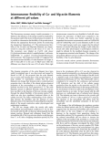
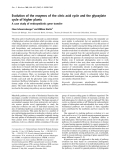
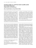
Fig. 1. (A) Surface model of M. albomyces laccase; a few of the last amino acids are represented as a purple worm (Protein Data Bank code: 2Q9O). (B) The C-terminus of M. albomyces (in purple) leading towards the trinuclear site. penetrates to the tunnel Coppers are represented as orange balls, water atoms as red balls, and dioxygen as a red stick.
The three-dimensional structure of MaL revealed that the C-terminus of the enzyme penetrates to a leading to the trinuclear site (Fig. 1). This tunnel unique feature has not been observed in any other published laccase crystal structures. Instead, in other
FEBS Journal 276 (2009) 6285–6300 ª 2009 The Authors Journal compilation ª 2009 FEBS
6286
M. Andberg et al. Function of C-terminus in M. albomyces laccase
from Neurospora crassa
acids of the mature protein [16]. Similar C-terminal processing has also been reported for other ascomycete [17], Podospori- laccases, na anserina [18], and Myceliophthora thermophila [19]. The processing site for all of these laccases is Asp-Ser- Gly-Leu. Interestingly, a similar type of sequence is found in the C-termini of other ascomycete laccases that do not undergo the C-terminal processing, e.g. Borytris cinerea, Cryptonectria parasitica, and Gau- emannomyces graminis var. tritici. The last four amino acids in these laccases are Asp, Ser, Gly, and Leu ⁄ Ile ⁄ Val. The C-terminal end seems to be conserved among all ascomycete laccases. The conserved C-terminus of ascomycete laccases might have a special role in the enzyme.
the L559A mutant
In order to analyze the role of the C-terminus, site- directed mutagenesis of MaL cDNA was performed, and the mutated proteins and wild-type enzyme were expressed in Trichoderma reesei and Saccharomyces cerevisiae. We report here the characterization of the C-terminal mutants and the three-dimensional structure expressed in S. cerevisiae of [Sc(L559A)].
Results
Production and characterization of MaL mutants expressed in T. reesei
a substantial effect on the production level in T. reesei, as very low expression levels were observed for the mutants according to the activity assays and western blot analysis, as compared with the production of wild-type rMaL (Fig. S1). The production level in the T. reesei culture supernatant of the two mutants was estimated to be 10 mgÆL)1, which was significantly lower than the production level of the pLLK13 wild- type rMaL (200 mgÆL)1). The activities on ABTS, 2,6- dimethoxyphenol (2,6-DMP) and syringaldazine in the culture supernatants of the mutants were considerably reduced. In fact, no activity could be detected for the Tr(delDSGL559) mutant, although amounts detectable by western blot analysis were expressed into the super- natant. The ABTS activity in the culture supernatant of the Tr(L559G) mutant was several hundred-fold lower than that of the wild-type rMaL. Comparison of the production level and the activity in the culture supernatant showed the specific activity of the mutants to be considerably lower than that of the wild-type enzyme. In addition to having low expression levels, the Tr(L559G) and Tr(delDSGL559) mutants were partly degraded (Fig. S1, lanes 2 and 3). Some laccase degradation products were also detected in the culture supernatants of the wild-type rMaL produced in the two strains, but the ratio of degraded laccase to full- length laccase was much higher in the mutant strains. Changes in the original C-terminus thus caused major defects in protein production in T. reesei as well as changes in the protein properties.
expressed
delDSGL559 mutant
The T. reesei strain producing the Tr(delDSGL559) protein was also cultivated in a laboratory-scale bioreactor (20 L), and the protein was purified from the culture supernatant by applying the procedure optimized for the wild-type rMaL [20]. The purifica- tion protocol contained three chromatographic steps: anion exchange chromatography, hydrophobic interac- tion chromatography and, finally, anion exchange chromatography with a high-resolution resin. The started to degrade during Tr(delDSGL559) mutant purification, and the mutant laccase could not be obtained from the T. reesei culture filtrate.
Production and characterization of MaL in S. cerevisiae
analyzed by
in T. reesei, S. cerevisiae was
Owing to the difficulties in producing the mutant laccases chosen as an expression host for the designed mutants. The full-length gene of MaL containing the C-terminal extension, i.e. the last 14 amino acids that are cleaved from the mature protein, was expressed in S. cerevisiae, and the protein was purified from this source. The
Two mutations were made in the MaL gene, and the mutated proteins were expressed in T. reesei. The mutated laccase constructs were produced by site- directed mutagenesis on the plasmid pLLK8, a T. ree- sei expression vector containing the cDNA coding for MaL between the cbh1 promoter and terminator. In the L559G mutant expressed in T. reesei [Tr(L559G)], Leu559 was replaced with a Gly to change the pro- cessing site to prevent C-terminal cleavage, and in in T. reesei the [Tr(delDSGL559)], an Asp at position 556 was replaced by a stop codon to delete the last four amino acids (Asp-Ser-Gly-Leu) of the mature MaL protein. The mutated laccases, as well as two wild-type recombinant MaLs (rMaLs) (produced by the pLLK13 and the cbh1-negative pMS176 strains), were expressed in T. reesei in shake flask cultures, and laccase pro- activity determination duction was with 2,2¢-azinobis(3-ethylbenzo-6-thiazolinesulfonic acid) (ABTS) and by immunoanalysis of culture superna- tants. For the mutant Tr(delDSGL559), a transformant in which the cbh1 gene had been replaced by the expression construct was found and used. The C-termi- nal mutations in Tr(L559G) and Tr(delDSGL559) had
FEBS Journal 276 (2009) 6285–6300 ª 2009 The Authors Journal compilation ª 2009 FEBS
6287
M. Andberg et al. Function of C-terminus in M. albomyces laccase
C-terminus of the purified laccase was analyzed by C-terminal sequencing to determine whether yeast is also able to process the C-terminus properly. MaL has previously been showed to be processed at its C-termi- nus during secretion, both in Melanocarpus and in Trichoderma [16,20]. The results from the sequencing clearly showed that the yeast was not able to process MaL correctly, and that the additional 14 amino acids were present in the protein. Therefore, all further work was performed with another construct, pMS175, where mature MaL cDNA, with a stop codon, was intro- duced after the C-terminal processing site [16].
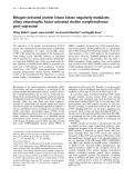
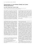
In lanes 1–3, a Coomasssie-stained gel
Fig. 2. SDS ⁄ PAGE gel and western blot analysis of purified ScMaL. The samples from a Resource Q run were separated in a 12% SDS ⁄ PAGE gel. is shown, and in lanes 5–7, the gel has been blotted using antibodies against MaL. The samples are rMaL (purified from T. reesei) in lanes 1 and 7, ScMaL pool I, containing the more heavily overgly- cosylated laccase, in lanes 2 and 5, and the less overglycosylated laccase ScMaL pool II in lanes 3 and 6.
The effects of using S. cerevisiae as expression host on the properties of M. albomyces laccase (ScMaL) were also studied. The conditions for production of ScMaL in shake flask cultures were optimized in terms of CuSO4 concentration in the medium, temperature, culture medium, and induction conditions. The opti- mal culture conditions were found to be as follows: synthetic complete medium (SC-URA) buffered to pH 6 with succinate and supplemented with 1 mm CuSO4 at 250 r.p.m. and 30 (cid:2)C. Yeast cells were grown on raffinose (20 gÆL)1), and a washing step prior to a change to induction medium containing galactose (20 gÆL)1) was shown to have a positive effect on lac- case production. The production level in shake flask cultures was about 4.5 nkatÆmL)1 (ABTS activity), roughly corresponding to 7 mgÆL)1 ScMaL, when calculated from the specific activity for ScMaL (610 nkatÆmg)1).
siae and T. reesei. Also, the temperature stability was for ScMaL and rMaL. The N-terminal similar sequencing verified correct processing of the yeast a-prepro sequence (KEX2 cleavage site). Removal of the glycans by enzymatic deglycosylation with endo- b-N-acetylglucosaminidase F1 slightly lowered the specific ABTS activity of ScMaL, but had no effect on the specific activity of rMaL. Deglycosylation of over- glycosylated ScMaL resulted in one pI isoform of the enzyme, in contrast to the several isoforms seen with the enzyme still having the glycans attached. The results confirmed that MaL can be expressed in S. cerevisiae and that the protein properties are com- parable to those of the wild-type laccases and the lac- case expressed heterologously in T. reesei.
Production and purification of the Sc(delDSGL559) and Sc(L559A) mutants
Two C-terminal mutants of MaL, Sc(delDSGL559) and Sc(L559A), were expressed in S. cerevisiae, and the proteins were purified to homogeneity from the yeast culture supernatant. In the Sc(delDSGL559) mutant, the last four amino acids (Asp-Ser-Gly-Leu) were deleted from the protein [equivalent to the Tr(del- DSGL559 mutant], and in the Sc(L559A) mutant, the C-terminal Leu was replaced with a smaller Ala, in order to prevent the hydrogen bonding of the carbox- ylate group to a side chain of His140. Production of the two mutated proteins in S. cerevisiae was carried in shake flasks using conditions optimized for out
ScMaL was heavily overglycosylated (Fig. 2). Iso- electric focusing combined with activity staining of the culture supernatant demonstrated that ScMaL had sev- eral pI forms (pI 3.5–5), whereas the rMaL produced in T. reesei only had one pI form (pI 4.0) (data not shown). Purification of ScMaL was performed in two chromatographic steps, including weak and strong anion exchange resins. Owing to the heterogeneous overglycosylation of ScMal, the laccase was fraction- ated into two separate pools: pool 1, containing the more heavily overglycosylated laccase; and pool 2, with less overglycosylated laccase showing a major laccase band at 100 kDa as analyzed by SDS ⁄ PAGE (Fig. 2). The ABTS activity for pool 1 (430 nkatÆmg)1) was consistently slightly lower than that for pool 2 (520 nkatÆmg)1). Pool 2 was therefore used for charac- terization. Table 1 presents a summary of the charac- teristics of purified ScMaL in comparison to the rMaL produced in T. reesei. The specific activity of ScMaL (520 nkatÆmg)1) was lower than the reported specific activity of rMaL (840 nkatÆmg)1). However, the Km values for ABTS showed practically no difference between the MaL preparations expressed in S. cerevi-
FEBS Journal 276 (2009) 6285–6300 ª 2009 The Authors Journal compilation ª 2009 FEBS
6288
M. Andberg et al. Function of C-terminus in M. albomyces laccase
Table 1. Characterization of the M. albomyces laccase produced in S. cerevisiae (ScMaL) and in T. reesei (rMaL).
Property ScMaL rMaL
Correctly processed 79 420 Da Yes (100 kDa + smear) Correctly processed 70 700 Da No (70 kDa) N-terminal amino acid sequence Molecular mass (MALDI-TOF) Overglycosylation (on SDS ⁄ PAGE and western blot)
Several isoforms, pI 3.5–5 4 400–610 nkatÆmg)1 260 lM 40 min One isoform, pI 4 One isoform, pI 4 3.5 640–840 nkatÆmg)1 280 lM 35 min None (one isoform), pI 4
Isoelectric point pH optimum (ABTS) Specific activity (ABTS, pH 4.5, 25 (cid:2)C) Km (ABTS, pH 4.5, 22 (cid:2)C) Thermostability, t1 ⁄ 2 at 65 (cid:2)C, pH 6 Effect of deglycosylation (endo-b-N-acetylglucosaminidase F1) on isoelectric point Eo (T1 copper center) 0.43 V 0.47 V
than that of
order to produce enough protein for crystallization purposes, the Sc(L559A) mutant was also produced in a laboratory-scale bioreactor (20 L), and the enzyme was purified to homogeneity. The protein yield was 22 mg, and the specific activity of the final Sc(L559A) sample was 184 nkatÆmg)1. Thus, the specific activity of the Sc(L559A) mutant was three-fold to four-fold lower the wild-type ScMaL (520 nkatÆmg)1).
Characterization of the Sc(delDSGL559) and Sc(L559A) mutants
ScMaL production. The total cultivation volume was 4 L for both mutated proteins. The culture superna- tant was concentrated prior to purification. Similarly to what was found for the Sc(delDSGL559) mutant expressed in T. reesei, laccase activity on ABTS sub- strate was not detectable in the Sc(delDSGL559) culture supernatant, or in the concentrated culture filtrate, although the expression of the mutant laccase was confirmed by western blot analysis. The result clearly confirms that the last four amino acids are essential for enzyme activity. In the culture supernatant of Sc(L559A), the activity on ABTS was 1.8 nkatÆmL)1, which was 2.5-fold lower than the activity for ScMaL (4.5 nkatÆmL)1).
The Sc(delDSGL559) mutant was purified in four subsequent chromatographic steps. As the Sc(del- DSGL559) mutant was not active in the culture super- natant or after the first purification steps, the pooling of the laccase-containing fractions was based on anti- body detection on dot blots. In the pooled fractions after the third hydrophobic interaction step, very low but detectable laccase activity on ABTS could be observed. The specific activities in two separate pools were 0.21 and 0.45 nkatÆmg)1. These values are over 1000-fold lower than the specific activity of the wild- type ScMaL (520 nkatÆmg)1). The activity results for the Sc(delDSGL559) mutant, together with the activity results for the corresponding mutant Tr(delDSGL559) (see above), clearly indicate the essential role of the last four amino acids for the function of MaL.
The Sc(L559A) mutant was also produced in shake flasks for the initial characterization studies. From 4 L shake flask cultures, overall 11.1-fold purification and activity recovery of 24% were achieved. Altogether, 8.7 mg of purified Sc(L559A) mutant was recovered, with a specific ABTS activity of 102 nkatÆmg)1. In
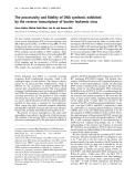
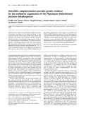
The two purified MaL mutant proteins expressed in yeast were characterized and compared with the wild- type ScMaL. In order to determine whether the muta- tions had affected the overall structure of the protein, the Sc(delDSGL559) and Sc(L559A) CD spectra of mutants were measured and compared with that of the wild-type laccase (ScMaL) (Fig. 3A). The general shapes of the spectra were the same for the Sc(del- DSGL559) and Sc(L559A) mutants and wild-type ScMaL, which suggests that no major changes in the conformation of the mutated enzymes had occurred. The thermal unfolding profiles measured with CD (Fig. 3B) were broad, with no clear folded–unfolded transition for ScMaL and the two mutants. The Sc(L559A) mutant starts to unfold at a lower tempera- ture as compared with ScMaL, suggesting slightly reduced thermal stability. The difference between the Sc(delDSGL559) mutant and ScMaL is even greater; the Sc(delDSGL559) mutant exhibited an unfolding behavior without any transition state, and started to unfold at relatively low temperatures (40–50 (cid:2)C). The Tm values were estimated from the graph to be 71 (cid:2)C, 69 (cid:2)C and 65 (cid:2)C for ScMaL, the Sc(L559A) mutant,
FEBS Journal 276 (2009) 6285–6300 ª 2009 The Authors Journal compilation ª 2009 FEBS
6289
M. Andberg et al. Function of C-terminus in M. albomyces laccase
ferrocyanide ⁄ ferricyanide
The redox potential of the mononuclear (T1) copper center for the Sc(L559A) mutant was measured using redox buffer the system in 20 mm Tris ⁄ HCl (pH 7.5). (E0,Fe = 0.433 V) [21] The laccase concentration used in the redox measure- ments was estimated from the 600 nm absorbance, using an extinction coefficient of 5700 m)1Æcm)1. The redox potential of the Sc(L559A) mutant was deter- mined to be 0.43 V, which is in agreement with the value measured for ScMaL and also for the rMaL expressed in T. reesei (0.47 V).
for
Fig. 3. (A) CD spectra of wild-type ScMaL and the Sc(delDSGL559) and Sc(L559A) mutants. The CD spectra of wild-type ScMaL (solid line) and the Sc(delDSGL559) (dotted line) and Sc(L559A) (double dotted line) mutants were recorded from 240 to 190 nm at 25 (cid:2)C in 10 mM sodium phosphate buffer (pH 7.1). (B) Temperature-induced unfolding of wild-type ScMaL and the Sc(delDSGL559) and Sc(L559A) mutants measured by CD spectroscopy. Changes in ellipticity of ScMaL and the Sc(delDSGL559) and Sc(L559A) mutants were recorded at 202 nm upon heating from 30 (cid:2)C to 90 (cid:2)C in 10 mM sodium phosphate buffer at pH 7.1. The data were smoothed with ORIGIN 7.5 (OriginLab).
As the deletion mutant Sc(delDSGL559) was practi- cally inactive, no detailed kinetic characterization could be performed, and the kinetic characterization was performed only with the Sc(L559A) mutant. For analysis of the purified Sc(L559A) mutant in more detail, three different substrates were used. The kinetic constants presented in Table 2 show that the mutation had a two-fold increased Km value on the nonphenolic substrate ABTS (Km = 900 lm) as compared with wild-type ScMaL (Km = 400 lm). The Sc(L559A) mutant also exhibited a four-fold decreased catalytic constant on ABTS (kcat = 394 min)1) as compared with ScMaL (kcat = 1686 min)1). Consequently, the specificity constant on ABTS dropped about 10-fold from 4.2 lm)1Æmin)1 for ScMaL to 0.44 lm)1Æmin)1 for the Sc(L559A) mutant. However, for the two phenolic substrates 2,6-DMP and syringaldazine, the L559A mutation did not greatly influence the catalytic (Table 2). The Km values the parameters Sc(L559A) mutant on 2,6-DMP and syringaldazine were 16 and 31 lm, and the corresponding Km values for ScMaL were 11 and 37 lm, respectively. The L559A mutation had decreased the turnover number on syringaldazine about two-fold (kcat = 1263 min)1) compared with wild-type ScMaL (kcat = 2410 as min)1). With 2,6-DMP, no significant changes in the kinetic parameters were observed.
inhibition constants
the trinuclear
site,
In order to determine whether the L559A mutation affects for sodium azide were also determined. Azide inhibits the
and the Sc(delDSGL559) mutant, respectively. Both mutants were expected to be stable at 25 (cid:2)C, and all of the following kinetic analyses were therefore performed at this temperature.
Table 2. Kinetic parameters for wild-type ScMaL and the Sc(L559A) mutant. ABTS activity was measured in 25 mM succinate buffer at pH 4.5 and 25 (cid:2)C, and 2,6-DMP and syringaldazine activities were measured in 40 mM MES buffer at pH 6 and 25 (cid:2)C. For determination of inhi- bition constants for the sodium azide, the enzyme was preincubated for 2 min with NaN3 prior to addition of substrate. The Ki value was obtained from Dixon plots. The error in all measurements was estimated to ± 15%. ND, not determined.
ABTS 2,6-DMP Syringaldazine
ScMaL Sc(L559A) ScMaL ScMaL Sc(L559A) Sc(L559A¢)
)1Æmin)1)
FEBS Journal 276 (2009) 6285–6300 ª 2009 The Authors Journal compilation ª 2009 FEBS
6290
400 1686 900 394 16 545 31 1263 0.44 4.2 7.9 85 11 612 48 29 34.3 55 37 2410 66 ND 41.3 ND Km (lM) kcat (min)1) kcat ⁄ Km (lM Ki (lM)
M. Andberg et al. Function of C-terminus in M. albomyces laccase
laccase activity by binding to the trinuclear centrer. In the crystal structure of ascorbate oxidase, azide is sug- gested to bind to one T3 copper [15], and in the crystal structure of BsL, between two T3 coppers [5]. Spectro- scopic findings have suggested that azide bridges the T2 copper and one of the T3 coppers [13,14]. The inhibition constants (Ki) of sodium azide for the Sc(L559A) mutant and ScMaL were determined using ABTS or 2,6-DMP as substrate. The results indicated that sodium azide was a mixed inhibitor with respect to both ABTS and 2,6-DMP; that is, the inhibitor binds at a location distinct from the reducing sub- strate-binding site. When the nonphenolic substrate ABTS was used, the Ki value of sodium azide was increased approximately 11-fold for the Sc(L559A) mutant as compared with that of the wild-type ScMaL, and was calculated (from Dixon plots) to be 85 lm (Table 2). The corresponding value for the wild-type enzyme was 7.9 lm. However, the difference in the Ki values for ScMaL and the Sc(L559A) mutant was only two-fold when the phenolic substrate 2,6-DMP was used.
Fig. 4. The pH optima of the wild-type (h) and Sc(L559A) mutant (d) laccases measured at 22 (cid:2)C, using ABTS (A) or 2,6-DMP (B) as substrate. The enzymes were incubated in McIlvaine’s buffer.
the pH activity profile of
Deglycosylation of ScMaL and the Sc(L559A) mutant
The pH optima of the purified Sc(L559A) mutant were determined using ABTS and 2,6-DMP as sub- strates. On ABTS, both wild-type ScMaL and the Sc(L559A) mutant had optimal activity at pH 4, but the pH activity profile of the Sc(L559A) mutant was more narrow than that of the wild-type enzyme, and had shifted to the alkaline side (Fig. 4). At pH 3, the relative laccase activity was 88% for ScMaL, whereas for the Sc(L559A) mutant it had dropped to below 3%. On 2,6-DMP, the Sc(L559A) mutant was similar to that of the wild-type the mutant having a slightly broader pH enzyme, activity in an alkaline pH range (Fig. 4).
The protein properties of wild-type ScMaL were com- parable to the properties of rMaL (Table 1), although ScMaL was heavily overglycosylated. Thus, the yeast was a suitable host for production of the MaL vari- ants for structural analysis. As the long glycan chains attached in yeast to the laccase protein most probably disturb crystallization, optimization of conditions for removing the N-glycans was performed. Enzymatic deglycosylation of ScMaL and the Sc(L559A) mutant was performed with endo-b-N-acetylglucosaminidase
the Sc(L559A) Table 3. The apparent half-life values, T1 ⁄ 2, of mutant and ScMaL for ABTS at 40 (cid:2)C, 50 (cid:2)C, and 60 (cid:2)C.
Sc(L559A) ScMaL
24 h 6 h > 10 min > 50 h 23 h 4.5 h T1 ⁄ 2 (40 (cid:2)C) T1 ⁄ 2 (50 (cid:2)C) T1 ⁄ 2 (60 (cid:2)C)
The stability of the purified Sc(L559A) mutant was also analyzed as a function of pH and temperature. The Sc(L559A) mutant remained stable within the pH range 5.5–8 after 330 h of incubation at 4 (cid:2)C (data not shown). At pH < 5, the enzyme started to lose its activity, the residual activity being 40% at pH 5, and 5% at pH 4 after 330 h. No activity was observed at pH 3 and pH 2 after 330 h. In addition, it was shown that the Sc(L559A) mutant was not stable at tempera- tures higher than 50 (cid:2)C during prolonged incubations (at pH 6). The thermal stability was clearly reduced in comparison to ScMaL. As an example, the half-life (T1 ⁄ 2) of wild-type ScMaL at 60 (cid:2)C was 4.5 h, whereas the half-life of the Sc(L559A) mutant at this tempera- ture was only a few minutes (Table 3). The results are consistent with the CD spectrum as a function of temperature, which also indicated lowered thermal stability of the Sc(L559A) mutant.
FEBS Journal 276 (2009) 6285–6300 ª 2009 The Authors Journal compilation ª 2009 FEBS
6291
M. Andberg et al. Function of C-terminus in M. albomyces laccase
F1 (Sigma-Aldrich, St. Louis, MO, USA), which is an enzyme suitable for deglycosylation of native proteins. Endo-b-N-acetylglucosaminidase F1 generates a trun- cated sugar molecule with one N-acetylglucosamine residue remaining attached to the Asn. The effect of deglycosylation was analysed by SDS ⁄ PAGE and activity measurements. A single band at 90 kDa was detected in the deglycosylated laccase samples, in con- trast to the major band at about 100 kDa with an additional smear of larger proteins observed for the nontreated enzyme (Fig. S2). The removal of the gly- cans reduced the laccase activity by approximately 5% (data not shown).
The
secondary structure of
comparison with
the
in
the deglycosylated ScMaL was also measured and compared with that of the nonglycosylated ScMaL by CD spectroscopy. The spectra of ScMaL and deglycosylated ScMaL showed very little difference, indicating no major changes in the protein fold (data not shown). The thermal stability of the enzymes was also analyzed by CD measurement. The results indicated that the ther- mostability of the deglycosylated enzyme was slightly improved glycosylated enzyme.
The three-dimensional structure of the Sc(L559A) mutant
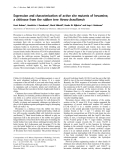
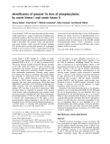
Fig. 5. (A) 2Fo ) Fc electron density map of the C-terminus in the crystal structure of the Sc(L559A) mutant (Protein Data Bank code: 3KDH). (B) Superimposition of the native enzyme (Protein Data Bank code: 2Q9O) (green) and the Sc(L559A) mutant structure (in blue).
In order to determine the structural effects of the the Sc(L559A) mutant was C-terminal mutation, crystallized for X-ray analysis. A crystal was diffracted to 2.4 A˚ , and the crystal structure was solved by molecular replacement. The electron density map clearly confirmed that the last residue of the mutant was an Ala instead of a Leu (Fig. 5A). Superimposi- tion of the rMaL (Protein Data Bank code: 2Q9O) and Sc(L559A) mutant (Protein Data Bank code: 3KDH) structures showed that an additional water molecule was present in the mutant structure (Fig. 5B). Owing to the lower steric limitations of the side chain of the Ala than of the Leu, water may occupy the space. Furthermore, the side chain of His140, which is coordinated to the T3 copper, rotated slightly and formed a hydrogen bond with the new water. In the structure of wild-type MaL, His140 was hydrogen- bonded to the carboxylate group of the C-terminus, with a distance of 3.1 A˚ . In the Sc(L559A) mutant, the distance between the ND1 atom of His140 and the ter- minal oxygen OXT atom of Ala559 increased to 4.0 A˚ . In addition, a nearby Asn109 adopted a different structure conformation in the Sc(L559A) mutant (Fig. 5B). The C-terminal mutation clearly affected the trinuclear site geometry.
In addition, the B-value of the T2 copper was clearly higher than the B-values of the two T3 coppers in the trinuclear site (Table 4). This was observed in both molecules in an asymmetric unit, thus verifying the phenomenon. Furthermore, no electron density was observed for the chloride ion in molecule A. A chlo- ride ion is coordinated to the T2 copper in the wild- type MaL [4]. In molecule B, some electron density was observed, but the refined chloride showed a very high B-value. On the basis of this structure solved at 2.4 A˚ resolution, it is impossible to say whether there is a hydroxide or chloride ion, but we decided to refine a chloride ion, because our near-atomic resolution structure has confirmed that MaL has a chloride ion in this position. It is likely that the occupancy of the chloride ion was less in molecule B and that it was totally lost in molecule A. Therefore, it seems that the
FEBS Journal 276 (2009) 6285–6300 ª 2009 The Authors Journal compilation ª 2009 FEBS
6292
M. Andberg et al. Function of C-terminus in M. albomyces laccase
Table 4. Distances and B-values of atoms at the trinuclear site. The Sc(L559A) mutant structure is compared with published native rMaL structures: a low-dose structure (Protein Data Bank: 2IH8), a high-dose structure (Protein Data Bank: 2IH9) and a near-atomic structure (Protein Data Bank: 2Q9O). A and B are two molecules in the asymmetric unit.
L559A mutant rMaL, low-dose structure rMaL, high-dose structure rMaL, near-atomic structure
A B A B A B A B
5.1 4.1 4.0 3.0 2.6 2.7 2.7 2.5 2.7 5.2 4.4 4.4 – 2.5 2.6 2.9 2.7 3.1 4.7 3.9 4.0 2.5 2.2 2.5 2.6 2.3 2.6 4.7 4.0 3.9 2.6 2.3 2.5 2.7 2.3 2.7 4.9 4.1 3.9 2.9 – 2.7 – 2.3 3.5 4.8 4.1 4.1 2.8 – 2.4 – 2.5 3.4 4.7 4.0 4.0 2.5 2.0 2.5 2.7 2.5 2.3 4.7 4.0 4.0 2.5 2.2 2.3 2.6 2.5 2.7
Distances (A˚ ) T3–T3¢ T3–T2 T3¢–T2 T2–Cl T3–O1 T3–O2 T3¢–O1 T3¢–O2 T2–O2 B-values (A˚ 2) T3 T3¢ T2 Cl O1 O2 26.0 15.5 48.0 49.9 1.0 1.1 23.0 25.9 58.1 – 9.9 8.3 18.6 15.9 15.9 22.7 33.2 28.2 22.5 17.0 20.1 27.0 32.0 27.8 19.1 16.4 21.4 31.9 11.2 – 18.3 18.7 22.1 32.1 13.8 – 11.9 11.7 10.1 15.2 24.4 26.0 12.3 11.7 10.5 15.4 23.9 25.3
C-terminal mutation on the binding of dioxygen. In addition, it should be noted that the B-values of oxygen atoms are very low, especially in molecule A.
Discussion
laccase
Sc(L559A) mutant at least partly loses its chloride ion and probably also the T2 copper, as the B-value of the T2 copper was rather high. On the basis of the anoma- lous signal, the estimated occupancy of the T2 copper was about 0.5. T2 copper depletion is known to be common for laccases. The first solved laccase structure lacked T2 copper [22], and several published structures seem to have only partial T2 occupation, on the basis of their high B-values. So far, MaL has been found to be particularly stable, and before this mutant structure, no signs of partial occupation of the T2 copper had been observed.
extension in the
The ascomycete M. albomyces produces a thermostable and alkaline that undergoes C-terminal processing. The processing site has been shown to be conserved, but the C-terminal extension is not present in all ascomycete-type laccases. The laccase sequences from Chaetomium globosum, P. arenaria, N. crassa, My. thermophila, Thielaviae arenaria and Magnapor- the grisea contain the extension (Fig. 6). Processing has been reported in the literature for only some laccases [16–19,23]. The amino acid preceding the C-terminal laccases undergoing processing has been shown to be a Leu. C-terminal processing has also been shown for the basidomycete laccase from Coprinus cinereus, but the processing is distinct from that of the ascomycete laccases, because the C-terminus of C. cinereus laccase does not contain the conserved ascomycete cleavage site [24].
The role of C-terminal processing of the ascomycete laccases is not known, but it has been suggested to be involved in the activation of the laccase [17,19]. Screening a My. thermophila laccase mutant library, Zumarraga et al. found a laccase variant with better kinetics than the parental type, with two mutated amino acids in the C-terminal extension. The mutations
Dioxygen was refined inside the trinuclear site of the Sc(L559A) mutant. Dioxygen has earlier been observed to bind to coppers in the trinuclear site, with all copper–oxygen distances being 2.2–2.6 A˚ [2] or slightly more close to one of the type 3 coppers as observed in a near-atomic resolution structure [4]. In the present Sc(L559A) mutant structure, dioxygen was again refined slightly differently. The copper–ligand distances are presented in Table 4. In molecule B, the distance between dioxygen and the T2 copper has extended to 3.1 A˚ . However, our previous studies with MaL have shown that the trinuclear site is sensitive to X-rays, and the observed structure may depend on the data collec- tion strategy or intensity of the beam [3]. Electrons are extracted in the mononuclear site by the T1 copper and further transferred to the trinuclear site, where dioxy- gen acts as a terminal electron acceptor. Therefore, it is difficult to draw any conclusions about the effect of the
FEBS Journal 276 (2009) 6285–6300 ª 2009 The Authors Journal compilation ª 2009 FEBS
6293
M. Andberg et al. Function of C-terminus in M. albomyces laccase
tritici Fig. 6. Multiple sequence alignment of the C-termini of some ascomycete laccases. Alignment of the C-terminal amino acid sequneces of MaL (Q70KY3) with the laccases or putative laccases of My. thermophila (MtL, from patent CN1157008), Th. arenaria (TaLcc1, from patent US2006063246), Ch. globosum (CgL1, XP_001228806; and CgL2, XP_001230068), Ma. grisea (MgL, XP_362544), P. anserina (PaL, P78722), (GgL, CAD10749), and Botryotinia fuckeliana (BfL, N. crassa (NcL, XP_956939), Cr. parasitica (CpL, Q03966), G. graminis var. AAK77953). The protein abbreviation and the protein accession numbers are in parentheses.
the mutation led to a slightly increased Km value without significantly altering the kcat.
in My. thermophila laccase (75% identical to MaL) resulted in a protein with disturbed T1 copper geome- try and reduced redox potential, as well as an altered trinuclear copper site, as shown by reduced oxygen uptake [25]. Surprisingly, the mutations in the C-termi- nal extension affected the protein properties, although the extension was cleaved from the mature protein. The mutations were suggested [25] to affect the folding of the protein during the post-translational processing, and thereby the function of the mature laccase.
the
temperatures. The crystal structure of
site.
Our structural analysis of wild-type MaL indicated that the last four amino acids of the mature protein penetrate the tunnel leading from the surface to the trinuclear site and form a plug [2]. C-terminal blocking might be a general feature of ascomycete laccases. In fact, we have determined a low-resolution structure (3.1 A˚ ) of Th. arenaria laccase (unpublished results), the C-terminus similarly and it clearly shows that blocks the tunnel leading towards the trinuclear site. This is strong evidence that C-terminal blocking is a common feature of ascomycete laccases.
specific activity of
The thermal stability of the yeast mutants Sc(del- DSGL559) and Sc(L559A) was determined by monitor- ing the CD spectrum as a function of temperature, the inactive Sc(del- because the kinetic stability of DSGL559) mutant was impossible to determine by activity measurements. Although the thermal unfolding profiles for the mutant and wild-type laccases were broad, with no clear folded–unfolded transition, it was evident that the mutants were less thermostable than the wild-type ScMaL. However, similarities between the far-UV CD spectra of the mutants and that of the wild-type ScMaL do not support any major conformational changes of the deletion mutant. The reduced thermostability of the Sc(L559A) mutant was also confirmed by residual activity measurements at the different Sc(L559A) mutant revealed T2 copper and chloride ion depletion in the trinuclear It has been observed that the first step in the denaturation of laccases, before actual denaturation, is the loss of one copper atom [26]. The clearly lowered protein stability of the Sc(L559A) mutant protein is probably due to T2 copper depletion.
constant of
The deletion of the last four amino acids in MaL dramatically affected the the enzyme, as the deletion mutant delDSGL559, expressed both in yeast and in T. reesei, was practically inactive. Also, the kinetic parameters were altered. The change of only one amino acid in the C-terminus of the lac- case, giving the Tr(L559G) and Sc(L559A) mutants, reduced the turnover of the mutant proteins. For the substrate ABTS, the the Michaelis Sc(L559A) mutant increased two-fold, and the turnover decreased four-fold, yielding an enzyme with 10-fold reduced catalytic efficiency, indicating that the last resi- due, Leu559, has a role in the catalysis of ABTS oxida- tion. The catalytic constants for the phenolic substrates 2,6-DMP and syringaldazine were not greatly affected by the L559A mutation. For the substrate 2,6-DMP,
A well-known laccase inhibitor, azide, has been shown to bind to the trinuclear site in the crystal structure of BsL [5], thus preventing binding of oxygen. We analyzed the azide inhibition and determined the inhibition constants for sodium azide, in order to see the effect of the mutation (L559A) on the binding properties in the trinuclear center. The inhibition of MaL by azide was determined to be mixed inhibition in which both specific and catalytic effects are present. Thus, azide can bind both to the free laccase and to the laccase–substrate complex. By comparing the inhibition constants for azide of wild-type ScMaL and
FEBS Journal 276 (2009) 6285–6300 ª 2009 The Authors Journal compilation ª 2009 FEBS
6294
M. Andberg et al. Function of C-terminus in M. albomyces laccase
Our results clearly show the critical role of the last amino acids in the C-terminus of MaL. Deletion of the four last amino acids strongly reduced the expression of the protein in T. reesei and S. cerevisiae. Further- the deletion resulted in practically inactive more, protein, although the CD spectra suggested that the secondary structure of the mutant was not significantly altered. Even a single amino acid change in the C-terminus of MaL (L559A) changed the catalytic properties of the laccase. The solved three-dimensional structure of the protein with the L559A mutation revealed conformational changes in the trinuclear site.
seen in the
trinuclear
changes
site of
Experimental procedures
Construction of the Tr(delDSGL559) and Tr(L559G) mutants for expression in T. reesei
the Sc(L559A) mutant, we found that the inhibition constants of azide were higher for the Sc(L559A) mutant than for ScMaL with both ABTS and the phe- nol substrate 2,6-DMP. This suggests that conforma- tional changes have taken place in the trinuclear site. The crystal structure of the Sc(L559A) mutant con- firmed that the geometry of the trinuclear site was dis- turbed. The His140, which is coordinated to the T3 copper, is slightly rotated as compared with the native enzyme, and it forms a hydrogen bond with a water molecule instead of a C-terminus. In addition, deple- tion of the T2 copper and chloride ion was observed. the The Sc(L559A) mutant may thus explain the more loose binding of azide to the trinuclear copper center. As the hydrogen bond between the C-terminus and His140 is the trinuclear site is broken, and the stability of decreased, the binding of azide to the trinuclear site may be decreased. The depletion of the T2 copper and chloride ion may also affect the azide binding. In fact, it has been shown that the T2 copper is crucial for dioxygen binding and that the depletion of the T2 copper inactivates the enzyme [13,22].
the Ki was
the
Notably, the effect of the mutation on the inhibi- tion constant (Ki) for azide was dependent on the sub- strate. Similarly to the specificity constant on ABTS, increased which was reduced 10-fold, 11-fold; however, changes with the phenolic substrates (2,6-DMP and syringaldazine) were not so dramatic. The different behavior of the nonphenolic ABTS and the phenolic substrates may be partly caused by pH. When ABTS was used, kinetic analyses were carried out at pH 4.5, whereas pH 6 was used for the phenolic substrates. The oxidation of ABTS does not involve proton transfer from the substrate to the enzyme, in contrast to what occurs with the phe- nolic substrates. When ABTS is used, many acidic amino acids around the trinuclear site (mainly Asp residues) can provide an immediate supply of protons for the reduction of dioxygen. Therefore, the mutation near the trinuclear site may have a more dramatic impact, especially at for the nonphenolic low pH, substrates.
Interestingly,
the conformational changes
Two mutants were produced by site-directed mutagenesis on the plasmid pLLK8, a T. reesei expression vector containing the cDNA coding for MaL between the cbh1 promoter and terminator [20]. The mutated fragments were generated by overlap extension PCR with 5¢-TCGCAGCA GCGCTTCGTGTT-3¢ and 5¢-GGGTTATGAACGGGAT GTTT-3¢ as upstream and downstream primers, respec- the Tr(L559G) mutant, tively, and for construction of the primers 5¢-ACCCCAAGATCGACTGGGCGG TAAG CGTCGCGCTGGGTGGAGGA-3¢ and 5¢-TCCTCCACC CAGCGGCGACGCTTACCGCCCGAGTCGATCTTGG GGT-3¢ were used as forward and reverse primers, respec- tively. For construction of the Tr(delDSGL559) mutant, the forward primer 5¢-CGAATCCCTACCCCAAGATCTGAT CGGGCCTGAAGCGTCGCCG-3¢ and the reverse primer 5¢-CGGCGACGCTTCAGGCCCGATCAGATCTTGGGG the mutagenesis TAGGGATTCG-3¢ were used. Briefly, was achieved by PCR with the use of specifically designed primers with the desired substitutions included in their sequence. Two independent PCR reactions were carried out with the mutagenic primer and an outer flanking primer to produce a forward and reverse fragment. The overlapping fragments containing the mutation were then fused together in a subsequent PCR reaction with the outside primers. The plasmid pLLK8 was digested with SacII and NsiI, and a fragment coding for the wild-type MaL was removed and replaced by the mutated fragments. The laccase mutant clones in plasmid pLLK8 were verified by sequencing to confirm that no other changes in the nucleotide sequence had occurred.
Production of the the Tr(delDSGL559) and Tr(L559G) mutants in T. reesei
The linearized expression constructs of the mutated laccases as well as the wild-type laccase construct pLLK13 [20],
in the trinuclear site seen in the three-dimensional structure of the mutant protein have a clear effect on the appar- ent affinity of the substrates, as seen from increased Km values (especially for ABTS). One may speculate that this effect would be caused by altered electron and ⁄ or proton transfer between the mononuclear site and the trinuclear site in the mutant protein, which might also indicate an interaction between the C-termi- nus and the mononuclear site.
FEBS Journal 276 (2009) 6285–6300 ª 2009 The Authors Journal compilation ª 2009 FEBS
6295
instructions. The transformants were grown on SC-URA [28] plates at 30 (cid:2)C for 3 days.
M. Andberg et al. Function of C-terminus in M. albomyces laccase
Production of the recombinant MaL and the Sc(delDSGL559) and Sc(L559A) mutants in S. cerevisiae
fragment
containing the full-length laccase cDNA between the cbh1 promoter and terminator sequences, were transformed into T. reesei RutC-30 essentially as described by Penttila¨ [27]. A MaL-producing strain in which the major cellulase gene cbh1 (Cel7A) of T. reesei was disrupted was also con- structed and transformed into T. reesei. The disruption plasmid construct was made by introducing a long cbh1 ter- minator into the MaL expression plasmid pLLK13. In the new construct pMS176, the laccase gene is between a long cbh1 promoter (2.3 kb) and a 0.8 kb termi- nator, followed by the long terminator fragment of 1.7 kb. The long promoter and terminator fragments are needed to improve the frequency of replacement of the cbh1 gene by homologous recombination. The purified transformants of mutated MaL, Tr(L559G) (VTT-D-03932) and Tr(del- DSGL559) (VTT-D-0393), as well as wild-type MaL pLLK13 [20] and pMS176, were cultured in shake flasks in minimal medium [27] supplemented with 40 gÆL)1 lactose, 20 gÆL)1 spent grain, 0.1 mm CuSO4 and 10 gÆL)1 sodium phthalate to buffer the medium to pH 6, for 7 days at 28 (cid:2)C and 200 r.p.m.
Construction of the Sc(delDSGL559) and Sc(L559A) mutants for expression in S. cerevisiae
The recombinant wild-type MaL (pMS175) [16] and mutant proteins Sc(delDSGL559) and Sc(L559A) were produced in S. cerevisiae under the inducible GAL1 promoter in shake flask cultures. The yeast cells were grown in SC-URA med- ium buffered to pH 6 with succinate and supplemented with 2 gÆL)1 raffinose and 1 mm CuSO4 for 2 days (to a D600 nm of approximately 7) at 30 (cid:2)C and 250 r.p.m. The well-grown inoculumn (8 · 50 mL) was used to inoculate 8 · 500 mL of fresh medium, and the cells were grown for an additional 1 day (D600 nm of 5–10), after which the cells were collected by centrifugation (5000 g for 10 min), washed with one vol- ume of sterile 0.9% NaCl solution, centrifuged again (5000 g for 10 min), and finally suspended in induction medium (one volume). The induction medium was similar to the inocula- tion medium, except that 2 gÆL)1 galactose was used instead of 2 gÆL)1 raffinose. After 3 days, the cells were removed by centrifugation (5000 g for 10 min), and the clear culture supernatant was collected and concentrated 10–20-fold by ultrafiltration (molecular mass cut-off of 10 000 Da).
The Sc(L559A) mutant was also produced in a 20 L laboratory-scale bioreactor. Culture was performed in synthetic complete medium (SC-URA) buffered to pH 6 with succinate and supplemented with 1 mm CuSO4. The yeast cells were grown on glucose (40 gÆL)1) for 2 days, and glu- cose was then fed into the culture for 3 days. The cells were collected by centrifugation (5000 g for 10 min) and washed with 0.9% sterile NaCl, and transferred into 12 L of induc- tion medium containing galactose (20 gÆL)1). After 100 h, the galactose had been used up, and more galactose was fed into the culture for an additional 60 h. The cells were removed by centrifugation (5000 g for 10 min), and the supernatant was collected and concentrated approximately 20-fold by ultrafiltration (molecular mass cut-off of 10 000 Da).
Purification of the ScMaL and the Sc(delDSGL559) and Sc(L559A) mutants from S. cerevisiae
All of the mutant laccase genes were cloned into the plas- mid vector pMS175 [16] containing the mature MaL sequence without the presequence and prosequence and the C-terminal extension of the native protein. The plasmid pMS175 is built on the S. cerevisiae expression vector pYES2 (Invitrogen, Carlsbad, CA, USA), and contains the a-factor prepro sequence of S. cerevisiae as a secretion sequence for improved yeast expression. The mutant laccase genes were created with Stratagene’s (La Jolla, CA, USA) QuickChange XL kit designed for large plasmids, according to the manufacturer’s instructions, with 18 cycles of PCR and transforming the PCR product to XL10 Gold ultra- competent Escherichia coli cells. Primers (Sigma-Aldrich) for construction of the Sc(L559A) mutant for PCR reac- tions were as follows: forward, 5¢-CCAAGATCGACTCG GGCGCTTAGCGTCGC-3¢; and reverse, 5¢-GCGACGCT AAGCGCCCGAGTCGATCTTGG-3¢. For construction of the Sc(delDSGL559) mutant, the primers used were as fol- forward, 5¢-ACCCCAAGATCTAATCGGGCCTG lows: TAGC-3¢; and reverse, 5¢-GCTACAGGCCCGATTAGAT CTTGGGGT-3¢. All of the laccase mutant clones in plas- mid pMS175 were sequenced to verify the mutations and to confirm that no other changes in the nucleotide sequence had occurred.
transformation kit
The concentrated culture supernatant, in 20 mm Tris ⁄ HCl (pH 7.5), of the wild-type or mutant MaL was applied to a weak anion exchange column (DEAE Sepharose FF) in 20 mm Tris ⁄ HCl (pH 7.5) and eluted with a linear 0–400 mm Na2SO4 gradient. Laccase-containing fractions were pooled and concentrated, and the buffer was changed to 20 mm Tris ⁄ HCl (pH 7.5) (Vivaspin; molecular mass cut-off of 10 000 Da). Typically, the laccase was further purified with a high-resolution anion exchange column (Resource Q). The bound proteins were eluted with a linear
The mutant plasmids were extracted and purified by Qiagen’s (Valencia, CA, USA) maxi-prep protocol and transformed into the yeast strain INVSc1 (MATa his3D1 leu2 trp1-289 ura3-52 ⁄ MATa his3D1 leu2 trp1-289 ura3-52; Invitrogen) with Gietz’s yeast (Tetra Link, Amherst, NY, USA), according to the manufacturer’s
FEBS Journal 276 (2009) 6285–6300 ª 2009 The Authors Journal compilation ª 2009 FEBS
6296
(pH 7.5). Pooling of
measurements were performed in triplicate. The reactions were started by the addition of substrate, and the rate of substrate oxidation was measured by monitoring the change in absorbance over 5 min, using a Varioskan kinetic plate (Thermo Electron Corporation, Waltham, MA, reader USA). The apparent kinetic parameters were obtained by curve fitting analysis using graphpad prism software 4.01 (GraphPad Software, Inc., San Diego, CA, USA).
in 20 mm Tris ⁄ HCl
Na2SO4 gradient (0–300 mm). The Sc(delDSGL559) mutant was further purified by hydrophobic interaction chromato- graphy. The sample was applied to a Phenyl Sepharose FF column pre-equilibrated with 700 mm Na2SO4 in 20 mm Tris ⁄ HCl (pH 7.5). Proteins were eluted with a 700–0 mm Na2SO4 gradient. Active fractions were concentrated and further loaded onto a gel filtration column (Sephacryl S-100) the Sc(delDSGL559) mutant-containing fractions was performed on the basis of antibody detection on dot blots. Purification of the laccases was followed by SDS ⁄ PAGE analysis.
M. Andberg et al. Function of C-terminus in M. albomyces laccase
Determination of protein concentration, SDS ⁄ PAGE, and western blot
For inhibitor studies, the enzyme was preincubated for 2 min at room temperature with various concentrations of sodium azide (NaN3) prior to addition of substrate. Dou- ble reciprocal plots (1 ⁄ V versus 1 ⁄ S) were used to analyze the type of inhibition. The inhibition constants were obtained by plotting the reciprocal rate (1 ⁄ V) against the inhibitor concentration (I) at different substrate values (S). This Dixon plot yields a series of straight lines that intersect at a unique point giving the negative inhibition constant ()Ki).
Thermal stability and pH dependency
e, of 115 720 m)1Æcm)1. The purity of
The protein concentration was determined using the Bio-Rad DC protein assay kit (Bio-Rad Laboratories Inc., Hercules, CA, USA), with BSA as standard or calculated from the UV absorbance at 280 nm, using a molar extinction the coefficient, enzymes was analyzed by SDS ⁄ PAGE (12% Tris ⁄ HCl Ready Gel; Bio-Rad, Laboratories Inc., Hercules, CA, USA), according to Laemmli [29]. The gels were stained with Coomassie brilliant blue for visualization of the protein bands. For western blot analysis, the proteins were trans- ferred to Hybond P poly(vinylidene difluoride) membranes (GE Healthcare Life Sciences, Uppsala, Sweden), and lac- case was detected with polyclonal antibodies raised against the native MaL and thereafter recognized by alkaline phos- phatase-conjugated goat anti-(rabbit IgG) as secondary antibody (Bio-Rad Laboratories Inc., Hercules, CA, USA).
The temperature stabilities of the wild-type enzyme and the Sc(L559A) mutant were determined by incubating 20 nkat of enzyme in 60 mm sodium citrate buffer (pH 6) at 40, 50 and 60 (cid:2)C. The stabilities of the laccases at different pH values were determined in McIlvaine buffer in the pH range 2.5–8.2, at 22 (cid:2)C and 4 (cid:2)C. The enzyme solutions were incubated for various time periods, and the residual activity was measured with 4.7 mm ABTS as substrate in 25 mm succinate buffer (pH 4.5). The pH optima of ScMaL and the Sc(L559A) mutant on 4.7 mm ABTS and 2 mm 2,6- DMP as substrates were determined in McIlvaine buffer (pH 2.5–8.2) at 22 (cid:2)C. The residual enzyme activities were measured with ABTS or 2,6-DMP as substrates, as described above.
Determination of laccase activities and kinetic constants
CD spectroscopy
laccase activities
(pH 6)
at
25 (cid:2)C at
The laccase activity was measured by monitoring the oxida- tion of 4.7 mm ABTS in 25 mm succinate buffer (pH 4.5) at 25 (cid:2)C. [30]. The activity was calculated by spectroscopic measurements at 436 nm, with an absorption coefficient (e) of 29 300 m)1Æcm)1. The for 2 mm 2,6-DMP and 0.06 mm syringaldazine were calculated by measuring the oxidation of these compounds in 40 mm 469 nm MES ⁄ NaOH buffer (e = 19 600 m)1Æcm)1) and 525 nm (e = 65 000 m)1Æcm)1) for 2,6-DMP and syringaldazine, respectively. Activities were expressed as nanokatals.
CD spectra were recorded on a JASCO model J-720 CD spectrometer equipped with a PTC-38WI Peltier thermally controlled cuvette holder. Far-UV (240–190 nm) CD mea- surements were performed with 2 lm enzyme in 10 mm sodium phosphate buffer (pH 7.1) at 25 (cid:2)C, using a 1 mm cell and a bandwith of 1 nm. Spectra were accumulated four times, and the values were corrected for buffer contri- butions. For comparison of the CD spectra, data smoothed by the Savitzky–Golay method were normalized by calcula- tions using the graphpad prism software.
Thermal unfolding curves were obtained by monitoring the 202 nm ellipticity as a function of temperature. The temperature was raised gradually at 1 (cid:2)CÆmin)1 from 30 (cid:2)C to 90 (cid:2)C. For comparison of the unfolding curves, the data measured at 202 nm were smoothed by an adjacent averag- ing procedure, prior to normalization by graphpad prism software.
Kinetic constants (Km and kcat values) for the different laccase proteins were determined using nonphenolic ABTS (pH 4.5) and phenolic 2,6-DMP and syringaldazine (pH 6.0) as substrates at 22 (cid:2)C. Eight different substrate concen- trations (0.06–4.7 mm, 0.006–1.7 mm and 0.001–0.12 mm for ABTS, 2,6-DMP, and syringaldazine, respectively) were used. Kinetic measurements were performed in microtiter the plates in a total reaction volume of 300 lL. All
FEBS Journal 276 (2009) 6285–6300 ª 2009 The Authors Journal compilation ª 2009 FEBS
6297
M. Andberg et al. Function of C-terminus in M. albomyces laccase
Redox titration
Mass analysis, N-terminal sequencing, and C-terminal sequencing
The redox potential of the mononuclear (T1) copper center for the mutated laccase was measured using the ferrocya- system (E0,Fe = 0.433 V) nide ⁄ ferricyanide redox buffer [21] in 20 mm Tris ⁄ HCl (pH 7.5). The laccase concentration used in the redox measurements was estimated from the 600 nm absorbance, using an extinction coefficient of 5700 m)1Æcm)1.
The mass analyses were performed on a MALDI-TOF Autoflex II (Bruker Daltonik GmbH, Bremen, Germany). N-terminal sequence analysis was performed according to Edman degradation chemistry, using a PE Biosystems Procise Sequencer (PE Biosystems, Foster City, CA, USA). C-terminal sequence analysis was carried out with a Procise C instrument at the Protein Analysis Center, Karolinska Institutet, Sweden.
Deglycosylation
using
endo-b-N-acetylglucosaminidase
Structure determination for the Sc(L559A) mutant
Deglycosylation of ScMaL and the Sc(L559A) mutant was F1 performed (Sigma, USA), according to the manufacturer’s instruc- tions. ScMaL and the Sc(L559A) mutant were incubated for 2 h at 37 (cid:2)C with endo-b-N-acetylglucosaminidase F1 in 50 mm sodum phosphate buffer (pH 5.5).
Table 5. Data collection and refinement statistics for the Sc(L559A) mutant. Rmeas = redundancy-independent R-factor. Values in paren- theses are for the highest-resolution shell.
Crystals of the Sc(L559A) mutant were grown by the vapor diffusion method at 20 (cid:2)C, using 15% PMME2000, 0.2 m ammonium sulfate, and 0.1 m sodium acetate buffer (pH 4.5). The protein concentration was 10 mgÆmL)1. Better- quality crystals were obtained with a microseeding method using 13% PMME2000 and an equilibrium time of tiny, with dimensions of about 4 h. Crystals were 0.1 · 0.1 · < 0.05 mm. The crystal was harvested and plunged into the liquid nitrogen, using 25% glycerol as cryoprotectant.
Data collection
C2 a = 173.5, b = 62.0, c = 125.7 A˚ , Space group Unit cell dimensions b = 99.92 2
1.365 A˚ 20–2.4 (2.5–2.4) 50 624 (5497) 97.4 (92.3) 13.8 (37.9) 11.8 (32.3) 9.0 (4.0)
48 093
Diffraction data were collected on a beamline X12 located at the DORIS storage ring at DESY, using a wave- length of 1.365 A˚ . The crystal was partly nonmerohedrally twinned, but the data-processing program xds was able to process it rather well and the data were scaled with xscale (Table 5). The space group was C2, with two molecules per asymmetric unit. The structure was solved by a molecular replacement method, using the coordinates of rMaL. The structure was refined by iterative cycles of manual fitting with o and positional refinements with cns. Refinements were carried out using an initial anisotropic B-factor and bulk solvent corrections. Data statistics are shown in Table 5. R-values of the final model are slightly high, but the electron density map was of good quality. The coordi- nates and structure factors of the Sc(L559A) mutant have been deposited in the Protein Data Bank as 3DKH.
2532
22.4 28.2
Acknowledgements
(Academy
Molecules per asymmetric unit Wavelength Resolution (A˚ ) Unique reflections Completeness (%) Rmeas (%) Rsym I ⁄ I(r) Refinement Number of reflections in working set Number of reflections in test set Rwork (%) Rfree (%) rmsd from restraint target values Bond lengths (A˚ ) Angle distances (A˚ ) Number of atoms Protein Water Copper Others Average B-factors (A˚ 2) Protein Water Copper Others 0.0119 1.5852 9858 8714 708 8 428 21.1 20.3 20.2 28.5 39.1
The authors wish to thank H. Boer for help with CD, P. Matikainen for help with the MALDI-TOF analysis, O. Liehunen and B. Hillebrandt-Chellaoui for excellent technical assistance, and B. Smit for fermentor culture. This work is a part of the research programme ‘VTT Industrial Biotechnology’ of Finland; Finnish Center of Excellence programme, 2000–2005, Project no. 64330). The study was performed with financial support from Tekes (Finnish Funding Agency for Technology and Innovation) and the Academy of
FEBS Journal 276 (2009) 6285–6300 ª 2009 The Authors Journal compilation ª 2009 FEBS
6298
structural studies of the fungal laccase from Cerrena maxima. J Biol Inorg Chem 11, 963–973.
11 Xu F (2001) Dioxygen reactivity of laccase. Dependence
on laccase source, pH, and anion inhibition. Appl Biochem Biotechnol 95, 125–133.
M. Andberg et al. Function of C-terminus in M. albomyces laccase
Finland. Structure determination work was supported by the Academy of Finland, Project 115085. Data collection at EMBL ⁄ DESY was supported by the European Community, Research Infrastructure Action, under FP6, ‘Structuring the European Research Area Programme’, contract number RII3-2004-506008. We also thank the staff of the beamline X12 EMBL ⁄ DESY Hamburg outstation.
12 Johannes C & Majcherczyk A (2000) Laccase activity tests and laccase inhibitors. J Biotechnol 78, 193–199. 13 Solomon EI, Sundaram UM & Machonkin TE (1996) Multicopper oxidases and oxygenases. Chem Rev 96, 2563–2606.
References
1 Kiiskinen L-L, Viikari L & Kruus K (2002) Purification
14 Solomon EI, Chen P, Metz M, Lee S-K & Palmer AE (2001) Oxygen binding, activation, and reduction to water by copper proteins. Angew Chem Int Ed 40, 4570–4590.
and characterisation of a novel laccase from the ascomycete Melanocarpus albomyces. Appl Microbiol Biotechnol 59, 198–204.
15 Messerschmidt A, Luecke H & Huber R (1993) X-ray structures and mechanistic implications of three func- tional derivatives of ascorbate oxidase from zucchini: reduced, peroxide and azide forms. J Mol Biol 230, 997–1014.
16 Kiiskinen L-L & Saloheimo M (2004) Molecular
2 Hakulinen N, Kiiskinen LL, Kruus K, Saloheimo M, Paananen A, Koivula A & Rouvinen J (2002) Crystal structure of a laccase from Melanocarpus albomyces with an intact trinuclear copper site. Nat Struct Biol 9, 601–605.
3 Hakulinen N, Kruus K, Koivula A & Rouvinen J
cloning and expression in Saccharomyces cerevisiae of a laccase gene from the ascomycete Melanocarpus albomyces. Appl Environ Microbiol 70, 137–144.
17 Germann U, Muller G, Hunziker P & Lerch K (1988)
(2006) A crystallographic and spectroscopic study on the effect of X-ray radiation on the crystal structure of Melanocarpus albomyces laccase. Biochem Biophys Res Commun 350, 929–934.
Characterization of two allelic forms of Neuros- pora crassa laccase. Amino- and carboxyl-terminal processing of a precursor. J Biol Chem 263, 885–896.
4 Hakulinen N, Andberg M, Kallio J, Koivula A, Kruus K & Rouvinen J (2008) A near atomic resolution struc- ture of a Melanocarpus albomyces laccase. J Struct Biol 162, 29–39.
18 Fernandez-Larrea J & Stahl U (1996) Isolation and characterization of a laccase gene from Podospora anserina. Mol Gen Genet 252, 539–551.
5 Bento I, Martins LO, Lopes GG, Carrondo MA &
Lindley PF (2005) Dioxygen reduction by multi-copper oxidases; a structural perspective. Dalton Trans 7, 3507– 3513.
6 Bento I, Peixoto C, Zaitsev VN & Lindley PF (2007)
19 Bulter T, Alcalde M, Sieber V, Meinhold P, Schlacht- bauer C & Arnold FH (2003) Functional expression of a fungal laccase in Saccharomyces cerevisiae by directed evolution. Appl Environ Microbiol 69, 987–995. 20 Kiiskinen L-L, Kruus K, Bailey M, Ylosmaki E,
Ceruloplasmin revisited: structural and functional roles of various metal cation-binding sites. Acta Crystallogr 63, 240–248.
Siika-aho M & Saloheimo M (2004) Expression of Melanocarpus albomyces laccase in Trichoderma reesei and characterization of the purified enzyme. Microbiology 150, 3065–3074.
21 Xu F, Shin W, Brown SH, Wahleithner JA, Sundaram
7 Piontek K, Antorini M & Choinowski T (2002) Crystal structure of a laccase from the fungus Trametes versicol- or at 1.90-A˚ resolution containing a full complement of coppers. J Biol Chem 277, 37663–37669.
UM & Solomon EI (1996) A study of a series of recom- binant fungal laccases and bilirubin oxidase that exhibit significant differences in redox potential, substrate speci- ficity, and stability. Biochim Biophys Acta 1292, 303–311.
22 Ducros V, Brzozowski AM, Wilson KS, Brown SH,
8 Bertrand T, Jolivalt C, Briozzo P, Caminade E, Joly N, Madzak C & Mougin C (2002) Crystal structure of a four-copper laccase complexed with an arylamine: insights into substrate recognition and correlation with kinetics. Biochemistry 41, 7325–7333.
O¨ stergaard P, Schneider P, Yaver DS, Pedersen AH & Davies GJ (1998) Crystal structure of the type-2 Cu depleted laccase from Coprinus cinereus at 2.2 A˚ resolu- tion. Nat Struct Biol 5, 310–316.
9 Garavaglia S, Teresa Cambria M, Miglio M, Ragusa S, Iacobazzi V, Palmieri F, D’Ambrosio C, Scaloni A & Rizzi M (2004) The structure of Rigidoporus lignosus laccase containing a full complement of copper ions, reveals an asymmetrical arrangement for the T3 copper pair. J Mol Biol 342, 1519–1531.
10 Lyashenko A, Bento I, Zaitsev V, Zhukhlistova N,
23 Berka R, Schneider P, Golightly E, Brown S, Madden M, Brown K, Halkier T, Mondorf K & Xu F (1997) Characterization of the gene encoding an extracellular laccase of Myceliophthora thermophila and analysis of the recombinant enzyme expressed in Aspergillus oryzae. Appl Environ Microbiol 63, 3151–3157.
Zhukova Y, Gabdoulkhakov A, Morgunova E, Voelter W, Kachalova G, Stepanova V et al. (2006) X-ray
FEBS Journal 276 (2009) 6285–6300 ª 2009 The Authors Journal compilation ª 2009 FEBS
6299
24 Yaver DS, Overjero MDC, Xu F, Nelson BA, Brown
30 Niku-Paavola M-L, Karhunen E, Salola P & Raunio V (1988) Ligninolytic enzymes of the white-rot fungus Phlebia radiata. Biochem J 254, 877–884.
M. Andberg et al. Function of C-terminus in M. albomyces laccase
Supporting information
KM, Halkier T, Bernauer S, Brown SH & Kauppinen S (1999) Molecular characterization of laccase genes from the basidiomycete Coprinus cinereus and heterologous expression of the laccase Lcc1. Appl Environ Microbiol 65, 4943–4948.
25 Zuma´ rraga M, Camarero S, Shleev SV, Martı´ nez-Arias A, Ballesteros A, Plou FJ & Alcalde M (2008) Altering the laccase functionality by in vivo assembly of mutant libraries with different mutational spectra. Proteins Struc, Funct Bioinf 71, 250–260.
deglycosylated
purified
of
The following supplementary material is available: Fig. S1. Western blot analysis of culture supernatants of T. reesei strains producing wild-type and mutant MaL. Fig. S2. SDS ⁄ PAGE Sc(L559A) mutant.
This supplementary material can be found in the
online version of this article.
26 Marjasvaara A, Kruus K & Vainiotalo P (2006) A lac- case study by electrospray ionization Fourier transform ion cyclotron resonance MS: copper depletion, glyco- forms and stability. J Mass Spectrom 41, 91–97. 27 Penttila¨ M, Nevalainen H, Ra¨ tto¨ M, Salminen E &
Knowles J (1987) A versatile transformation system for the cellulolytic filamentous fungus Trichoderma reesei. Gene 61, 155–164.
28 Sherman F (1991) Getting started with yeast. Methods
Enzymol 194, 3–21.
Please note: As a service to our authors and readers, this journal provides supporting information supplied by the authors. Such materials are peer-reviewed and may be re-organized for online delivery, but are not copy-edited or typeset. Technical support issues arising from supporting information (other than missing files) should be addressed to the authors.
29 Laemmli UK (1970) Cleavage of structural proteins
during the assembly of the head of bacteriophage T4. Nature 227, 680–685.
FEBS Journal 276 (2009) 6285–6300 ª 2009 The Authors Journal compilation ª 2009 FEBS
6300