
Open Access
Available online http://ccforum.com/content/11/5/R109
Page 1 of 8
(page number not for citation purposes)
Vol 11 No 5
Research
Continuous control of endotracheal cuff pressure and tracheal
wall damage: a randomized controlled animal study
Saad Nseir1, Alexandre Duguet2, Marie-Christine Copin3, Julien De Jonckheere4, Mao Zhang5,
Thomas Similowski2 and Charles-Hugo Marquette6
1Intensive Care Unit, Calmette Hospital, University Hospital of Lille, boulevard du Pr Leclercq, 59037 Lille cedex, France
2Intensive Care Unit, Department of Respiratory Diseases, Public Hospitals of Paris, La Pitié-Salpêtrière Hospital, 47-83 boulevard de l'Hôpital,
75013 Paris, France
3Department of Pathology, Biology and Pathology Center, University Hospital of Lille, Lille 2 University, 1 place de Verdun, 59045 Lille, France
4Institut de Technologie Médicale, EA1049, CHRU de Lille, Pavillon Vancostenobel, 2 avenue Oscar Lambret, 59037 Lille cedex, France
5Department of Emergency Medicine, Zhejiang University, School of Medicine and Research Institute of Emergency Medicine, Zhejiang University,
Hangzhou, China
6Respiratory Disease Department, Calmette Hospital, University Hospital of Lille, boulevard du Pr Leclercq, 59037 Lille cedex, France
Corresponding author: Saad Nseir, s-nseir@chru-lille.fr
Received: 29 Jun 2007 Revisions requested: 8 Aug 2007 Revisions received: 12 Sep 2007 Accepted: 3 Oct 2007 Published: 3 Oct 2007
Critical Care 2007, 11:R109 (doi:10.1186/cc6142)
This article is online at: http://ccforum.com/content/11/5/R109
© 2007 Nseir et al., licensee BioMed Central Ltd.
This is an Open Access article distributed under the terms of the Creative Commons Attribution License (http://creativecommons.org/licenses/by/
2.0), which permits unrestricted use, distribution, and reproduction in any medium, provided the original work is properly cited.
Abstract
Background Intubation is frequently performed in intensive care
unit patients. Overinflation of the endotracheal tube cuff is a risk
factor for tracheal ischemia and subsequent complications.
Despite manual control of the cuff pressure, overinflation of the
endotracheal cuff is common in intensive care unit patients. We
hypothesized that efficient continuous control of the
endotracheal cuff pressure using a pneumatic device would
reduce tracheal ischemic lesions in piglets ventilated for 48
hours through a high-volume, low-pressure endotracheal tube.
Materials and methods Twelve piglets were intubated and
mechanically ventilated for 48 hours. Animals were randomized
to manual control of the endotracheal cuff pressure (n = 6) or to
continuous control of the endotracheal cuff pressure using a
pneumatic device (n = 6). In the two groups, we inflated the
endotracheal cuff with 50 ml air for 30 minutes, eight times daily.
This hyperinflation of the endotracheal cuff aimed at mimicking
high-pressure periods observed in intubated critically ill patients.
In all animals, the cuff pressure and the airway pressure were
continuously recorded for 48 hours. After sacrifice of the study
animals, the trachea was removed and opened longitudinally for
gross and histological examination. A pathologist evaluated the
slides without knowledge of treatment group assignment.
Results The cuff pressure was significantly lower in piglets with
the pneumatic device than in piglets without the pneumatic
device (median (interquartile range), 18.6 (11–19.4) cmH2O
versus 26 (20–56) cmH2O, P = 0.009). No significant
difference was found in the percentage of time spent with a cuff
pressure <15 cmH2O and that with a cuff pressure between 30
and 50 cmH2O. The percentage of time between 15 and 30
cmH2O of cuff pressure, however, was significantly higher in
piglets with the pneumatic device than in piglets without the
pneumatic device (98% (95–99%) versus 65% (44–80%), P =
0.002). In addition, the percentage of time with cuff pressure
>50 cmH2O was significantly lower in piglets with the
pneumatic device than in piglets without the pneumatic device
(0% versus 19% (12–41%), P = 0.002).
In all animals, hyperemia and hemorrhages were observed at the
cuff contact area. Histological examination showed no
difference in tracheal lesions between animals with and without
the pneumatic device. These lesions included deep mucous
ulceration, squamous metaplasia and intense mucosal
inflammation. No cartilage lesions were observed.
Conclusion The pneumatic device provided effective
continuous control of high-volume, low-pressure endotracheal
cuff pressure in piglets mechanically ventilated for 48 hours. In
the present model, however, no significant difference was found
in tracheal mucosal lesions of animals with or without a
pneumatic device. Further studies are needed to determine the
impact of continuous control of cuff pressure over a longer
duration of mechanical ventilation.
ICU = intensive care unit.

Critical Care Vol 11 No 5 Nseir et al.
Page 2 of 8
(page number not for citation purposes)
Introduction
Endotracheal intubation is frequently performed in intensive
care unit (ICU) patients [1]. The endotracheal tube cuff is
responsible for tracheal mucosal lesions that are visible at the
cuff contact area a few hours after intubation [2-5]. These
lesions may result in serious complications such as tracheal
stenosis and tracheal ruptures [6-8]. According to the results
of studies using a low-volume, high-pressure endotracheal
cuff, the prevalence of postintubation and post-tracheotomy
stenosis varies from 10% to 19% in ICU patients [9,10]. More
recent studies using a high-volume, low-pressure cuff, how-
ever, showed that clinically significant stenosis was less com-
mon (1‰–1%) [11,12]. Hyperinflation of the endotracheal
tube cuff is the most frequent risk factor for tracheal ischemia
and subsequent complications in these patients [13].
Complications related to insufficient cuff inflation have never-
theless been reported, including leaking of the tidal volume
and microaspiration of secretions and subsequent ventilator-
associated pneumonia [14]. In most ICUs, the endotracheal
cuff pressure is never checked [15-18]. In these ICUs, car-
egivers frequently overinflate the tube cuff to prevent gas leak
and pulmonary aspiration [15,18].
High-volume, low-pressure endotracheal tubes have signifi-
cantly reduced the frequency of ischemic tracheal lesions.
Even when high-volume, low-pressure endotracheal tubes are
used, however, ischemic tracheal lesions may occur [19]. An
endoscopic study performed in 40 patients undergoing sur-
gery showed that obstruction of mucosal blood flow occurred
at a lateral wall pressure above 30 cmH2O [20].
Based on recent recommendations, the cuff pressure should
be maintained around 25 cmH2O in critically ill intubated and
mechanically ventilated patients [21,14]. Although manual
measurement of the cuff pressure could reduce overinflation
and underinflation frequency, manual measurement may not
provide effective control of the cuff pressure. As shown by
Duguet and colleagues [22], despite manual control of the
endotracheal pressure with a portable manometer according
to the French Society of Critical Care Medicine recommenda-
tions, the percentage of time the cuff pressure was >30
cmH2O was 29 ± 25% and the percentage of time the cuff
pressure was <15 cmH2O was 15 ± 17%.
Several devices enabling efficient continuous control of cuff
pressure have been recently described [22,23]. The pneu-
matic device is a simple mechanical device that continuously
maintains the cuff pressure during mechanical ventilation with
minimal human resources [22].
We hypothesized that efficient continuous control of the
endotracheal cuff pressure using a pneumatic device would
reduce tracheal ischemic lesions in piglets ventilated for 48
hours through a high-volume, low-pressure endotracheal tube.
Methods
This study was conducted in the experimental intensive care
unit at Lille II University. All animals were treated according to
the guidelines of the Department of Experimental Research of
Lille University and according to the Guide for the Care and
Use of Laboratory Animals (NIH Publication Number 93-23,
revised 1985).
Animal preparation
Healthy, bred, domestic Largewhite-Landrace piglets, weigh-
ing 22 ± 2 kg, were anesthetized using propofol 3 mg/kg and
were orotracheally intubated with a 7.0 Hi-Lo Lanz™ Malinck-
rodt tube (Malinckrodt Inc, Argyle, NY, USA). Anesthesia was
maintained with a continuous infusion of midazolam 0.3 mg/
kg/hour, pancuronium 0.3 mg/kg/hour and fentanyl 0.3 μg/kg/
hour. The femoral artery was cannulated with a 3 F polyethyl-
ene catheter (Plastimed, St Leu la Forêt, France) for pressure
monitoring. An 8 F suprapubic urinary catheter (Vesicoset;
Angiomed, Karlsruhe, Germany) was placed in the bladder
transabdominally. Animals were mechanically ventilated in the
prone position in a volume-controlled mode with a Cesar type
1 ventilator (Taema, Antony, France). The ventilatory parame-
ters consisted of a tidal volume of 15 ml/kg, a respiratory rate
of 15 breaths/minute, an expiratory ratio of 0.5 and zero end-
expiratory pressure. Inspired gases were humidified using a
conventional humidifier (MR290; Fisher Paykel, Auckland,
New Zealand), and an initial fraction of inspired oxygen of 0.21
was used. All animals were sacrificed 48 hours after starting
mechanical ventilation.
Device for control of endotracheal cuff pressure
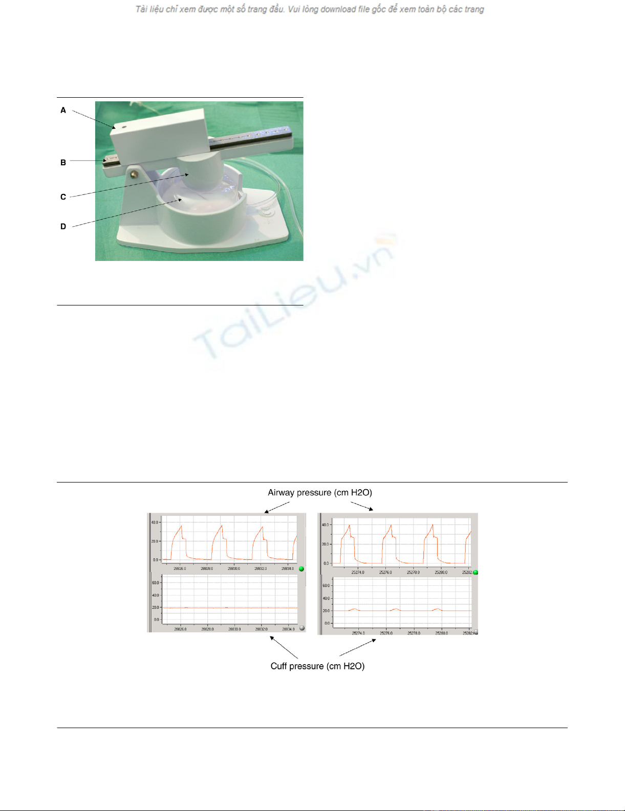
The Nosten® device (Leved, St-Maur, France) is amechanical
appliance that does not require a power supply (Figure 1).
Asterile single-use 200 ml cylindrical cuff encased in arigid
compartment is connected to the endotracheal cuff with plas-
tic tubing (internal diameter 3 mm, length 2 m). Aweight
mounted on an articulated arm constantly exerts pressure on
this cuff. This pressure can be adjusted by moving another
weight along the arm to modulate the corresponding force,
allowing the user to obtain the desired cuff pressure. Any var-
iation is immediately cancelled out by the disproportion
between the volumes of the two cuffs [22]. The device pro-
vides effective continuous control of endotracheal cuff pres-
sure in mechanically ventilated ICU patients [22].
Study protocol
Twelve animals were randomly assigned (1:1) to one of the
two study groups. In the interventional study group, the
endotracheal cuff was connected to the continuous cuff pres-
sure control device, the mobile weight of which was moved
along the articulated arm to obtain acuff pressure of 22
cmH2O. In the standard care group, the cuff pressure was
managed according to the French Society of Intensive Care
recommendations [24]; namely, a target cuff pressure at 22
cmH2O with cuff pressure checks twice a day at fixed intervals
Available online http://ccforum.com/content/11/5/R109
Page 3 of 8
(page number not for citation purposes)
and after each intervention on the endotracheal tube (manual
portable manometer, Hi-Lo™; Tyco Healthcare, Hazelwood,
Mo, USA).
In all animals, the cuff pressure and the airway pressure were
continuously recorded at adigitizing frequency of 100 Hz for
48 hours (Physiotrace®; Estaris, Lille, France) (Figure 2) [25].
The connection between the pressure transducer and the
endotracheal cuff was identical in the two groups, with athree-
way stopcock of which the third port was either closed or con-
nected to the pneumatic device. During each experiment, two
piglets were randomized to the standard care group or to the
pneumatic device group. Continuous recording of the cuff
pressure and the respiratory pressure was performed simulta-
neously in the two animals. Connections were checked every
3 hours.
In the two groups, we inflated the endotracheal cuff with 50 ml
air for 30 minutes eight times daily. This hyperinflation of the
endotracheal cuff aimed at mimicking high-pressure periods
observed in intubated critically ill patients [22]. After each
period of hyperinflation, the cuff pressure was readjusted as
described above. Hyperinflation periods represented 16% of
the total duration of mechanical ventilation (8 hours out of the
total 48 hours).
Postmortem evaluation
After sacrifice of the study animals, the trachea was removed
and opened longitudinally for gross examination. Full-thick-
ness samples of two contiguous tracheal rings were collected
and were placed in formalin for later histological examination.
The first sample was taken from the mid-cuff contact area, and
the second sample was taken distally beyond the endotra-
cheal tube. The proximal limit of cuff contact with mucosa was
easily recognized in all animals by visual examination of the tra-
cheal mucosa (Figure 3). The pathologist evaluated the slides
without knowledge of treatment group assignment. Tracheal
lesions were graded as: Grade I lesions including squamous
metaplasia, few inflammatory cells, and edema; as Grade II
lesions including mucous ulceration and normal subcartilagi-
nous tissue; or Grade III lesions including mucous ulceration
and a dense inflammatory reaction from the surface tissue to
the subcartilaginous tissue [7].
Figure 1
Photograph of the pneumatic devicePhotograph of the pneumatic device. A, mobile mass; B, arm; C, fixed
mass; D, 200 ml cuff connected to the external control cuff of the
endotracheal tube.
Figure 2
Continuous recording of cuff and airway pressures in piglets with and without the pneumatic deviceContinuous recording of cuff and airway pressures in piglets with and without the pneumatic device. Left: continuous recording of the cuff pressure
and the airway pressure in a piglet with the pneumatic device – the cuff pressure was constant despite variations of airway pressure. Right: continu-
ous recording of the cuff pressure and the airway pressure in a piglet without the pneumatic device – the cuff pressure decreased and increased
with airway pressure variations.
Critical Care Vol 11 No 5 Nseir et al.
Page 4 of 8
(page number not for citation purposes)
Statistical analysis
SPSS software (SPSS, Chicago, IL, USA) was used for data
analysis. In each animal, we measured the time spent with a
cuff pressure below 15 cmH2O, a pressure between 15 and
30 cmH2O, a pressure between 30 and 50 cmH2O, and with
a cuff pressure over 50 cmH2O. Qualitative variables were
described as the number (percentage), and quantitative varia-
bles were described as the median (interquartile range). The
distribution of quantitative values was tested for normality
using the Shapiro–Wilk test. Proportions were compared
using the chi-square test or the Fisher exact test where appro-
priate. The Student t test or the Mann–Whitney U test was
used for quantitative variables, as appropriate. Differences
were considered significant if P < 0.05. We expected grade II
or grade III tracheal lesions would occur in all control animals.
Inclusion of 12 animals (six in each group) was required to
detect a difference of 60% in the rate of animals with grade II
or grade III tracheal lesions (two-sided α = 0.05, power =
0.80).
Results
The mean arterial pressure (100 (85–110) mmHg versus 100
(89–115) mmHg), the diastolic arterial pressure (70 (61–80)
mmHg versus 68 (59–78) mmHg) and the heart rate (101
(90–115) beats/min versus 98 (89–112) beats/min) were
similar (P > 0.2) in animals with the pneumatic device and in
animals without the pneumatic device.
The mean airway pressure was similar in piglets with or without
the pneumatic device (11.3 (11–12.5) cmH2O versus 12.4
(10.4–13.2) cmH2O, P = 0.5). The cuff pressure was signifi-
cantly lower in piglets with the pneumatic device than in pig-
lets without the pneumatic device (18.6 (11–19.4) cmH2O
versus 26 (20–56) cmH2O, P = 0.009). During overinflation
periods, the cuff pressure was significantly lower in piglets
with the pneumatic device than in piglets without the pneu-
matic device (23 (20–25) cmH2O versus 76 (63–82) cmH2O,
P < 0.001). No significant difference was found in the percent-
age of time spent with a cuff pressure <15 cmH2O and the
percentage of time with a cuff pressure between 30 and 50
cmH2O. The percentage of time between 15 and 30 cmH2O
cuff pressure, however, was significantly higher in piglets with
the pneumatic device than in piglets without the pneumatic
device. In addition, the percentage of time >50 cm H2O cuff
pressure was significantly lower in piglets with the pneumatic
device than in piglets without the pneumatic device (Table 1).
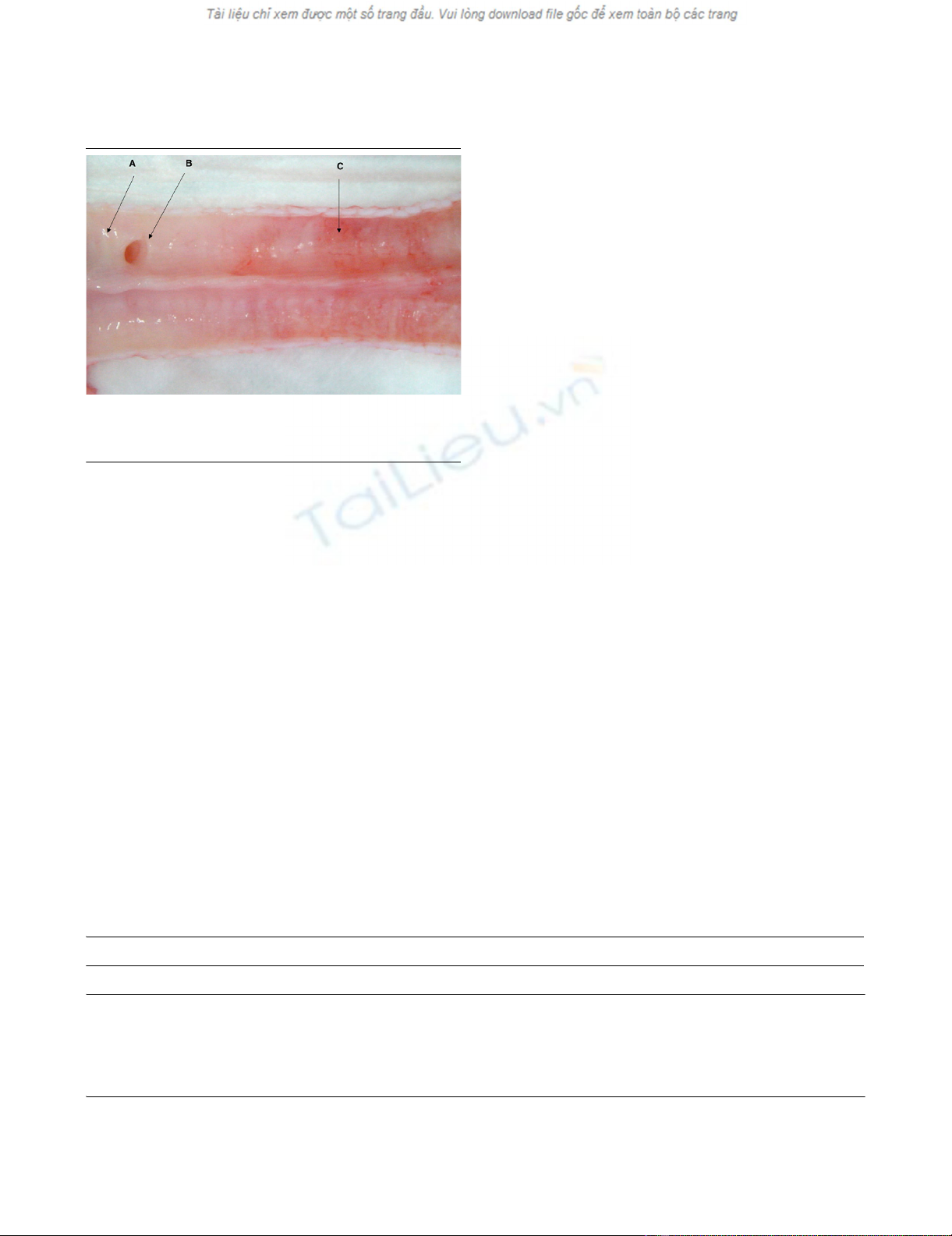
Macroscopic examination showed no lesions on the tracheal
mucosa distal to the endotracheal tube. In all animals, how-
ever, hyperemia and hemorrhages were observed at the cuff
contact area (Figure 3).
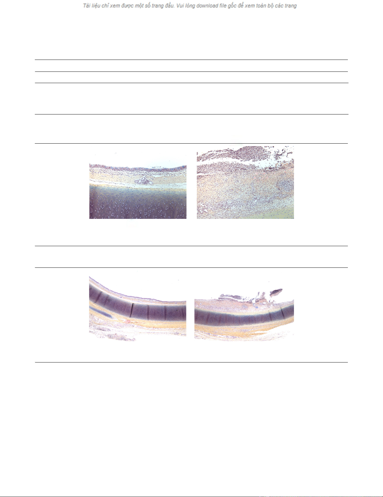
Histological examination showed no difference in tracheal
lesions between animals with or without the pneumatic device.
Although no lesions were observed in samples taken distally
beyond the endotracheal tube, grade I and grade II lesions
were observed in all animals in samples taken from the cuff
contact area (Table 2). These lesions included deep mucous
ulceration, including fibrin and polynuclear cells, squamous
metaplasia and intense mucosal inflammation. Neither
cartilage lesion nor inflammation expanding to the subcartilag-
inous tissue was observed (Figures 4 and Figure 5).
Figure 3
Gross examination of longitudinally opened tracheaGross examination of longitudinally opened trachea. A, no lesions on
tracheal mucosa distal to the endotracheal tube; B, origin of the tra-
cheal bronchi; C, hyperemia and hemorrhages at the cuff contact area.
Table 1
Endotracheal cuff pressure in animals with and without the pneumatic device
Animals with the pneumatic device (n = 6) Animals without the pneumatic device (n = 6) P value
Percentage of time at <15 cmH2O 1.4 (0.02–4.3) 0.3 (0.02–22.9) 0.910
Percentage of time at 15–30 cmH2O 98 (95–99) 65.8 (44–80) 0.002
Percentage of time at 30–50 cmH2O 0.01 (0–0.02) 0.3 (0–0.95) 0.315
Percentage of time at >50 cmH2O 0 19.8 (12–41) 0.002
Results presented as the median (interquartile range).
Available online http://ccforum.com/content/11/5/R109
Page 5 of 8
(page number not for citation purposes)
Discussion
In piglets ventilated for 48 hours through a high-volume, low-
pressure endotracheal tube, the pneumatic device enabled an
effective continuous control of the endotracheal cuff pressure.
This effective control of cuff pressure did not, however, result
in any difference with regard to tracheal mucosal damage.
Continuous recording of the cuff pressure in study animals
confirmed that the pneumatic device was efficient at continu-
ous cuff pressure regulation. The high volume of the pneu-
matic-device cuff (200 ml) explains how the injection of 50 ml
air did not result in endotracheal cuff overinflation in animals
with the pneumatic device, since the endotracheal cuff and the
pneumatic-device cuff were connected during inflation peri-
ods. In a previous prospective study, the efficacy of the pneu-
matic device in maintaining constant endotracheal cuff
pressure was evaluated in nine consecutive mechanically ven-
tilated critically ill patients [22]. The cuff pressure was contin-
uously registered for 24 hours during standard care and for 24
hours with the regulatory device. The authors reported a
significant reduction in the coefficient of variation of cuff pres-
sure in patients during the period of mechanical ventilation
with the pneumatic device. Other devices are available for cuff
pressure control [23,26-28]; however, the device used in the
Table 2
Distribution of histological tracheal lesions
Animals with the pneumatic device (n = 6) Animals without the pneumatic device (n = 6)
Grade I lesions 6 (100) 6 (100)
Grade II lesions 6 (100) 6 (100)
Grade III lesions 0 (0) 0 (0)
Results presented as n (%).
Figure 5
Histological examination (1 × 10) of tracheal samplesHistological examination (1 × 10) of tracheal samples. Left: sample taken distally beyond the endotracheal tube showing moderate inflammation.
Right: sample taken from the cuff contact area with localized ulceration, including fibrin and polynuclear cells, squamous metaplasia and intense
mucosal inflammation.
Figure 4
Histological examination (1 × 2.5) of tracheal samplesHistological examination (1 × 2.5) of tracheal samples. Left: sample taken distally beyond the endotracheal tube, no visible lesions. Right: sample
taken from the cuff contact area with localized ulceration.


![Vaccine và ứng dụng: Bài tiểu luận [chuẩn SEO]](https://cdn.tailieu.vn/images/document/thumbnail/2016/20160519/3008140018/135x160/652005293.jpg)


%20--%3e%3cdefs%3e%3cstyle%3e%20.st0%20{%20fill:%20%23fff;%20}%20.st1%20{%20fill:%20%237800fa;%20}%20%3c/style%3e%3c/defs%3e%3cpath%20class='st1'%20d='M117.78,12.18H43.11c2.9,3.47,4.65,7.94,4.65,12.82,0,5.6-2.3,10.66-6.01,14.29h76.02l7.22-13.56-7.22-13.56Z'/%3e%3cg%3e%3cpath%20class='st0'%20d='M53.58,26.17h-.59v-1.46h.59v-4.96h2.83c1.78,0,2.67.94,2.67,2.82v5.76c0,1.87-.89,2.81-2.67,2.81h-2.83v-4.96ZM55.36,21.37v3.34h1.1v1.46h-1.1v3.34h1.01c.61,0,.91-.37.91-1.1v-5.93c0-.74-.3-1.1-.91-1.1h-1.01Z'/%3e%3cpath%20class='st0'%20d='M65.99,31.14h-1.8l-.31-2.07h-2.19l-.31,2.07h-1.64l1.82-11.39h2.62l1.82,11.39ZM65.28,18.04c-.25.46-.51.77-.75.94-.21.15-.47.22-.79.22-.26,0-.57-.07-.92-.22l-.38-.15c-.14-.05-.26-.07-.37-.07-.3,0-.53.18-.71.54l-.91-.68c.25-.46.51-.77.75-.94.21-.14.48-.21.79-.21.26,0,.57.07.92.21l.38.15c.14.05.26.07.37.07.3,0,.53-.18.71-.54l.91.68ZM61.91,27.52h1.73l-.87-5.76-.87,5.76Z'/%3e%3cpath%20class='st0'%20d='M74.53,26.89v1.52c0,1.91-.89,2.86-2.67,2.86s-2.67-.95-2.67-2.86v-5.93c0-1.91.89-2.86,2.67-2.86s2.67.95,2.67,2.86v1.11h-1.69v-1.22c0-.75-.31-1.12-.93-1.12s-.93.37-.93,1.12v6.15c0,.74.31,1.11.93,1.11s.93-.37.93-1.11v-1.63h1.69Z'/%3e%3cpath%20class='st0'%20d='M81.4,31.14h-1.8l-.31-2.07h-2.19l-.31,2.07h-1.64l1.82-11.39h2.62l1.82,11.39ZM75.9,19.2l1.52-1.91h1.71l1.51,1.91h-1.61l-.76-.95-.75.95h-1.61ZM77.32,27.52h1.73l-.87-5.76-.87,5.76ZM83.1,15.99l-1.76,1.91h-1.26l1.17-1.91h1.86Z'/%3e%3cpath%20class='st0'%20d='M84.86,19.75c1.78,0,2.67.94,2.67,2.82v1.48c0,1.87-.89,2.81-2.67,2.81h-.85v4.28h-1.79v-11.39h2.64ZM84.01,21.37v3.86h.85c.58,0,.87-.36.87-1.08v-1.71c0-.71-.29-1.07-.87-1.07h-.85Z'/%3e%3cpath%20class='st0'%20d='M93.51,19.75c1.78,0,2.67.94,2.67,2.82v1.48c0,1.87-.89,2.81-2.67,2.81h-.85v4.28h-1.79v-11.39h2.64ZM92.66,21.37v3.86h.85c.58,0,.87-.36.87-1.08v-1.71c0-.71-.29-1.07-.87-1.07h-.85Z'/%3e%3cpath%20class='st0'%20d='M98.8,31.14h-1.79v-11.39h1.79v4.88h2.03v-4.88h1.83v11.39h-1.83v-4.88h-2.03v4.88Z'/%3e%3cpath%20class='st0'%20d='M105.36,24.55h2.46v1.62h-2.46v3.34h3.09v1.63h-4.88v-11.39h4.88v1.63h-3.09v3.18ZM108.17,17.29l-1.76,1.91h-1.26l1.17-1.91h1.86Z'/%3e%3cpath%20class='st0'%20d='M112.2,19.75c1.78,0,2.67.94,2.67,2.82v1.48c0,1.87-.89,2.81-2.67,2.81h-.85v4.28h-1.79v-11.39h2.64ZM111.35,21.37v3.86h.85c.58,0,.87-.36.87-1.08v-1.71c0-.71-.29-1.07-.87-1.07h-.85Z'/%3e%3c/g%3e%3ccircle%20class='st1'%20cx='25'%20cy='25'%20r='20'/%3e%3cpath%20class='st0'%20d='M32.78,19.27c2.92,0,4.43,2.55,5.28,5.33l.71,2.17c.14.38-.33.75-.71.75h-5.61c.19-.33.24-.71.09-1.08l-.75-2.45c-.43-1.32-.99-2.64-1.79-3.77.75-.57,1.65-.94,2.78-.94h0ZM25,18.38c3.25,0,4.9,2.78,5.89,5.89l.76,2.45c.14.42-.33.8-.8.8h-11.69c-.42,0-.94-.38-.8-.8l.75-2.45c.99-3.11,2.64-5.89,5.89-5.89h0ZM25,11.35c1.74,0,3.11,1.37,3.11,3.11s-1.37,3.11-3.11,3.11-3.11-1.41-3.11-3.11,1.41-3.11,3.11-3.11h0ZM17.27,19.27c1.08,0,1.98.38,2.73.94-.8,1.13-1.37,2.45-1.74,3.77l-.8,2.45c-.14.38-.05.75.09,1.08h-5.56c-.42,0-.9-.38-.75-.75l.71-2.17c.9-2.78,2.41-5.33,5.33-5.33h0ZM17.27,12.91c1.51,0,2.78,1.27,2.78,2.83s-1.27,2.83-2.78,2.83-2.83-1.27-2.83-2.83,1.27-2.83,2.83-2.83h0ZM32.78,12.91c1.56,0,2.78,1.27,2.78,2.83s-1.23,2.83-2.78,2.83-2.83-1.27-2.83-2.83,1.27-2.83,2.83-2.83h0ZM27.07,28.56v.09c0,.57-.24,1.08-.61,1.46h0v.05c-.38.33-.9.57-1.46.57s-1.08-.24-1.46-.61h0c-.38-.38-.61-.9-.61-1.46v-.09h1.41v.09c0,.19.05.38.19.47v.05c.09.09.28.19.47.19s.38-.09.47-.19v-.05c.14-.09.24-.28.24-.47t-.05-.09h1.41ZM30.99,28.56v.09c0,1.65-.66,3.16-1.74,4.24-1.08,1.08-2.59,1.79-4.24,1.79s-3.16-.71-4.24-1.79l-.05-.05c-1.04-1.08-1.7-2.55-1.7-4.2v-.09h1.41v.09c0,1.27.47,2.4,1.27,3.25h.05c.85.85,1.98,1.37,3.25,1.37s2.4-.52,3.25-1.37c.85-.8,1.37-1.98,1.37-3.25v-.09h1.37ZM34.99,28.56v.09c0,2.78-1.13,5.28-2.92,7.07-1.79,1.79-4.29,2.92-7.07,2.92s-5.23-1.13-7.07-2.92c-1.79-1.79-2.92-4.29-2.92-7.07v-.09h1.41v.09c0,2.4.94,4.53,2.5,6.08,1.56,1.56,3.72,2.5,6.08,2.5s4.52-.94,6.08-2.5c1.56-1.56,2.5-3.68,2.5-6.08v-.09h1.41Z'/%3e%3c/svg%3e)