
Open Access
Available online http://ccforum.com/content/12/1/R3
Page 1 of 9
(page number not for citation purposes)
Vol 12 No 1
Research
Diagnostic utility of B-type natriuretic peptide in critically ill
patients with pulmonary edema: a prospective cohort study
Joseph E Levitt1*, Ajeet G Vinayak2*, Brian K Gehlbach3, Anne Pohlman3, William Van Cleve4,
Jesse B Hall3 and John P Kress3
1Division of Pulmonary and Critical Care Medicine, Stanford University Medical Center, 300 Pasteur Drive, MC 5236, Stanford, CA 94305, USA
2University of Virginia Health Systems, PO 800546, Charlottesville, VA 22908, USA
3University of Chicago Hospitals, 5841 S. Maryland Avenue, MC 6026, Chicago, IL 60637, USA
4University of Washington School of Medicine, Pediatric Residency Program, Children's Hospital and Regional Medical Center, 4800 Sand Point
Way NE, PO Box 5371/G-0061, Seattle, WA 98105-0371, USA
* Contributed equally
Corresponding author: Joseph E Levitt, jlevitt@stanford.edu
Received: 21 Jun 2007 Revisions requested: 24 Jul 2007 Revisions received: 21 Sep 2007 Accepted: 14 Jan 2008 Published: 14 Jan 2008
Critical Care 2008, 12:R3 (doi:10.1186/cc6764)
This article is online at: http://ccforum.com/content/12/1/R3
© 2008 Levitt et al.; licensee BioMed Central Ltd.
This is an open access article distributed under the terms of the Creative Commons Attribution License (http://creativecommons.org/licenses/by/2.0),
which permits unrestricted use, distribution, and reproduction in any medium, provided the original work is properly cited.
Abstract
Introduction Distinguishing pulmonary edema due to acute lung
injury (ALI) or the acute respiratory distress syndrome (ARDS)
from hydrostatic or cardiogenic edema is challenging in critically
ill patients. B-type natriuretic peptide (BNP) can effectively
identify congestive heart failure in the emergency room setting
but, despite increasing use, its diagnostic utility has not been
validated in the intensive care unit (ICU).
Methods We performed a prospective, blinded cohort study in
the medical and surgical ICUs at the University of Chicago
Hospitals. Patients were eligible if they were admitted to the ICU
with respiratory distress, bilateral pulmonary edema and a
central venous catheter suggesting either high-pressure
(cardiogenic) or low-pressure (ALI/ARDS) pulmonary edema.
BNP levels were measured within 48 hours of ICU admission
and development of pulmonary edema and onward up to three
consecutive days. All levels were drawn simultaneously with the
measurement of right atrial or pulmonary artery wedge pressure.
The etiology of pulmonary edema – cardiogenic or ALI/ARDS –
was determined by three intensivists blinded to BNP levels.
Results We enrolled a total of 54 patients (33 with ALI/ARDS
and 21 with cardiogenic edema). BNP levels were lower in
patients with ALI/ARDS than in those with cardiogenic edema
(496 ± 439 versus 747 ± 476 pg/ml, P = 0.05). At an accepted
cutoff of 100 pg/ml, specificity for the diagnosis of ALI/ARDS
was high (95.2%) but sensitivity was poor (27.3%). Cutoffs at
higher BNP levels improved sensitivity at considerable cost to
specificity. Invasive measures of filling pressures correlated
poorly with initial BNP levels and subsequent day BNP values
fluctuated unpredictably and without correlation with
hemodynamic changes and net fluid balance.
Conclusion BNP levels drawn within 48 hours of admission to
the ICU do not reliably distinguish ALI/ARDS from cardiogenic
edema, do not correlate with invasive hemodynamic
measurements, and do not track predictably with changes in
volume status on consecutive daily measurements.
Introduction
Early implementation of a lung protective ventilation strategy
can improve survival from acute lung injury and the acute res-
piratory distress syndrome (ALI/ARDS) [1]. However, a recent
survey of intensive care units (ICUs) found that a lack of phy-
sician recognition of ALI/ARDS was a major barrier to the ini-
tiation of lung-protective ventilation [2]. Attributing pulmonary
edema to volume overload or congestive heart failure may
explain some of this underdiagnosis. The American–European
Consensus Conference definition of ALI/ARDS requires the
exclusion of left atrial hypertension [3]. However, advanced
age and comorbidities can make this difficult in critically ill
ALI = acute lung injury; ARDS = acute respiratory distress syndrome; AUC = area under curve; BNP = B-type natriuretic peptide; CHF = congestive
heart failure; ICU = intensive care unit; LVD = left ventricular dysfunction; PCWP = pulmonary capillary wedge pressure; RAP = right atrial pressure;
ROC = receiver operating characteristic.

Critical Care Vol 12 No 1 Levitt et al.
Page 2 of 9
(page number not for citation purposes)
patients. Pulmonary artery catheters reliably measure left atrial
pressure, but placement can be time-consuming and a recent
multicenter randomized trial found no benefit with their routine
use in ALI/ARDS [4]. Echocardiography provides noninvasive
assessment of left ventricular dysfunction but requires an
experienced operator and is limited by lack of universal acces-
sibility and added cost.
B-type natriuretic peptide (BNP), a rapidly-assayed, serum
biomarker, has been found to be effective in distinguishing
congestive heart failure (CHF) from other causes of dyspnea
in the emergency or urgent care setting [5-7]. Ease, low cost,
and objectivity have led to widespread incorporation of BNP
into the clinical evaluation of CHF. Anecdotal experience also
suggests an increasing use of BNP by physicians in the ICU;
however, although extrapolation to other clinical settings is
tempting, appropriate validation is lacking.
Jefic and colleagues found that levels of BNP correlated with
severity of left ventricular dysfunction but did not reliably dis-
tinguish high from low pulmonary capillary wedge pressure
(PCWP) causes of respiratory failure in critically ill patients [8].
In addition, BNP levels can be markedly, but similarly,
increased in both cardiogenic and septic shock despite signif-
icant differences in hemodynamic measures [9-11]. Con-
versely, Rana and colleagues found that a BNP level of less
than 250 pg/ml had a high specificity for ALI/ARDS and was
comparable to measuring PCWP and superior to troponin lev-
els and echocardiography for distinguishing between ALI/
ARDS and cardiogenic edema [12].
There are many possible explanations for these discrepancies.
Coexisting cardiac and other organ dysfunction, rapid
changes in volume status, variable bioavailability [13] and
burst synthesis of BNP [14,15] may all confound interpretation
of BNP levels in critically ill patients. Given the potential for
confounding by coexisting or overlapping conditions of lung
injury and hydrostatic pulmonary edema, we performed a pro-
spective clinical trial of the diagnostic utility of BNP in selected
patients with convincing evidence of either ALI/ARDS or car-
diogenic pulmonary edema.
Materials and methods
Patients
This prospective, blinded cohort study was approved by the
Institutional Review Board and performed in the medical and
surgical ICUs at the University of Chicago Hospitals. Patients
were eligible for enrollment on the following criteria: if they
were admitted to an ICU; if they had a chest radiograph con-
sistent with bilateral pulmonary edema on the morning of
enrollment, if they had a partial pressure of arterial oxygen/frac-
tion of inspired oxygen (PaO2/FiO2) ratio of less than 300; and
if they had a pulmonary artery catheter or a central venous
catheter and current echocardiogram. Enrollment and first
BNP sampling were required within 48 hours of the first qual-
ifying chest radiograph performed in an ICU.
To aid in definitive classification, only patients identified during
screening by a study physician as having clear clinical evi-
dence of high-pressure (cardiogenic) or low-pressure (ALI/
ARDS) pulmonary edema were enrolled, with the exclusion of
ambiguous, intermediate cases. In addition to clinical history,
enrollment to the cardiogenic edema cohort required either (1)
a PCWP of more than 20 mmHg or (2) a right atrial pressure
(RAP) of more than 14 mmHg with a current echocardiogram
documenting (on final report by readers blinded to patient's
study classification and BNP level) new or worsening left ven-
tricular systolic or diastolic dysfunction (LVD). Echocardio-
grams were required during the current admission up to
enrollment. LVD was considered 'new' in patients without a
previous history of CHF or with a previous echocardiogram
documenting normal left ventricular function and 'worsened'
only when a previous echocardiogram was available for direct
comparison. Conversely, enrollment to the ALI/ARDS cohort
required a PCWP of less than 16 mmHg or a RAP of less than
10 mmHg and no echocardiographic evidence of new or
worsening LVD. Invasive hemodynamic pressure tracings
were recorded simultaneously with blood sampling for BNP
levels. Readings were taken at end-expiration using airway
pressure waveform tracings as recommended by the ARDS
Clinical Trials Network [4].
Final classification as ALI/ARDS or cardiogenic edema was
done independently by a jury of three experienced critical care
attending physicians blinded to BNP results and to the
patient's enrollment cohort. Jurors reviewed information on
clinical course and response to treatment up to discharge in
addition to daily waveform tracings of invasive pressure meas-
urements, echocardiogram reports, and chest radiographs.
Discrepant cases were classified by majority opinion.
Patients with renal failure requiring dialysis, patients with
intracranial hemorrhage or elevated intracranial pressure,
patients with a history of cardiac surgery within 2 months,
patients on a nesiritide infusion, pregnant women, and patients
with persistent symptoms for greater than 2 weeks before
admission were excluded.
Procedures
Informed consent was obtained from each patient or surrogate
decision maker. Baseline characteristics that were collected
included the following: patient demographics, serum creati-
nine, Acute Physiology and Chronic Health Evaluation II
(APACHE II) severity of illness score [16], lung injury score
[17], requirement for vasoactive drugs (dobutamine, milrinone,
vasopressin, norepinephrine, or dopamine) at the time of blood
draw on day 1, and need for mechanical ventilation (noninva-
sive positive pressure ventilation or mechanical ventilation by
means of an endotracheal tube or tracheostomy). A presence

Available online http://ccforum.com/content/12/1/R3
Page 3 of 9
(page number not for citation purposes)
of right heart dysfunction was defined as a mean pulmonary
artery pressure of more than 20 mmHg or echocardiographic
evidence of mild or worsening pulmonary hypertension with
right ventricular dysfunction or dilatation [18].
Measurement of BNP occurred immediately after enrollment
(within 48 hours of qualifying chest radiograph and ICU admis-
sion) and then daily for a total of 3 days. Subsequent samples
were not available for patients who were transferred from the
ICU, who had discontinuation of invasive venous monitoring or
who were started on dialysis or a nesiritide infusion during the
3-day study period. Waveform tracings from central venous
and pulmonary artery catheters were recorded simultaneously
with the time of blood draws. Blood samples were collected in
tubes containing potassium EDTA and were measured with a
rapid fluorescence immunoassay (Triage; Biosite Diagnostics,
San Diego, CA, USA) [5,6].
Statistical analysis
Data were analysed with GraphPad Prism (GraphPad, San
Diego, CA, USA) software. A Student's t test or Mann–Whit-
ney U test was used to assess differences between continu-
ous variables as appropriate. Dichotomous, categorical
variables were analyzed by Fisher exact or χ2 tests. Correlation
between continuous variables was assessed by Pearson cor-
relation coefficients. Data are presented as means ± standard
deviations and medians with interquartile ranges where appro-
priate. Despite a positive skew in distribution of BNP levels,
similar results were found between analyses of log-trans-
formed and raw BNP values, and only comparisons of raw
BNP values are reported. Receiver operating characteristic
(ROC) curves generated by Analyse-It Clinical Laboratory
(Leeds, UK) were used to assess the utility of BNP as a diag-
nostic tool.
Results
Fifty-four patients were enrolled in the study. On completion of
adjudication by the three intensivists, 21 and 33 patients were
classified as cardiogenic and ALI/ARDS, respectively. Base-
line characteristics of cardiogenic and ALI/ARDS groups are
presented in Table 1. There were no significant differences in
Table 1
Baseline characteristics and invasive hemodynamics by edema classification
Characteristic ALI/ARDS CHF P
n33 21
Age, yr 60 ± 3 59 ± 5 0.81
Female sex, n (percentage) 21 (64) 10 (48) 0.25
Weight (kg) 74.7 ± 4.9 91.7 ± 6.8 0.04
Race, n (percentage)
Black 16 (48) 10 (48)
Caucasian 16 (48) 11 (52) 0.67
Hispanic, non-black 1 (4) 0 (0)
APACHE II score 20.7 ± 1.1 20.2 ± 1.2 0.77
Lung injury score 2.6 ± 0.1 2.6 ± 0.2 1.0
Creatinine, mg/dl 1.2 ± 0.1 2.2 ± 0.3 <0.01
Vasoactive druga use, n
(percentage)
15 (45) 11 (52) 0.25
Mechanical ventilation, n
(percentage)
24 (72) 11 (52) 0.13
RHDb, n (percentage) 16 (48) 15 (71) 0.10
LVDc, n (percentage) 4 (12) 20 (95) <0.01
RAP, mmHg 5.9 ± 6.3 15.2 ± 5.7 <0.0001
PCWP, mmHg (n = 5 and 9) 6.8 ± 2.5 21.4 ± 5.5 <0.0001
ALI, acute lung injury; ARDS, acute respiratory distress syndrome; CHF, congestive heart failure; APACHE II, Acute Physiology and Chronic
Health Evaluation II severity of illness; RAP, right atrial pressure; PCWP, pulmonary capillary wedge pressure. Where errors are shown, results are
means ± SD.
aVasoactive drugs include dobutamine, milrinone, norepinephrine, phenylephrine, vasopressin or dopamine; bechocardiographic evidence of right
ventricular dilatation, dysfunction and/or pulmonary hypertension or pulmonary artery catheter readings of mean pulmonary artery pressure ≥ 20
mmHg;cechocardiographic evidence of left ventricular dysfunction.
Critical Care Vol 12 No 1 Levitt et al.
Page 4 of 9
(page number not for citation purposes)
age, sex, race, lung injury score, frequency of right heart dys-
function or need for mechanical ventilation. Mean weight and
serum creatinine levels were higher in the cardiogenic edema
cohort. LVD was present in 20 of 21 (one patient met PCWP
criteria without echocardiographic evidence of LVD) patients
with cardiogenic edema. Four patients with ALI/ARDS had
LVD that was deemed stable (two patients) or slightly
improved (two patients) by echocardiography. None of these
four patients had an increased RAP or PCWP. Mean RAP (5.9
± 6.3 versus 15.2 ± 5.7 mmHg, P < 0.0001) and PCWP (6.8
± 2.5 versus 21.4 ± 5.5 mmHg, p < 0.0001) were significantly
lower in the ALI/ARDS cohort.
Jury decisions were unanimous in 50 of 54 cases (92.6%).
The remaining four judgments made on majority rule were split
evenly between CHF and ALI/ARDS groups, so that 31 of 33
(93.9%) ALI/ARDS and 19 of 21 (90.5%) CHF cases were
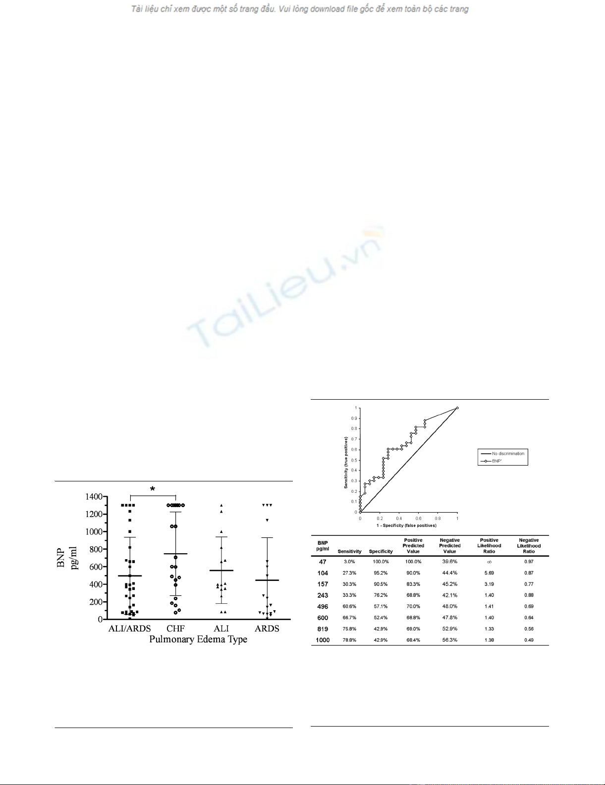
judged unanimously. Baseline BNP levels (median [interquar-
tile range]) were higher in patients with cardiogenic edema
(600 pg/ml [352 to 1,300] versus 369 pg/ml [87 to 709], P =
0.045) (Figure 1). There was no difference in BNP values
between patients with ALI (n = 15) and ARDS (n = 18) (398
pg/ml [344 to 782] versus 202 pg/ml [68 to 657], P = 0.15).
The utility of BNP measurements in distinguishing ALI/ARDS
(disease positive) from cardiogenic edema (disease negative)
was assessed with the ROC curve analysis (Figure 2). The
area under the curve (AUC) is 0.67 (95% confidence interval
0.52 to 0.81). Using a cutoff of BNP < 100 pg/ml (established
in emergency department patients) [5-7] to diagnose ALI/
ARDS, the specificity was 95.2% but the sensitivity was only
27.3%. Given the slightly greater prevalence of ALI/ARDS in
our cohort, there were actually more ALI/ARDS patients with
BNP values above this cutoff (false negatives) than cardio-
genic edema patients (true negatives). At a cutoff of less than
250 pg/ml (suggested by Rana and colleagues [12]), specifi-
city and sensitivity were 76.2% and 33.3%, respectively.
Higher cutoff levels improved sensitivity but at considerable
cost to specificity (Figure 2).
Results of subgroup analyses are summarized in Table 2.
Exclusion of patients with a serum creatinine greater than 3.0
mg/dl slightly increased the difference in mean BNP values
between the cardiogenic and ALI/ARDS groups and the AUC
of the corresponding ROC curve (0.67 to 0.70). Conversely,
separate evaluation excluding the four ALI/ARDS patients with
evidence of LVD and the four patients who did not receive
unanimous adjudication decreased differences in mean BNP
values between ALI/ARDS and cardiogenic edema groups
and had no effect on the AUC of the corresponding ROC
curves.
Correlations of invasive measurements of filling pressures
(RAP and PCWP) with BNP levels are shown in Figure 3. A
significant relationship exists between RAP and BNP, but the
correlation is poor (R2 = 0.11). In addition, no significant rela-
tionship was found between changes in subsequent day BNP
levels and the associated change in RAP or PCWP (Figure 3).
Serial measurements of BNP revealed no significant
Figure 1
Dot-plot of initial B-type natriuretic peptide value classified by edema typeDot-plot of initial B-type natriuretic peptide value classified by edema
type. Bold line and whiskers represent mean and ± 1 standard devia-
tion. *, P = 0.05 for the difference in B-type natriuretic peptide (BNP)
levels between patients with acute lung injury/acute respiratory distress
syndrome (ALI/ARDS) and patients with congestive heart failure. There
is no difference between patients with ALI and patients with ARDS (P
= 0.47).
Figure 2
Receiver operating characteristics of the diagnostic utility of B-type natriuretic peptideReceiver operating characteristics of the diagnostic utility of B-type
natriuretic peptide. True positives are patients with acute lung injury/
acute respiratory distress syndrome, and true negatives are patients
with congestive heart failure. Area under curve = 0.67 (95% confi-
dence interval 0.52 to 0.81). The table provides the corresponding sen-
sitivity, specificity, predictive values and likelihood ratios of
representative B-type natriuretic peptide (BNP) values.

Available online http://ccforum.com/content/12/1/R3
Page 5 of 9
(page number not for citation purposes)
difference in either the direction (number of subjects whose
BNP value increased versus decreased) or the magnitude of
change (mean change in each edema class) in BNP levels
between the ALI/ARDS and cardiogenic groups (Table 3).
Finally, changes in BNP levels did not correlate with net fluid
balance for the previous 24 hours.
Discussion
In this prospective, blinded cohort study, we found that BNP
levels did not reliably distinguish ALI/ARDS from cardiogenic
causes of pulmonary edema despite efforts to exclude patients
with possible overlapping conditions. In addition, BNP levels
correlated poorly with simultaneous invasive measures of RAP
and PCWP. Serial measurements over a 3-day period did not
improve performance because changes in BNP levels did not
correlate with changes in invasive measures of filling pres-
sures and did not differ in direction or magnitude between
patients with ALI/ARDS and those with cardiogenic edema.
Our results are similar to those of other investigators who
found that BNP levels did not discriminate between cardio-
genic and septic shock [9-11] and between high and low
PCWP causes of pulmonary edema [8]. This may be due to
increased levels of BNP related to myocardial dysfunction of
sepsis or direct effect of inflammatory mediators on myocytes
[19,20]. In addition, BNP levels are known to be elevated in
ARDS, in part as a result of acute right heart dysfunction
[21,22]. Right heart dysfunction was a common occurrence in
our cohort (48% and 71% of the ALI/ARDS and CHF cohorts,
respectively). Increased stretch of the right ventricle and right
atrium may be a source of BNP release in critically ill patients,
independently of left ventricular filling pressures. In addition, in
the previous studies of shock, there were significant differ-
ences in PCWP values between cardiac and non-cardiac eti-
ologies; however, the 'low' PCWP values were markedly
abnormal (means of 16 ± 4 and 18 ± 7 mmHg, respectively)
[10,11].
We sought to avoid this confounder by including only ALI/
ARDS patients with a PCWP of less than 16 mmHg and car-
diogenic edema patients with a PCWP of more than 20
mmHg. In our study, mean RAP and PCWP were 5.9 ± 5.7
and 6.8 ± 2.5 mmHg, respectively, in the ALI/ARDS patients,
in contrast with 15.2 ± 5.7 and 21.4 ± 5.5 mmHg in the CHF
Table 2
Mean BNP values and receiver operating characteristic analysis by subgroup
Patients nBNPb (pg/ml) PAUC
ALI/ARDS CHF ALI/ARDS CHF
All 33 21 369 (87–709) 600 (352–1,300) 0.04 0.67 (0.52–0.81)
Serum creatinine < 3.0 mg/dl 32 16 359 (86–665) 653 (419–1,300) 0.02 0.70 (0.55–0.86)
Unanimous jury 31 19 369 (86–665) 653 (419–1,300) 0.05 0.67 (0.52–0.82)
Excluding the four ALI/ARDS with LVDa29 21 394 (87–864) 600 (352–1,300 0.06 0.67 (0.52–0.82)
BNP, B-type natriuretic peptide; ALI, acute lung injury; ARDS, acute respiratory distress syndrome; CHF, congestive heart failure; AUC, area
under curve. P values are for comparisons of BNP values between ALI/ARDS and CHF patients.
aLeft ventricular dysfunction on recent echocardiogram (stable or improved in all four patients); bmedian (interquartile range).
Table 3
Serial BNP measurements by edema classification
Period Direction of BNP change n (ΔBNP, pg/ml) P
ALI/ARDS CHF
Days 1 to 2 Increase 17 (254 ± 302) 5 (228 ± 287)
Decrease 9 (-246 ± 178) 8 (-252 ± 208) 0.17b
Alla26 (73 ± 339) 17 (-52 ± 290) 0.21c
Days 2 to 3 Increase 9 (143 ± 200) 5 (396 ± 132)
Decrease 11 (-191 ± 187) 7 (-160 ± 142) 1.0b
Alla24 (-34 ± 231) 15 (57 ± 281) 0.28c
BNP, B-type natriuretic peptide; ALI, acute lung injury; ARDS, acute respiratory distress syndrome; CHF, congestive heart failure. The table shows
an analysis of changes in BNP levels in direction (increase or decrease) and magnitude (ΔBNP) from day 1 to day 2 and from day 2 to day 3,
classified by edema type. Where errors are shown, results are means ± SD.
aValues for some patients remained above the upper limit of the assay (1,300 pg/ml) and were consider unchanged. bχ2 comparing the proportion
of subjects with an increase in BNP by edema type; ct test of magnitude of BNP change by edema type.


