
Open Access
Available online http://ccforum.com/content/11/5/R112
Page 1 of 10
(page number not for citation purposes)
Vol 11 No 5
Research
Improved survival of children with sepsis and purpura: effects of
age, gender, and era
Martine Maat1, Corinne MP Buysse2, Marieke Emonts1, Lodewijk Spanjaard3, Koen FM Joosten2,
Ronald de Groot4 and Jan A Hazelzet2
1Department of Paediatrics, Division of Infectious Diseases and Immunology, Erasmus MC-Sophia Children's Hospital, University Medical Center, Dr.
Molewaterplein 60, 3015 GJ Rotterdam, The Netherlands
2Department of Paediatrics, Division of Paediatric Intensive Care, Erasmus MC-Sophia Children's Hospital, University Medical Center, Dr.
Molewaterplein 60, 3015 GJ Rotterdam, The Netherlands
3Netherlands Reference Laboratory for Bacterial Meningitis, Department of Medical Microbiology, Academic Medical Center Amsterdam,
Meibergdreef 15, 1100 DD Amsterdam, The Netherlands
4Department of Paediatrics, University Medical Center St. Radboud, Geert Grooteplein 10, 6500 HB Nijmegen, The Netherlands
Corresponding author: Jan A Hazelzet, j.a.hazelzet@erasmusmc.nl
Received: 18 Jun 2007 Revisions requested: 18 Jul 2007 Published: 18 Oct 2007
Critical Care 2007, 11:R112 (doi:10.1186/cc6161)
This article is online at: http://ccforum.com/content/11/5/R112
© 2007 Maat et al; licensee BioMed Central Ltd.
This is an open access article distributed under the terms of the Creative Commons Attribution License (http://creativecommons.org/licenses/by/2.0),
which permits unrestricted use, distribution, and reproduction in any medium, provided the original work is properly cited.
Abstract
Background To gain insight into factors that might affect results
of future case-control studies, we performed an analysis of
children with sepsis and purpura admitted to the paediatric
intensive care unit (PICU) of Erasmus MC-Sophia Children's
Hospital (Rotterdam, The Netherlands).
Methods Between 1988 and 2006, all 287 children
consecutively admitted with sepsis and purpura were included
in various sepsis studies. Data regarding age, gender, ethnicity,
serogroup of Neisseria meningitidis, severity, therapy, and
survival were collected prospectively. These data were pooled
into one database and analyzed retrospectively.
Results The case fatality rate (CFR) from sepsis and purpura
was 15.7%. During the study period, survival improved
significantly. Younger age was significantly associated with
more severe disease and a higher CFR. Children under the
median age of 3.0 years had an increased risk of case fatality
(odds ratio 4.3, 95% confidence interval 2.1 to 9.2; p < 0.001).
Gender was not associated with CFR. However, males did have
higher Paediatric Risk of Mortality scores, fewer PICU-free days,
and more presence of shock. The course of sepsis and purpura
was not related to ethnic origin. A causative organism was
isolated in 84.3% of cases. N. meningitidis was the major
organism (97.5%). Although N. meningitidis serogroup B was
observed more often in younger children, serogroups were not
associated with severity or survival. During the study period, the
use of inotropic agents and corticosteroids changed
substantially (less dopamine and more dobutamine,
norepinephrine, and corticosteroids).
Conclusion Age and gender are determinants of severity of
paediatric sepsis and purpura. Survival rates have improved
during the last two decades.
Introduction
Sepsis and purpura in children is a clinically distinct disease
entity caused by high concentrations of microbes and their
products. Since the introduction of a vaccine against Haemo-
philus influenzae type b, more than 90% of the cases of sep-
sis and purpura in the Western world have been caused by
Neisseria meningitidis [1-3]. The resulting disease entity is
referred to as meningococcal sepsis.
Meningococcal sepsis in children develops when the initial
host response to the infection becomes inappropriately ampli-
fied and dysregulated. Clinically, the onset is often insidious.
After the development of the first petechiae, the patient rapidly
deteriorates and may subsequently develop shock, dissemi-
nated intravascular coagulation (DIC), and ultimately organ
failure. The severity of these symptoms requires immediate
therapy [4,5]. Despite recent advances in therapy, the case
CFR = case fatality rate; CI = confidence interval; CRP = C-reactive protein; DIC = disseminated intravascular coagulation; PDR = predicted death
rate; PICU = paediatric intensive care unit; PRISM = Paediatric Risk of Mortality; rs = Spearman correlation coefficient.
Critical Care Vol 11 No 5 Maat et al.
Page 2 of 10
(page number not for citation purposes)
fatality rate (CFR) remains high and ranges from 4% to 40%
[1,6-8]. The incidence of disease is highest among young chil-
dren (0 to 4 years old) and adolescents [1-3]. In The Nether-
lands, meningococcal sepsis occurs in 4.5 per 100,000
inhabitants (2001). Due to the sudden increase in the inci-
dence of meningococcal disease in 2001, a national vaccina-
tion campaign against serogroup C meningococci (2002) was
implemented among children from 1 to 18 years of age [9,10].
In recent years, many studies have focused on the elucidation
of the pathogenesis of sepsis. However, much about the epi-
demiology of sepsis in children is still unknown. In this paper,
we seek to describe the epidemiology of sepsis and purpura
in children referred to the paediatric intensive care unit (PICU)
of Erasmus MC-Sophia Children's Hospital in Rotterdam, The
Netherlands. The aim of this study was to analyze the variation
in severity and survival of children with respect to age, gender,
ethnicity, and serogroup of N. meningitidis.
Materials and methods
The study was conducted in accordance with the Declaration
of Helsinki. Permission for the study was obtained from the
medical ethics committee of Erasmus MC.
Participants
All children admitted with sepsis and purpura (and/or
petechiae) to the PICU of the Erasmus MC-Sophia Children's
Hospital since 1988 were included. A vast majority of the chil-
dren were previously included in Rotterdam-based sepsis
studies [11-16]. Data regarding the remaining children with
sepsis and purpura were derived from PICU admission
records. Informed consent was obtained from parents or legal
guardians of all children who were included in this study. Chil-
dren were considered to have sepsis when they presented
with tachycardia, tachypnea, and a body temperature of less
than 36°C or greater than 38.5°C (rectal) [17]. Prospective
data on all children were collected at various time points in the
course of the disease. Both laboratory parameters and dis-
ease severity scoring systems, like Paediatric Risk of Mortality
(PRISM) score and predicted death rate (PDR) based on the
Rotterdam score, were selected as markers of severity of dis-
ease [18-20]. Additionally, presence of DIC and presence of
shock were recorded as markers of severity [17,19,21]. The
number of PICU-free days was determined on day 28 after
admission using the date of admission and the date of dis-
charge. A non-survivor had 0 PICU-free days. All laboratory
parameters, obtained at baseline from an arterial blood sam-
ple, were collected within 4 hours after admission to the PICU.
Ethnicity was determined by checking patient information, and
if it was not specified, first and last names were checked and
ethnicity was determined by means of the combined name
method [22]. Ethnicity was categorized into Dutch Caucasian,
Turkish, Moroccan, Hindustani, African descent, and other.
Serogrouping of N. meningitidis isolates was performed at the
Netherlands Reference Laboratory for Bacterial Meningitis
Amsterdam using immunodiffusion with polyclonal antisera
[23].
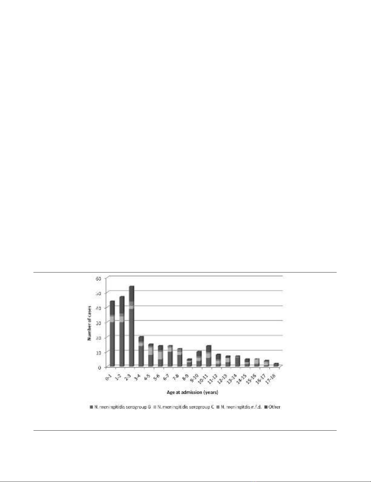
Figure 1
Distribution of age at admission in children with sepsis and purpuraDistribution of age at admission in children with sepsis and purpura. The children are subdivided according to causative organism. N. meningitidis,
Neisseria meningitidis. Not further defined (n.f.d.).
Available online http://ccforum.com/content/11/5/R112
Page 3 of 10
(page number not for citation purposes)
Statistical analyses
Retrospectively, severity and survival of children with sepsis
and purpura with respect to age, gender, causative organism,
and ethnicity were analyzed by means of SPSS 11.01 (SPSS
Inc., Chicago, IL, USA) Clinical and laboratory parameters
were included in the analysis only if they were determined in at
least 90% of all children.
Mann-Whitney U test, Student t test, chi-square test, and
Spearman correlation (rs) were used when appropriate. When
necessary, variables were log-transformed to obtain an
approximately normal distribution. For these variables, geo-
metric mean values and their 95% confidence intervals (CIs)
are depicted in the text and tables. P values of less than or
equal to 0.05 were considered statistically significant.
Results
Between August 1988 and June 2006, 287 children with sep-
sis and purpura were admitted to the PICU of the Erasmus
MC-Sophia Children's Hospital. The overall CFR was 15.7%
(45 children died). The median age at admission was 3.0 years
(range 0.1 to 17.9 years) (Figure 1). Of the 287 children, 155
(54%) were male and 132 (46%) were female. The male-to-
female ratio was 1.2. The majority of the children were Dutch
Caucasians (73.8%). Laboratory parameters present at base-
line in more than 90% of the children were base excess, lac-
tate, C-reactive protein (CRP), fibrinogen, platelet count,
leukocytes, and glucose.
Survival
Severity of illness was significantly less in survivors when com-
pared with non-survivors, both in disease severity scoring sys-
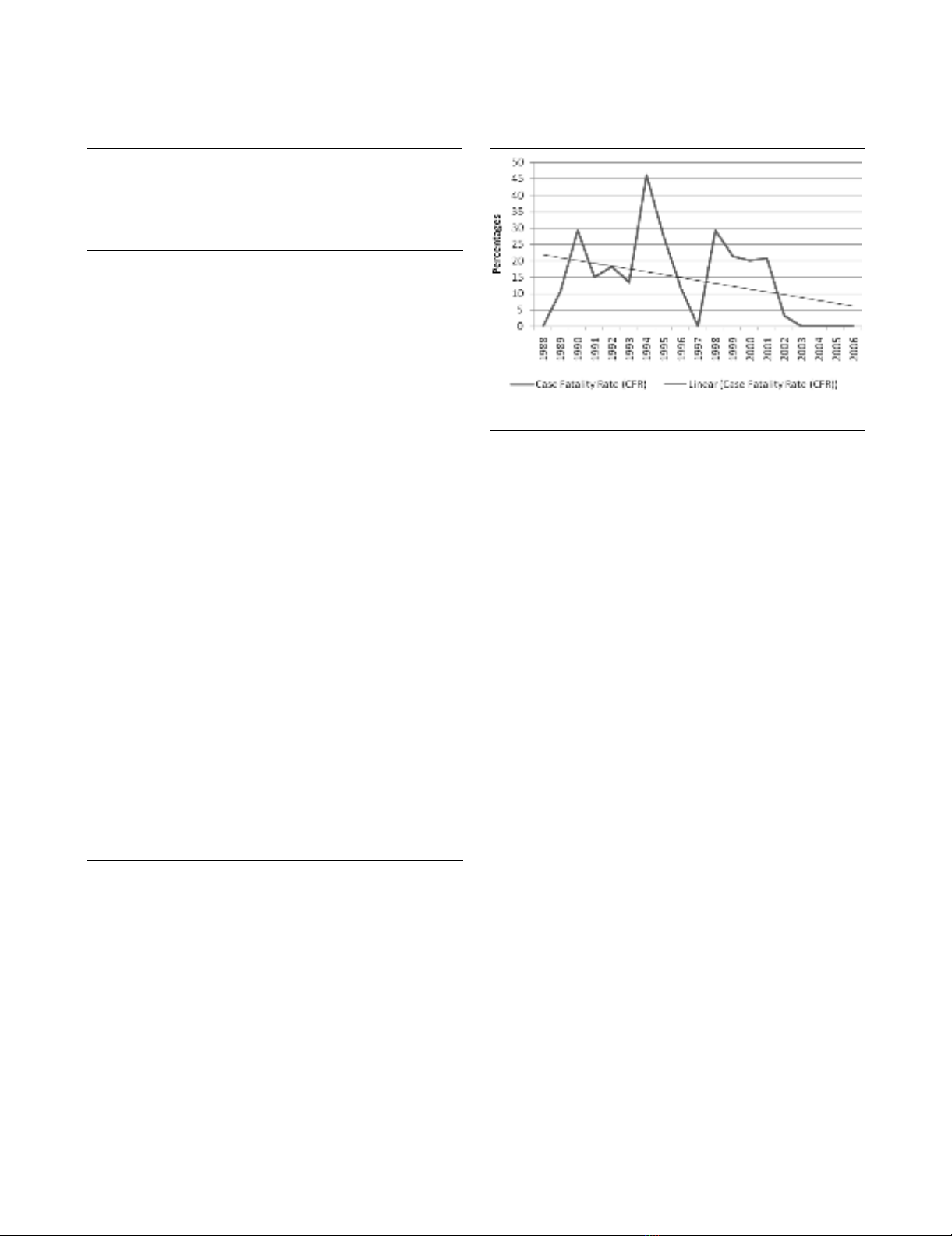
tems and laboratory parameters (Table 1). Survival was
significantly correlated with year of admission (p ≤ 0.05, rs
0.128), indicating that survival has improved significantly dur-
ing the study period (Figure 2). Gender did not differ between
survivors and non-survivors (p = 0.15). The vast majority of
fatal cases died of refractory septic shock (75.6%).
Age
Age was significantly correlated with PRISM score (p < 0.001,
rs -0.317), PDR (p < 0.001, rs -0.321), presence of DIC (p <
0.001, rs -0.245), base excess (p < 0.001, rs 0.313), CRP (p
< 0.05, rs 0.161), fibrinogen (p < 0.001, rs 0.301), leukocyte
count (p < 0.001, rs 0.284), thrombocyte count (p < 0.01, rs
0.184), and glucose levels (p < 0.001, rs 0.296). This indi-
cates that younger children had higher PRISM scores, higher
PDR, more presence of DIC, lower base excess, lower CRP,
Table 1
Comparison of disease characteristics between non-survivors
and survivors
SurvivorsaNon-survivorsa
Total number of children (%) 242 45
(84.3) (15.7)
Male-to-female ratio 1.1 1.7
Number of children with DIC (%) 174b32b
(75) (97)
Neisseria meningitidis serogroup
B (%) 147 (74.2) 28 (73.7)
C (%) 37 (18.7) 7 (18.4)
PRISM score 14c23c
(1 to 37) (8 to 44)
Predicted death rate (%)d3.1c87.4c
(0 to 100) (1.1 to 100.0)
Base excess (mmol/L) -7c-13c
(-23 to 4.4) (-28 to 0.6)
Lactate (mmol/L) 3.7c6.6c
Geometric mean, 95% CI 3.4 to 4.3 5.8 to 7.4
C-reactive protein (mg/L) 106c53c
(10 to 334) (6 to 226)
Fibrinogen (g/L) 2.8c0.9c
(0.3 to 6.8) (0.2 to 5.4)
Platelet count (×103/μL) 126c47c
(15 to 475) (13 to 202)
Leukocytes (×103/μL) 10.6c4.7c
Geometric mean, 95% CI 9.5 to 11.9 3.7 to 6.0
Glucose (mmol/L) 6.3c4.3c
Geometric mean, 95% CI 5.9 to 6.8 3.6 to 5.3
aResults represent median (min-max) unless stated otherwise. bp <
0.01.cp < 0.001. dPredicted death rate was based on the Rotterdam
score. CI, confidence interval; DIC, disseminated intravascular
coagulation; PRISM, Paediatric Risk of Mortality.
Figure 2
Case fatality rate (CFR) and CFR trend line during the study periodCase fatality rate (CFR) and CFR trend line during the study period.
Critical Care Vol 11 No 5 Maat et al.
Page 4 of 10
(page number not for citation purposes)
lower fibrinogen, lower leukocyte count, lower thrombocyte
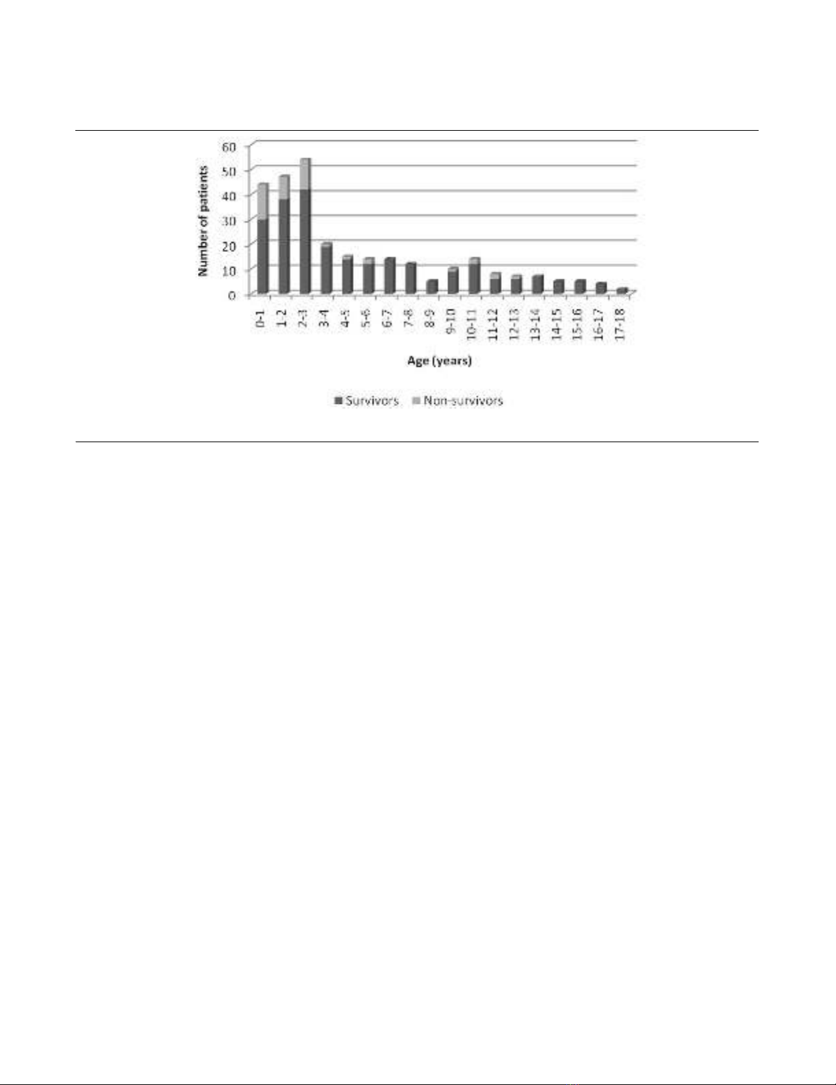
count, and lower glucose levels on admission. The median age
of children was 3.0 years (range 0.1 to 17.9 years). Children
3.0 years old or younger had a higher CFR (odds ratio 4.3,
95% CI 2.1 to 9.2; p < 0.001) (Figure 3).
Gender
The median age did not differ significantly between males (2.8
years) and females (3.5 years) (p = 0.16). Male patients had
significantly fewer PICU-free days (p = 0.04) and higher
PRISM scores (p = 0.02) than females. Shock was slightly
more common in males than in females (89% versus 80%; p
= 0.04). CFR and other markers of severity of disease did not
differ between males and females. Because males had higher
PRISM scores but no increased CFR, we analyzed the differ-
ent variables determining the PRISM score. Of these variables,
only a trend for lower glucose levels in males compared with
females was observed (p = 0.06).
Ethnicity
The majority of the children were Dutch Caucasians (n = 211,
73.5%). Of the remaining 76 children, 12 were Turkish (4.2%),
16 were Moroccan (5.6%), 3 were Hindustani (1.1%), 7 were
of African descent (2.5%), 7 were designated other (2.5%),
and in 31 children ethnicity could not be determined (10.8%).
No differences with respect to severity of disease or case
fatality were found between the different ethnic groups.
Causative organism
A causative organism could be determined in 242 children
(84.3%), with N. meningitidis being the major causative
organism (n = 236, 97.5%) (Figure 4). Of these 236, 175
(74.2%) were N. meningitidis serogroup B, 44 (18.6%) were
serogroup C, and in 17 (7.2%) the serogroup was not deter-
mined (Table 2). Streptococcus pneumoniae was the
causative organism in 3 children, Staphylococcus aureus in 1,
and H. influenzae in 2. Of the remaining 45 children, 43 had
clinical features of meningococcal sepsis [3].
For logistic reasons, the causative organism could not be
determined in 2 children. No differences with respect to sur-
vival, disease severity scoring systems, and presence of shock
were observed between N. meningitidis serogroups B and C.
However, the median age of children with sepsis and purpura
due to serogroup B was lower than that of the serogroup C-
infected children (2.8 and 6.0 years, respectively; p < 0.001)
(Table 3). The distribution of serogroup, serotype, and sero-
subtype of N. meningitidis in the positive cultures is depicted
in Table 2.
Meningococcal C vaccination campaign and therapy
In 2001 and 2002, a sudden increase was noted in the inci-
dence of meningococcal infection in The Netherlands. This
was caused mainly by serogroup C N. meningitidis. The imple-
mentation of the meningococcal C vaccination campaign in
July 2002 resulted in a sharp decline in the number of cases
caused by serogroup C (Figure 4). Since 2003, there has not
been a case of sepsis and purpura due to N. meningitidis
serogroup C in our hospital. Parallel to this, the incidence of
serogroup B has declined and is returning to the incidence
level of before 1989. Before the national meningococcal C
vaccination, 248 children in our study population were admit-
ted with sepsis and purpura; since the vaccination campaign,
39 children have been admitted.
Figure 3
Distribution of age at admission among survivors and non-survivors of sepsis and purpuraDistribution of age at admission among survivors and non-survivors of sepsis and purpura.

Available online http://ccforum.com/content/11/5/R112
Page 5 of 10
(page number not for citation purposes)
Remarkably, since the implementation of meningococcal C
vaccination, no deaths have occurred in children with sepsis
and purpura admitted to our PICU. The median age of the chil-
dren did not differ significantly before and after vaccination
(3.2 and 2.5 years, respectively; p = 0.23) (Table 4). Glucose
levels were significantly lower in the patient group before the
vaccination campaign compared with the patient group after
(p < 0.05). Children admitted before the vaccination campaign
had significantly fewer PICU-free days and more presence of
DIC (both p < 0.05). The PRISM score was not significantly
different between patient groups before and after the menin-
gococcal C vaccination campaign. In addition, since 2002,
treatment of children with meningococcal sepsis at our PICU
has changed due to the implementation of international guide-
lines [8]. After the vaccination campaign, more children were
treated with corticosteroids (18 [9.3%] before versus 15
[42.9%] after; p < 0.001) and more children were mechani-
cally ventilated (128 [51.8%] before versus 28 [71.8%] after;
p < 0.05) (Table 3). In addition, year of admission was signifi-
cantly correlated with the use of dobutamine (p < 0.001, rs
0.262), dopamine (p < 0.001, rs -0.218), norepinephrine (p <
0.001, rs 0.329), and corticosteroids (p < 0.001, rs 0.245) but
not with the use of epinephrine. This indicates that during the
study period the use of dobutamine, norepinephrine, and cor-
Table 2
Incidence of serogroup, serotype, and serosubtype of Neisseria meningitidis
Serogroup Serotype Serosubtype Number Percentage
B1P1.441.8
P1.16 4 1.8
NT 3 1.4
Other 1 0.5
2A 3 1.4
4P1.457 26
P1.6 3 1.4
P1.7 3 1.4
P1.9 4 1.8
P1.10 4 1.8
P1.15 5 2.3
NT 28 12.8
Other 13 5.9
NT P1.1 6 2.7
P1.4 8 3.7
NT 8 3.7
Other 4 1.8
Other 17 7.8
C2AP1.2125.5
P1.5 9 4.1
P1.7 1 0.5
NT 7 3.2
2B P1.1 1 0.5
P1.2 7 3.2
4P1.43 1.4
NT 2 0.9
Other 2 0.9
NT, non-typable.


![Vaccine và ứng dụng: Bài tiểu luận [chuẩn SEO]](https://cdn.tailieu.vn/images/document/thumbnail/2016/20160519/3008140018/135x160/652005293.jpg)


%20--%3e%3cdefs%3e%3cstyle%3e%20.st0%20{%20fill:%20%23fff;%20}%20.st1%20{%20fill:%20%237800fa;%20}%20%3c/style%3e%3c/defs%3e%3cpath%20class='st1'%20d='M117.78,12.18H43.11c2.9,3.47,4.65,7.94,4.65,12.82,0,5.6-2.3,10.66-6.01,14.29h76.02l7.22-13.56-7.22-13.56Z'/%3e%3cg%3e%3cpath%20class='st0'%20d='M53.58,26.17h-.59v-1.46h.59v-4.96h2.83c1.78,0,2.67.94,2.67,2.82v5.76c0,1.87-.89,2.81-2.67,2.81h-2.83v-4.96ZM55.36,21.37v3.34h1.1v1.46h-1.1v3.34h1.01c.61,0,.91-.37.91-1.1v-5.93c0-.74-.3-1.1-.91-1.1h-1.01Z'/%3e%3cpath%20class='st0'%20d='M65.99,31.14h-1.8l-.31-2.07h-2.19l-.31,2.07h-1.64l1.82-11.39h2.62l1.82,11.39ZM65.28,18.04c-.25.46-.51.77-.75.94-.21.15-.47.22-.79.22-.26,0-.57-.07-.92-.22l-.38-.15c-.14-.05-.26-.07-.37-.07-.3,0-.53.18-.71.54l-.91-.68c.25-.46.51-.77.75-.94.21-.14.48-.21.79-.21.26,0,.57.07.92.21l.38.15c.14.05.26.07.37.07.3,0,.53-.18.71-.54l.91.68ZM61.91,27.52h1.73l-.87-5.76-.87,5.76Z'/%3e%3cpath%20class='st0'%20d='M74.53,26.89v1.52c0,1.91-.89,2.86-2.67,2.86s-2.67-.95-2.67-2.86v-5.93c0-1.91.89-2.86,2.67-2.86s2.67.95,2.67,2.86v1.11h-1.69v-1.22c0-.75-.31-1.12-.93-1.12s-.93.37-.93,1.12v6.15c0,.74.31,1.11.93,1.11s.93-.37.93-1.11v-1.63h1.69Z'/%3e%3cpath%20class='st0'%20d='M81.4,31.14h-1.8l-.31-2.07h-2.19l-.31,2.07h-1.64l1.82-11.39h2.62l1.82,11.39ZM75.9,19.2l1.52-1.91h1.71l1.51,1.91h-1.61l-.76-.95-.75.95h-1.61ZM77.32,27.52h1.73l-.87-5.76-.87,5.76ZM83.1,15.99l-1.76,1.91h-1.26l1.17-1.91h1.86Z'/%3e%3cpath%20class='st0'%20d='M84.86,19.75c1.78,0,2.67.94,2.67,2.82v1.48c0,1.87-.89,2.81-2.67,2.81h-.85v4.28h-1.79v-11.39h2.64ZM84.01,21.37v3.86h.85c.58,0,.87-.36.87-1.08v-1.71c0-.71-.29-1.07-.87-1.07h-.85Z'/%3e%3cpath%20class='st0'%20d='M93.51,19.75c1.78,0,2.67.94,2.67,2.82v1.48c0,1.87-.89,2.81-2.67,2.81h-.85v4.28h-1.79v-11.39h2.64ZM92.66,21.37v3.86h.85c.58,0,.87-.36.87-1.08v-1.71c0-.71-.29-1.07-.87-1.07h-.85Z'/%3e%3cpath%20class='st0'%20d='M98.8,31.14h-1.79v-11.39h1.79v4.88h2.03v-4.88h1.83v11.39h-1.83v-4.88h-2.03v4.88Z'/%3e%3cpath%20class='st0'%20d='M105.36,24.55h2.46v1.62h-2.46v3.34h3.09v1.63h-4.88v-11.39h4.88v1.63h-3.09v3.18ZM108.17,17.29l-1.76,1.91h-1.26l1.17-1.91h1.86Z'/%3e%3cpath%20class='st0'%20d='M112.2,19.75c1.78,0,2.67.94,2.67,2.82v1.48c0,1.87-.89,2.81-2.67,2.81h-.85v4.28h-1.79v-11.39h2.64ZM111.35,21.37v3.86h.85c.58,0,.87-.36.87-1.08v-1.71c0-.71-.29-1.07-.87-1.07h-.85Z'/%3e%3c/g%3e%3ccircle%20class='st1'%20cx='25'%20cy='25'%20r='20'/%3e%3cpath%20class='st0'%20d='M32.78,19.27c2.92,0,4.43,2.55,5.28,5.33l.71,2.17c.14.38-.33.75-.71.75h-5.61c.19-.33.24-.71.09-1.08l-.75-2.45c-.43-1.32-.99-2.64-1.79-3.77.75-.57,1.65-.94,2.78-.94h0ZM25,18.38c3.25,0,4.9,2.78,5.89,5.89l.76,2.45c.14.42-.33.8-.8.8h-11.69c-.42,0-.94-.38-.8-.8l.75-2.45c.99-3.11,2.64-5.89,5.89-5.89h0ZM25,11.35c1.74,0,3.11,1.37,3.11,3.11s-1.37,3.11-3.11,3.11-3.11-1.41-3.11-3.11,1.41-3.11,3.11-3.11h0ZM17.27,19.27c1.08,0,1.98.38,2.73.94-.8,1.13-1.37,2.45-1.74,3.77l-.8,2.45c-.14.38-.05.75.09,1.08h-5.56c-.42,0-.9-.38-.75-.75l.71-2.17c.9-2.78,2.41-5.33,5.33-5.33h0ZM17.27,12.91c1.51,0,2.78,1.27,2.78,2.83s-1.27,2.83-2.78,2.83-2.83-1.27-2.83-2.83,1.27-2.83,2.83-2.83h0ZM32.78,12.91c1.56,0,2.78,1.27,2.78,2.83s-1.23,2.83-2.78,2.83-2.83-1.27-2.83-2.83,1.27-2.83,2.83-2.83h0ZM27.07,28.56v.09c0,.57-.24,1.08-.61,1.46h0v.05c-.38.33-.9.57-1.46.57s-1.08-.24-1.46-.61h0c-.38-.38-.61-.9-.61-1.46v-.09h1.41v.09c0,.19.05.38.19.47v.05c.09.09.28.19.47.19s.38-.09.47-.19v-.05c.14-.09.24-.28.24-.47t-.05-.09h1.41ZM30.99,28.56v.09c0,1.65-.66,3.16-1.74,4.24-1.08,1.08-2.59,1.79-4.24,1.79s-3.16-.71-4.24-1.79l-.05-.05c-1.04-1.08-1.7-2.55-1.7-4.2v-.09h1.41v.09c0,1.27.47,2.4,1.27,3.25h.05c.85.85,1.98,1.37,3.25,1.37s2.4-.52,3.25-1.37c.85-.8,1.37-1.98,1.37-3.25v-.09h1.37ZM34.99,28.56v.09c0,2.78-1.13,5.28-2.92,7.07-1.79,1.79-4.29,2.92-7.07,2.92s-5.23-1.13-7.07-2.92c-1.79-1.79-2.92-4.29-2.92-7.07v-.09h1.41v.09c0,2.4.94,4.53,2.5,6.08,1.56,1.56,3.72,2.5,6.08,2.5s4.52-.94,6.08-2.5c1.56-1.56,2.5-3.68,2.5-6.08v-.09h1.41Z'/%3e%3c/svg%3e)