Roles of conserved arginines in ATP-binding domains of AAA+ chaperone ClpB from Thermus thermophilus Takashi Yamasaki1, Yosuke Nakazaki1, Masasuke Yoshida2 and Yo-hei Watanabe1
1 Department of Biology, Faculty of Science and Engineering, Konan University, Okamoto, Kobe, Japan 2 Department of Molecular Biosciences, Kyoto Sangyo University, Motoyama-Kamigamo, Japan
Keywords AAA+; aggregation; arginine finger; chaperone; ClpB
Correspondence Y-h. Watanabe, Department of Biology, Faculty of Science and Engineering, Konan University, Okamoto 8-9-1, Kobe 658-8501, Japan Fax ⁄ Tel: +81 78 435 2514 E-mail: ywatanab@center.konan-u.ac.jp
(Received 7 March 2011, revised 30 April 2011, accepted 6 May 2011)
doi:10.1111/j.1742-4658.2011.08167.x
ClpB, a member of the expanded superfamily of ATPases associated with diverse cellular activities (AAA+), forms a ring-shaped hexamer and coop- erates with the DnaK chaperone system to reactivate aggregated proteins in an ATP-dependent manner. The ClpB protomer consists of an N-termi- nal domain, an AAA+ module (AAA-1), a middle domain, and a second AAA+ module (AAA-2). Each AAA+ module contains highly conserved WalkerA and WalkerB motifs, and two arginines (AAA-1) or one arginine (AAA-2). Here, we investigated the roles of these arginines (Arg322, Arg323, and Arg747) of ClpB from Thermus thermophilus in the ATPase cycle and chaperone function by alanine substitution. These mutations did not affect nucleotide binding, but did inhibit the hydrolysis of the bound ATP and slow the threading of the denatured protein through the central pore of the T. thermophilus ClpB ring, which severely impaired the chaper- one functions. Previously, it was demonstrated that ATP binding to the AAA-1 module induced motion of the middle domain and stabilized the ClpB hexamer. However, the arginine mutations of the AAA-1 module destabilized the ClpB hexamer, even though ATP-induced motion of the middle domain was not affected. These results indicated that the three argi- nines are crucial for ATP hydrolysis and chaperone activity, but not for ATP binding. In addition, the two arginines in AAA-1 and the ATP- induced motion of the middle domain independently contribute to the sta- bilization of the hexamer.
Structured digital abstract l TClpB binds to TClpB by molecular sieving (View Interaction 1, 2)
Introduction
The expanded superfamily of ATPases associated with diverse cellular activities (AAA+) are involved in a including membrane variety of
cellular activities,
fusion, DNA replication, protein degradation, and dis- aggregation. Members of the AAA+ family contain one or more highly conserved AAA+ modules, and
Abbreviations AAA, ATPase associated with diverse cellular activities; AAA+, expanded superfamily of ATPases associated with diverse cellular activities; ABD-F, 7-fluorobenz-2-oxa-1,3-diazole-4-sulfonamide; AMP-PNP, adenosine 5¢-(b,c-imido)triphosphate; ATPcS, adenosine 5¢-O-(thiotriphosphate); FITC, fluorescein isothiocyanate; G6PDH, glucose-6-phosphate dehydrogenase; Mant-ADP, 2¢(3¢)-O-N¢-methylaniloyl- aminoadenosine-5¢-diphosphate; P1, position 1; P2, position 2; T BAP, Thermus thermophilus BAP; TCEP, tris-(2-carboxyethyl)phosphine hydrochloride; TClpA, Thermus thermophilus ClpA; TClpB, Thermus thermophilus ClpB; TClpP, Thermus thermophilus ClpP; T DnaJ, Thermus thermophilus DnaJ; TDnaK, Thermus thermophilus DnaK; TGrpE, Thermus thermophilus GrpE.
FEBS Journal 278 (2011) 2395–2403 ª 2011 The Authors Journal compilation ª 2011 FEBS
2395
T. Yamasaki et al.
Roles of conserved arginine of ClpB chaperone
domain
contains
AAA+ modules (AAA-1 and AAA-2 from the N-ter- minus), and a middle domain (Fig. 1A) [18]. The N-terminal domain is a highly mobile globular a-heli- cal domain. The middle domain is an 85-A˚ coiled-coil structure tethered to the a-helical domain of the AAA-1 module, and is only found in ClpB ⁄ Hsp104. Like other AAA+ proteins, ClpB ⁄ Hsp104 forms a ring-shaped hexamer, and its stability is influenced by salt and pro- tein concentrations, temperature, and bound nucleotide [4,19–24].
shows
most of them form ring-shaped oligomers [1,2]. The AAA+ module consists of a RecA-like nucleotide- binding domain and an a-helical domain. The RecA- like the WalkerA motif the (GXXGXGKT, where X is any amino acid), WalkerB motif (hhhhDE, where h is a hydrophobic residue), and conserved arginines called the arginine fingers. The roles of arginine fingers have been explored in several AAA+ proteins [3–8]. The arginine finger is typically located in the subunit interface, and interacts with the c-phosphate of ATP bound to the adjacent subunit; it is thought to participate in cataly- sis by stabilizing the transition state during ATP hydrolysis. Some AAA+ modules have two potential arginine fingers at position 2 (P2) and position 1 (P1) from the N-terminus [3]. Whereas the P1 arginine is highly conserved in most AAA+ proteins, the P2 argi- nine is conserved only in members of subfamilies of the AAA+ family, ATPases associated with diverse cellular activities (AAA) family, and several of the Clp ⁄ Hsp100 family members. However, little is known about the differences between the roles of P1 and P2 arginines.
The molecular chaperone ClpB ⁄ Hsp104 is a member of the Clp ⁄ Hsp100 subfamily of the AAA+ family, and is essential for the survival of bacteria and yeast during severe thermal stress [9,10]. ClpB ⁄ Hsp104 coop- erates with the DnaK ⁄ Hsp70 chaperone system in the solubilization and reactivation of aggregated proteins by utilizing ATP hydrolysis [11–17]. The ClpB ⁄ Hsp104 two monomer consists of an N-terminal domain,
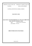
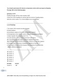
The AAA-1 and AAA-2 of ClpB ⁄ Hsp104 contain two conserved arginines (P1 and P2) and one con- served arginine (P1), respectively (Fig. 1B,C). The crys- structure of ClpB from Thermus thermophilus tal (TClpB) that Arg322 (P2 of AAA-1) and Arg747 (P1 of AAA-2) are directed towards the c-phosphate of adenosine 5¢-(b,c-imido)triphosphate (AMP-PNP) that is bound to the neighboring subunit, whereas Arg323 (P1 of AAA-1) faces away from it (Fig. 1B) [18]. Previously, investigation of the effects of alanine substitutions in Escherichia coli ClpB showed that the alanine mutation of the P1 arginine of AAA- 1, R332A, decreased the chaperone activity and desta- bilized the hexamer, and that the alanine mutation of the P1 arginine of AAA-2, R756A, decreased the AT- Pase and chaperone activities [4]. Recently, the effects of alanine substitution of the P2 arginine of AAA-1, Arg322, of TClpB on nucleotide binding and ATPase were also investigated [25]. However, the exact roles of these arginines in the individual AAA+ modules are unclear.
Fig. 1. Positions of the conserved arginines of TClpB. (A) Structure of TClpB. The N-terminal domain (green), the first AAA+ module (AAA-1) (blue), the second AAA+ module (AAA-2) (red) and the middle domain (yellow) are shown. (B) Close-up views of the subunit interface of AAA-1. AMP-PNP is bound to the left subunit (blue), and conserved arginines, Arg322 (P2) (yellow) and Arg323 (P1) (magenta), of the right subunit (cyan) are shown as sticks. (C) The subunit interface of AAA-2. AMP-PNP is bound to the left subunit (red), and conserved arginines, Arg747 (green), of the right subunit (pink) are shown as sticks.
FEBS Journal 278 (2011) 2395–2403 ª 2011 The Authors Journal compilation ª 2011 FEBS
2396
T. Yamasaki et al.
Roles of conserved arginine of ClpB chaperone
Disaggregation of proteins by ClpB requires stable hexamer formation and threading of aggregated sub- strate proteins through the central pore of ClpB [26]. The ATP-induced motion of the middle domain stabi- lizes the ClpB hexamer, and is important for the disag- gregation process [18,27]. However, the relationships between these processes and the conserved arginines have not been clarified. Here, we used TClpB to exam- ine the roles of the conserved arginines, Arg322 (P2) and Arg323 (P1) of AAA-1, and Arg747 (P1) of AAA-2, and determined that the arginines of TClpB are not involved in nucleotide binding but are crucial for ATP hydrolysis, substrate threading, and disaggregation. In addition, the arginines of AAA-1 are important for stabilization of the hexameric form, but not for the ATP-induced motion of the middle domain.
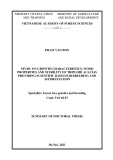
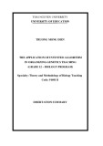
Fig. 2. Mant-ADP binding to the TClpB mutants. The increases in fluorescence after mixing the indicated concentrations of Mant- ADP with wild-type (open circles), 1KT ⁄ AA (open squares), 2KT ⁄ AA (open triangles), 1R ⁄ A(P1)–2KT ⁄ AA (filled circles), 1R ⁄ A(P2)– 2KT ⁄ AA (filled squares) and 1KT ⁄ AA-2R ⁄ A (filled triangles) TClpB the Mant-ADP concentrations. Theoretical were plotted against curves are also shown.
Results
The conserved arginines are not involved in nucleotide binding
Table 1. Dissociation constants of nucleotides for TClpB mutants. Standard deviations are shown.
TClpB
Kd for Mant-ADP (lM)
Kd for ADP (lM)
Kd for ATP (lM)
reported that
0.71 ± 0.13
8.08 ± 0.17 4.36 ± 0.07 9.44 ± 0.55 13.5 ± 5.1 41.2 ± 24.5 8.34 ± 0.37
31.1 ± 1.7a 30.7 ± 0.4a 56.4 ± 15.9a 133 ± 73 96.4 ± 26.6 81.9 ± 29.6
1.00 ± 0.09 Wild type 0.30 ± 0.15 1KT ⁄ AA 2KT ⁄ AA 11.0 ± 1.1 1R ⁄ A(P1)–2KT ⁄ AA 14.5 ± 0.4 1R ⁄ A(P2)–2KT ⁄ AA 25.8 ± 4.9 1KT ⁄ AA–2R ⁄ A
a The values may only represent a lower limit, because of the intrin- sic ATPase activity of TClpB.
were similar to those for the corresponding single Wal- kerA mutants. These results indicated that the three arginines are not involved in nucleotide binding to the corresponding AAA+ module.
The conserved arginines are indispensable for ATP hydrolysis in the corresponding AAA+ module
(Table 1).
Increased
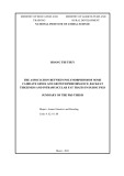
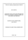
We next measured the ATPase activities of the argi- nine mutants of TClpB with or without 0.1 mgÆmL)1 j-casein. At 55 (cid:2)C, wild-type TClpB hydrolyzed ATP at a rate of approximately 60 min)1, and the addition of j-casein stimulated the rate approximately 1.7-fold (Fig. 3A). The ATPase activities of 1R ⁄ A(P1) and 1R ⁄ A(P2) were approximately six-fold lower, and that of 2R ⁄ A was approximately 40-fold lower, than that of the wild type (Fig. 3A). However, for all mutants, activity j-casein
stimulated ATPase
significantly
To examine the roles of conserved arginines, we gener- ated three mutants of TClpB: R322A [1R ⁄ A(P2)], R323A [1R ⁄ A(P1)], and R747A (2R ⁄ A). In addition, we made double mutants by combining these arginine mutations in AAA-1 or AAA-2 and the WalkerA mutations, replacement of Lys-Thr with Ala-Ala, in AAA-2 (2KT ⁄ AA) or AAA-1 (1KT ⁄ AA), respectively. the 1KT ⁄ AA and Previously, we 2KT ⁄ AA mutants could not bind nucleotide to the AAA-1 and AAA-2 modules, respectively [23]. Nucleo- tide binding to the domain with the arginine mutation was estimated by measuring nucleotide binding to the double mutants 1R ⁄ A(P1)–2KT ⁄ AA, 1R ⁄ A(P2)– 2KT ⁄ AA, and 1KT ⁄ AA–2R ⁄ A. The fluorescence inten- sity increased when 2¢(3¢)-O-N¢-methylaniloyl-aminoad- (Mant-ADP) bound TClpB. enosine-5¢-diphosphate The extent of the changes in fluorescence intensity at 440 nm that was induced by wild-type and mutant TClpB were plotted against the concentrations of Mant-ADP (Fig. 2), and the apparent Kd values were fluorescence was calculated observed in all double mutants, and the Kd values were 14.5 lm [1R ⁄ A(P1)–2KT ⁄ AA], 25.8 lm [1R ⁄ A(P2)– 2KT ⁄ AA], and 0.71 lm [1KT ⁄ AA–2R ⁄ A]. These Kd values were similar to those of corresponding single WalkerA mutants: 2KT ⁄ AA (11.0 lm) and 1KT ⁄ AA (0.30 lm) (Table 1). We also measured the decreases in Mant-ADP fluorescence by adding Mg-ADP or Mg- ATP, and calculated the apparent Kd values of ADP and ATP for the TClpB mutants (Table 1). The Kd values of ADP and ATP for these double mutants
FEBS Journal 278 (2011) 2395–2403 ª 2011 The Authors Journal compilation ª 2011 FEBS
2397
T. Yamasaki et al.
Roles of conserved arginine of ClpB chaperone
that
sequence
and tested
threading
their
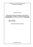
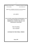
binds T. thermophilus ClpP acid (TClpP), YNVGPAIGFTSKEVDTESPLKA, instead of the Leu714–Val735 sequence. ClpP is a barrel- shaped protease that degrades the substrate proteins that are translocated by bound ClpA. By the use of TBAP, the threading activity of TClpB could be esti- mated by the degradation of a-casein in the presence of TClpP [28]. We combined TBAP with the arginine activities. mutations Degradation of fluorescein isothiocyanate (FITC)- labeled a-casein was monitored by increased fluores- cence intensities, and the initial rates of degradation were calculated (Fig. 4A,B). At 55 (cid:2)C, TBAP and TClpP degraded casein at a rate of 0.14 s)1. All three combined mutants could degrade casein in cooperation with TClpP, but the rates were low: 0.07 s)1 [TBAP– 1R ⁄ A(P1)], 0.04 s)1 [TBAP–1R ⁄ A(P2)], and 0.05 s)1 (TBAP–2R ⁄ A).
The chaperone activities of the arginine mutants
Fig. 3. ATPase activities of the TClpB mutants. ATPase activities of wild-type and mutant TClpB with 3 mM ATP were measured at 55 (cid:2)C in the absence (open bars) or presence (filled bars) of 0.1 mgÆmL)1 j-casein. ATPase activities are expressed as turn- over ⁄ monomer TClpB. (A) ATPase activities of the wild-type and the single arginine mutants of TClpB. The measurements were per- formed at 0.3 lM TClpB monomer. (B) ATPase activities of the Wal- kerA mutants and the combined WalkerA and arginine mutants. The measurements were performed at 1.5 lM TClpB monomer. The error bars represent the standard deviation.
By using glucose-6-phosphate dehydrogenase (G6PDH) and a-glucosidase as substrate proteins, we tested the chaperone activities of the arginine mutants of TClpB. G6PDH and a-glucosidase were aggregated by incuba- tion at 72 (cid:2)C for 8 min and 73 (cid:2)C for 10 min, respec- in the presence of 3 mm ATP and 1 mm tively, dithiothreitol. Subsequently, T. thermophilus DnaK (TDnaK), T. thermophilus DnaJ (TDnaJ), T. thermo- philus GrpE (TGrpE) and wild-type or mutant TClpB was added, and the reaction mixtures were incubated at 55 (cid:2)C for 90 min. The recovered activities of and G6PDH and a-glucosidase were measured, these the activities of expressed as percentages of enzymes before heat treatment. Whereas wild-type TClpB reactivated approximately 64% of heat-aggre- gated G6PDH, the yields of G6PDH that were reacti- vated by 1R ⁄ A(P1), 1R ⁄ A(P2) and 2R ⁄ A were only about 10% (Fig. 5A). Although the reactivation yields were low, a similar tendency was observed in the case of a-glucosidase (Fig. 5B).
The conserved arginines in AAA-1 are important for stabilizing the hexameric form
(Fig. 3A). To elucidate the effects of the arginine mutations on the ATPase activity in each AAA+ module, we measured the ATPase activities of 1R ⁄ A(P1)–2KT ⁄ AA, 1R ⁄ A(P2)–2KT ⁄ AA, and 1KT– the single WalkerA AA-2R ⁄ A (Fig. 3B). Whereas mutants, 1KT ⁄ AA and 2KT ⁄ AA, showed significant ATPase activities, all three double mutants showed no significant ATPase activity with or without j-casein. These results indicated that the three arginines play a crucial role in ATP hydrolysis in each AAA+ module.
The threading activities of the arginine mutants
T. thermophilus BAP (TBAP) is a TClpB mutant that has part of the T. thermophilus ClpA (TClpA) amino
We examined the hexamerization properties of 1R ⁄ A(P1), 1R ⁄ A(P2) and 2R ⁄ A by using gel filtration chromatography. Gel filtration analyses were per- formed at 55 (cid:2)C in the presence of 2 mm ATP, because stable hexamerization of TClpB is dependent on high temperature and ATP. When the elution buffer con- tained 150 mm KCl, the elution times of 1R ⁄ A(P1) and 1R ⁄ A(P2) were slightly delayed as compared with
FEBS Journal 278 (2011) 2395–2403 ª 2011 The Authors Journal compilation ª 2011 FEBS
2398
T. Yamasaki et al.
Roles of conserved arginine of ClpB chaperone
(thin line), T BAP–1R ⁄ A(P2)
Fig. 5. Chaperone activities of the TClpB mutants. G6PDH (A) or a-glucosidase (B) (final concentration, 0.2 lM monomers) was incu- bated at 72 (cid:2)C for 8 min (G6PDH) or at 73 (cid:2)C for 10 min (a-glucosi- dase) in the presence of 3 mM ATP. The temperature was shifted to 55 (cid:2)C, and T DnaK (0.6 lM), T DnaJ (0.2 lM), TGrpE (0.1 lM) and TClpB or its mutant (0.05 lM hexamer) was added immediately to the solution. After incubation for 90 min at 55 (cid:2)C, the activity of the recovered enzyme was measured. The recovery is shown as the percentage of the activity before heat inactivation. The error bars represent the standard deviation.
Fig. 4. Threading activities of the TClpB mutants. (A) FITC-labeled a-casein (3 lM) was incubated with 0.05 lM T BAP (thick line), TBAP–1R ⁄ A(P1) (dashed line) and TBAP–2R ⁄ A (dotted line) as hexamer in the presence of 0.5 lM TClpP and 3 mM ATP at 55 (cid:2)C, and changes in fluorescence inten- sity were monitored. The excitation and emission wavelengths were 490 and 520 nm, respectively. (B) The initial rates of degrada- tion of the FITC-labeled a-casein calculated from changes in fluores- cence intensity are shown as turnover ⁄ hexamer TClpB. The error bars represent the standard deviation.
the wild type (Fig. 6A). At a higher concentration of KCl (300 mm), these delays increased, particularly in the case of 1R ⁄ A(P2) (Fig. 6B). In both conditions, the elution profiles of 2R ⁄ A were same as that of the wild type.
The conserved arginines in AAA-1 are not involved in the nucleotide-induced motion of the middle domain
The middle domain of TClpB is an 85 A˚ coiled-coil that extends to the outside of the hexamer [18,29]. Nucleotide binding to AAA-1 causes the middle domain to lean towards AAA-1. This motion stabilizes the hexameric form of TClpB. This motion can be detected by the change in fluorescence intensity of 7-fluorobenz-2-oxa-1,3-diazole-4-sulfonamide (ABD-F), a fluorescent probe that is conjugated to the cysteine
introduced at position 419, which is at the edge of the middle domain [27]. We prepared three TClpB mutants, A419C, 1KT ⁄ AA–A419C, and 1R ⁄ A(P2)– A419C, with ABD-F-labeled cysteines. The labeling yields of these mutants were 90–110%. Following the addition of 3 mm ATP, the fluorescence intensity of the ABD-F-labeled A419C mutant decreased to 46% in the presence of 150 mm KCl (Fig. 7A,D). However, in the case of ABD-F-labeled 1KT ⁄ AA–A419C, the decrease in fluorescence intensity was marginal (to 80%) (Fig. 7B,D). These results were consistent with a [27]. The fluorescence intensity of previous report ABD-F-labeled 1R ⁄ A(P2)–A419C decreased similarly to that of the wild type (to 49%) with the addition of ATP (Fig. 7C,D). Similar decreases in fluorescence intensity were observed when ADP and adenosine 5¢-O-(thiotriphosphate) (ATPcS) were added (Fig. 7D). results were observed in the presence of Similar
FEBS Journal 278 (2011) 2395–2403 ª 2011 The Authors Journal compilation ª 2011 FEBS
2399
T. Yamasaki et al.
Roles of conserved arginine of ClpB chaperone
Discussion
Previously, the effects of mutations of the P1 arginines in AAA-1 (R332A) and in AAA-2 (R756A) on chaper- one activity, ATPase activity and hexamerization prop- erties were investigated in E. coli ClpB [4]. However, as ClpB possesses two AAA+ modules, the roles of the arginines in individual AAA+ modules were not elucidated. The role of the other conserved arginine (P2) in AAA-1 was also unclear. Here, we investigated in detail the roles of these three arginines in TClpB. By combining an arginine mutation with the WalkerA mutation in the other AAA+ module, we tested the roles of the arginines in ATP binding and hydrolysis in each module. The arginine mutations did not affect nucleotide binding to the mutated AAA+ module (Fig. 2; Table 1) but inhibited hydrolysis of the bound ATP (Fig. 3A,B). These results suggested that these ar- ginines act as arginine fingers, as observed in other AAA+ proteins, such as FtsH and p97 [5,7,30].
Fig. 6. Stabilities of hexameric structure of the TClpB mutants. The wild-type and mutant TClpB were analyzed by gel filtration chroma- tography in the presence of 2 mM ATP at 55 (cid:2)C. The elution buffer contained 150 mM (A) or 300 mM (B) KCl. In both panels, the elu- tion profiles of the wild-type, 1R ⁄ A(P1), 1R ⁄ A(P2) and 2R ⁄ A mutants, from top to bottom, are shown. The arrows indicate the calculated retention time that corresponds to 577, 385, and 192 kDa (hexamer, tetramer and dimer of 96.2-kDa TClpB), respectively.
combination of
these
the
In all three arginine mutants, the rates of the thread- ing of a-casein, a model denatured protein, decreased to 25–50% (Fig. 4A,B), and the chaperone activities were severely impaired (Fig. 5A,B). These results sug- gested that the AAA+ modules independently contrib- ute to substrate threading, but effective disaggregation requires two motors. Although the ATPase activity of 2R ⁄ A was signifi- cantly lower than those of 1R ⁄ A(P1) and 1R ⁄ A(P2), the threading and chaperone activities of 2R ⁄ A were comparable to those of 1R ⁄ A(P1) and 1R ⁄ A(P2). These differential effects might be caused by the differ- ences in stability of the hexameric structures of these mutants (Fig. 6A,B).
found that
the hexameric
Fig. 7. Nucleotide-induced fluorescence changes of ABD-F-labeled TClpB mutants. Fluorescence spectra of ABD-F-labeled TClpB- A419C (A), 1KT ⁄ AA-A419C (B) and 1R ⁄ A(P2)-A419C (C) in the absence (solid lines) or presence (dotted lines) of 3 mM ATP. The excitation wavelength was 390 nm. (D) Relative fluorescence inten- sities at 512 nm of ABD-F-labeled TClpB in the absence or pres- ence of 3 mM ATP, 3 mM ADP, or 3 mM ATPcS. The intensities in the absence of nucleotide were considered to be 100%. (A–D) The buffer contained 150 mM KCl. (E) The same experiment as in (D) was performed, with 300 mM KCl. The error bars represent the standard deviation.
Consistent with a previous report on E. coli ClpB [4], the hexameric structure of 1R ⁄ A(P1) was slightly unstable, whereas that of 2R ⁄ A was stable. In addi- tion, we structure of 1R ⁄ A(P2) was more unstable than that of 1R ⁄ A(P1) (Fig. 6A,B). According to the crystal structure of TClpB, the P2 arginine of AAA-1 is located near the c-phosphate of AMP-PNP bound to the neighboring subunit, whereas the P1 arginine of AAA-1 faces away from it (Fig. 1B) [18]. This structural difference might cause a difference in the degree of contribution to the stabilization of the hexamer. Previously, it was shown that ATP binding to AAA-1 caused a leaning motion of the middle domain towards AAA-1, and that this motion stabilized the hexameric form of TClpB [27]. Although the ATP-induced motion of the middle domain was observed for 1R ⁄ A(P2) even in the pres- ence of 300 mm KCl (Fig. 7E), the hexameric structure of this mutant was not stable. These results suggested
300 mm KCl (Fig. 7E). Together with the results of the gel filtration analysis, these results indicated that the middle domain of 1R ⁄ A(P2) leans towards AAA-1 upon nucleotide binding to AAA-1, regardless of the stability of the hexameric form.
FEBS Journal 278 (2011) 2395–2403 ª 2011 The Authors Journal compilation ª 2011 FEBS
2400
T. Yamasaki et al.
Roles of conserved arginine of ClpB chaperone
tion and the emission wavelengths were 360 and 440 nm, respectively. The apparent dissociation constants of Mant- ADP, ADP and ATP for TClpB (monomer) were calcu- lated by fitting the data as described previously [23]. The data were analyzed with kaleidagraph 4.1 (Synergy Soft- ware, Reading, PA, USA).
Measurement of ATPase activity
contained
that both ATP-induced motion of the middle domain and the conserved arginines, especially the P2 arginine, in AAA-1 independently contributed to the stabiliza- tion of the hexamer. This model also explained the previous observation that ADP binding to AAA-1 induced motion of the middle domain but did not sta- bilize the hexamer [23,27]. As arginine has an extended and flexible side chain with a positively charged guani- dine group, this positive charge can interact with nega- tively charged groups, especially phosphate groups. By interacting with the c-phosphate of ATP, the arginines in AAA-1 might discriminate ATP from ADP and sta- bilize the hexamer only when ATP is bound to the neighboring subunit.
Experimental procedures
The ATPase activities of wild-type or mutant TClpB were measured spectrophotometrically with an ATP-regenerating 2.5 mm phosphoenolpyruvate, system that 0.2 mm NADH, 50 lgÆmL)1 pyruvate kinase, 50 lgÆmL)1 lactate dehydrogenase and 3 mm ATP at 55 (cid:2)C as described previously [28]. j-Casein (0.1 mgÆmL)1) was also added to the reaction mixture, if needed. The changes in absorbance at 340 nm were monitored in a V-650 spectrophotometer (Jasco).
Proteins
Measurement of threading activity
from Roche
(TDnaJ), pMGE3 (TGrpE)
10% trichloroacetic analyzed acid and
G6PDH from Bacillus stearothermophilus was purchased from Unitika (Tokyo, Japan), a-glucosidase from B. stearo- thermophilus, a-casein, and j-casein from Sigma (St Louis, MO, USA), and rabbit pyruvate kinase and hog lactate (Basel, Switzerland). The dehydrogenase recombinant plasmid pMCB1 [13], which contains the T. thermophilus ClpB gene, was used for the mutagenesis template. Site-directed mutagenesis was performed by using the overlap extension PCR method with Ex Taq DNA polymerase (Takara, Otsu, Japan) [31,32]. The mutations were confirmed by DNA sequence analysis. TClpB and its mutants were expressed in E. coli BL21(DE3), and purified as described previously [27]. TDnaK, TDnaJ, TGrpE, and TClpP were expressed in E. coli BL21(DE3) with pMDK6 (TDnaK), pMDJ10 and pET23a–TClpP (TClpP) vectors, respectively, and purified as described previously [28,33–36]. The concentrations of substrate proteins were expressed as monomers, and those of T. thermophilus chaperones were expressed as monomers for TDnaK and TDnaJ, dimer for TGrpE, and 14-mer for TClpP. TClpB and its mutants were expressed as mono- mers or hexamers, as indicated.
Measurement of nucleotide binding
FITC was purchased from Dojindo (Kumamoto, Japan). a-Casein (100 lm) was incubated in 50 mm Mops ⁄ NaOH (pH 7.5), 150 mm KCl, 5 mm MgCl2 and 6 mm FITC for 1 h at 25 (cid:2)C. Unreacted FITC was removed with a PD10 gel filtration column (GE Healthcare, Little Chalfont, UK). The labeling yield was 186%. Fluorescence measurements were performed with an FP-6500 fluorometer. The excita- tion and emission wavelengths were 490 and 520 nm, [50 mm Mops ⁄ NaOH respectively. The assay mixture (pH 7.5), 150 mm KCl, 5 mm MgCl2, 3 mm ATP, 0.5 lm TClpP, and 3 lm FITC-labeled casein] was preincubated at 55 (cid:2)C for 2 min. Subsequently, monitoring of the fluores- cence intensity of this mixture was commenced, and the TClpB mutant was then added (the final concentration was 0.05 lm hexamer). The time when the TClpB mutant was added was set as time zero. After incubation for 30 min, the proteins in the reaction mixture were precipitated by using by SDS ⁄ PAGE (14%). The bands of any remaining a-casein were visualized by staining with Coomassie Brilliant Blue, and quantified by molecular imager fx pro plus (BioRad, Tokyo, Japan). There was a linear correlation between the fluorescence intensity and the amount of degraded FITC- labeled casein, and a calibration curve was constructed. The initial rates of casein degradation were calculated by using the calibration curve.
Reactivation of heat-aggregated proteins
FEBS Journal 278 (2011) 2395–2403 ª 2011 The Authors Journal compilation ª 2011 FEBS
2401
The chaperone activities of TClpB mutants were measured as previously described [28]. G6PDH and a-glucosidase from B. stearothermophilus were used as substrate proteins. G6PDH activity was assayed at 55 (cid:2)C by monitoring the Mant-ADP, a fluorescent nucleotide analog, was purchased from Invitrogen (Carlsbad, CA, USA). Nucleotide binding was detected as described previously [23], by monitoring the increase in fluorescence of Mant-ADP after incubation with TClpB mutants. The displacement of Mant-ADP by ADP or ATP was detected as described previously [23], by monitoring the decrease in fluorescence after addition of Mg-ADP or Mg-ATP. All measurements were performed at 55 (cid:2)C. Fluorescence measurements were performed with a FP-6500 fluorometer (Jasco, Tokyo, Japan). The excita-
T. Yamasaki et al.
Roles of conserved arginine of ClpB chaperone
associated with the assembly, operation, and disassem- bly of protein complexes. Genome Res 9, 27–43.
2 Ogura T & Wilkinson AJ (2001) AAA+ superfamily ATPases: common structure – diverse function. Genes Cells 6, 575–597. absorbance at 340 nm in the assay solution [100 mm (pH 8.8), 40 mm MgCl2, 1 mm NADP+, and Tris ⁄ HCl 3 mm glucose 6-phosphate]. Similarly, a-glucosidase activity was assayed at 55 (cid:2)C by monitoring the absorbance at 405 nm in the assay solution [50 mm sodium phosphate (pH 6.8) and 2 mm p-nitrophenyl-a-d-glucopyranoside].
Gel filtration analysis
3 Ogura T, Whiteheart SW & Wilkinson AJ (2004) Con- served arginine residues implicated in ATP hydrolysis, nucleotide-sensing, and inter-subunit interactions in AAA and AAA+ ATPases. J Struct Biol 146, 106–112. 4 Mogk A, Schlieker C, Strub C, Rist W, Weibezahn J & Bukau B (2003) Roles of individual domains and con- served motifs of the AAA+ chaperone ClpB in oligo- merization, ATP hydrolysis, and chaperone activity. J Biol Chem 278, 17615–17624.
5 Wang Q, Song C, Irizarry L, Dai R, Zhang X & Li CC (2005) Multifunctional roles of the conserved Arg resi- dues in the second region of homology of p97 ⁄ valosin- containing protein. J Biol Chem 280, 40515–40523. 6 Yakushiji Y, Nishikori S, Yamanaka K & Ogura T Gel filtration analysis of TClpB mutants was performed as previously described [28], with an HPLC gel filtration col- umn (TSK G-3000SWXL; Tosoh, Tokyo, Japan). Wild- type or mutant TClpB (1.73 lm as hexamer) was eluted at a flow rate of 0.5 mLÆmin)1 at 55 (cid:2)C, and monitored spec- trophotometrically at 290 nm. The elution buffer contained 150 mm or 300 mm KCl, as indicated. The molecular mass standards were thyroglobulin (669 kDa), ferritin (440 kDa), DnaKJ complex from T. thermophilus (319 kDa), catalase (232 kDa), G6PDH from B. stearothermophilus (212 kDa), and aldolase (158 kDa).
Detection of the motion of the middle domain
(2006) Mutational analysis of the functional motifs in the ATPase domain of Caenorhabditis elegans fidgetin homologue FIGL-1: firm evidence for an intersubunit catalysis mechanism of ATP hydrolysis by AAA ATP- ases. J Struct Biol 156, 93–100.
7 Briggs LC, Baldwin GS, Miyata N, Kondo H, Zhang X & Freemont PS (2008) Analysis of nucleotide binding to P97 reveals the properties of a tandem AAA hexa- meric ATPase. J Biol Chem 283, 13745–13752. 8 Zhao C, Matveeva EA, Ren Q & Whiteheart SW
(2010) Dissecting the N-ethylmaleimide-sensitive factor: required elements of the N and D1 domains. J Biol Chem 285, 761–772. 9 Sanchez Y & Lindquist SL (1990) HSP104 required for induced thermotolerance. Science 248, 1112–1115.
ABD-F was purchased from Invitrogen. Tris-(2-carboxyeth- yl)phosphine hydrochloride (TCEP) was purchased from Sigma. TClpB mutants (4 mgÆmL)1) were incubated with 1 mm ABD-F in 50 mm Mops ⁄ NaOH (pH 7.5), 150 mm KCl, 5 mm MgCl2 and 5 mm TCEP at 55 (cid:2)C for 2 h. Unre- acted ABD-F and TCEP were removed with an HPLC gel filtration column (G-3000SWXL). The amount of Cys- ABD was determined spectrophotometrically by using an extinction coefficient (e384 nm) of 7800 m)1Æcm)1 [37]. ABD- F-labeled ClpB mutants (0.1 mgÆmL)1) in 50 mm Mops ⁄ NaOH (pH 7.5), 5 mm MgCl2 and 150 or 300 mm KCl in the presence or absence of nucleotide were incubated for more than 2 min at 55 (cid:2)C. Fluorescence was measured with an FP-6500 fluorometer (excitation, 390 nm; emission, 400– 600 nm). 10 Thomas JG & Baneyx F (1998) Roles of the Escherichia coli small heat shock proteins IbpA and IbpB in ther- mal stress management: comparison with ClpA, ClpB, and HtpG in vivo. J Bacteriol 180, 5165–5172. 11 Parsell DA, Kowal AS, Singer MA & Lindquist S
Acknowledgements
(1994) Protein disaggregation mediated by heat-shock protein Hsp104. Nature 372, 475–478.
12 Glover JR & Lindquist S (1998) Hsp104, Hsp70, and Hsp40: a novel chaperone system that rescues previ- ously aggregated proteins. Cell 94, 73–82.
13 Motohashi K, Watanabe Y, Yohda M & Yoshida M (1999) Heat-inactivated proteins are rescued by the DnaK.J-GrpE set and ClpB chaperones. Proc Natl Acad Sci USA 96, 7184–7189. 14 Goloubinoff P, Mogk A, Zvi AP, Tomoyasu T &
This work was supported by the Naito Foundation, the Sumitomo Foundation, the Inamori Foundation, (B) Number a Grant-in-Aid for Young Scientists 21770151 (to Y. Watanabe) and a Grant-in-Aid for Scientific Research on Priority Area Number 19058004 (to M. Yoshida and Y. Watanabe) from the Ministry of Education, Culture, Sports, Science and Technology of Japan.
References
FEBS Journal 278 (2011) 2395–2403 ª 2011 The Authors Journal compilation ª 2011 FEBS
2402
Bukau B (1999) Sequential mechanism of solubilization and refolding of stable protein aggregates by a bichap- erone network. Proc Natl Acad Sci USA 96, 13732– 13737. 15 Zolkiewski M (1999) ClpB cooperates with DnaK, 1 Neuwald AF, Aravind L, Spouge JL & Koonin EV (1999) AAA+: a class of chaperone-like ATPases DnaJ, and GrpE in suppressing protein aggregation.
T. Yamasaki et al.
Roles of conserved arginine of ClpB chaperone
A novel multi-chaperone system from Escherichia coli. J Biol Chem 274, 28083–28086. 16 Mogk A, Tomoyasu T, Goloubinoff P, Rudiger S, the ClpB chaperone induces motion of the long coiled- coil, stabilizes the hexamer, and activates NBD2. J Biol Chem 280, 24562–24567.
Roder D, Langen H & Bukau B (1999) Identification of thermolabile Escherichia coli proteins: prevention and reversion of aggregation by DnaK and ClpB. EMBO J 18, 6934–6949. 28 Watanabe YH, Nakazaki Y, Suno R & Yoshida M (2009) Stability of the two wings of the coiled-coil domain of ClpB chaperone is critical for its disaggrega- tion activity. Biochem J 421, 71–77.
29 Lee S, Choi JM & Tsai FT (2007) Visualizing the ATPase cycle in a protein disaggregating machine: structural basis for substrate binding by ClpB. Mol Cell 25, 261–271. 17 Krzewska J, Langer T & Liberek K (2001) Mitochon- drial Hsp78, a member of the Clp ⁄ Hsp100 family in Saccharomyces cerevisiae, cooperates with Hsp70 in protein refolding. FEBS Lett 489, 92–96.
18 Lee S, Sowa ME, Watanabe YH, Sigler PB, Chiu W, Yoshida M & Tsai FT (2003) The structure of ClpB: a molecular chaperone that rescues proteins from an aggregated state. Cell 115, 229–240.
30 Karata K, Inagawa T, Wilkinson AJ, Tatsuta T & Ogura T (1999) Dissecting the role of a conserved motif (the second region of homology) in the AAA family of ATPases. Site-directed mutagenesis of the ATP-dependent protease FtsH. J Biol Chem 274, 26225–26232. 31 Higuchi R, Krummel B & Saiki RK (1988) A general 19 Parsell DA, Kowal AS & Lindquist S (1994) Saccharo- myces cerevisiae Hsp104 protein. Purification and char- acterization of ATP-induced structural changes. J Biol Chem 269, 4480–4487.
method of in vitro preparation and specific mutagenesis of DNA fragments: study of protein and DNA interac- tions. Nucleic Acids Res 16, 7351–7367. 20 Zolkiewski M, Kessel M, Ginsburg A & Maurizi MR (1999) Nucleotide-dependent oligomerization of ClpB from Escherichia coli. Protein Sci 8, 1899–1903. 21 Schlee S, Groemping Y, Herde P, Seidel R & Reinstein
32 Ho SN, Hunt HD, Horton RM, Pullen JK & Pease LR (1989) Site-directed mutagenesis by overlap extension using the polymerase chain reaction. Gene 77, 51–59. 33 Motohashi K, Yohda M, Endo I & Yoshida M (1996) A novel factor required for the assembly of the DnaK and DnaJ chaperones of Thermus thermophilus. J Biol Chem 271, 17343–17348.
J (2001) The chaperone function of ClpB from Thermus thermophilus depends on allosteric interactions of its two ATP-binding sites. J Mol Biol 306, 889–899. 22 Krzewska J, Konopa G & Liberek K (2001) Importance of two ATP-binding sites for oligomerization, ATPase activity and chaperone function of mitochondrial Hsp78 protein. J Mol Biol 314, 901–910. 23 Watanabe YH, Motohashi K & Yoshida M (2002)
34 Motohashi K, Yohda M, Odaka M & Yoshida M (1997) K+ is an indispensable cofactor for GrpE stimulation of ATPase activity of DnaK · DnaJ complex from Thermus thermophilus. FEBS Lett 412, 633–636. 35 Watanabe YH & Yoshida M (2004) Trigonal DnaK– Roles of the two ATP binding sites of ClpB from Thermus thermophilus. J Biol Chem 277, 5804–5809. 24 Akoev V, Gogol EP, Barnett ME & Zolkiewski M
DnaJ complex versus free DnaK and DnaJ: heat stress converts the former to the latter, and only the latter can do disaggregation in cooperation with ClpB. J Biol Chem 279, 15723–15727. 36 Mizutani T, Nemoto S, Yoshida M & Watanabe YH (2004) Nucleotide-induced switch in oligomerization of the AAA+ ATPase ClpB. Protein Sci 13, 567–574. 25 Werbeck ND, Zeymer C, Kellner JN & Reinstein J (2011) Coupling of oligomerization and nucleotide binding in the AAA+ chaperone ClpB. Biochemistry 50, 899–909.
(2009) Temperature-dependent regulation of Thermus thermophilus DnaK ⁄ DnaJ chaperones by DafA protein. Genes Cells 14, 1405–1413.
FEBS Journal 278 (2011) 2395–2403 ª 2011 The Authors Journal compilation ª 2011 FEBS
2403
26 Weibezahn J, Tessarz P, Schlieker C, Zahn R, Maglica Z, Lee S, Zentgraf H, Weber-Ban EU, Dougan DA, Tsai FT et al. (2004) Thermotolerance requires refold- ing of aggregated proteins by substrate translocation through the central pore of ClpB. Cell 119, 653–665. 27 Watanabe YH, Takano M & Yoshida M (2005) ATP binding to nucleotide binding domain (NBD)1 of 37 Toyo’oka T & Imai K (1985) Isolation and character- ization of cysteine-containing regions of proteins using 4-(aminosulfonyl)-7-fluoro-2,1,3-benzoxadiazole and high-performance liquid chromatography. Anal Chem 57, 1931–1937.