* Corresponding author.
E-mail addresses:Shafigh@iiau.ac.ir (M. Shafigh)
© 2013 Growing Science Ltd. All rights reserved.
doi: 10.5267/j.esm.2013.08.003
Engineering Solid Mechanics 1 (2013) 43-56
Contents lists available at GrowingScience
Engineering Solid Mechanics
homepage: www.GrowingScience.com/esm
Determining the biomechanical properties of human intracranial blood vessels through biaxial
tensile test and fitting them to a hyperelastic model
Mohammad Shafigha*, Nasser Fatouraeeb and Amir Saied Seddighic
aDepartment of Biomedical Engineering, Science and Research Branch, Islamic Azad University, Tehran, Iran
bBiological Fluid Mechanics Research Laboratory, Biomechanics Department, Faculty of the Biomedical Engineering, Amirkabir University of
Technology (Tehran Polytechnic),Tehran, Iran
cFunctional Neurosurgery Research Center, Shohadae Tajrish Hospital, Shahid Beheshti University of Medical Sciences, Tehran, Iran
A R T I C L E I N F O A B S T R A C T
Article history:
Received January 20, 2013
Received in Revised form
July, 2, 2013
Accepted 6 August 2013
Available online
7 August 2013
Understanding mechanical properties of healthy and unhealthy cerebral vessels is a key element
in the development of their science and the relevant clinical diagnosis, prevention and
treatment. Thirteen healthy samples were obtained from 23 middle cerebral arteries. The
changes of force and deformation until the vessel rupture were recorded using a biaxial device.
Thereafter, the stress-strain curve was plotted and fitted with a hyperelastic five-parameter
Mooney-Rivlin model and the model parameters (C1, C2, C3, C4, and C5) were determined
according to the best fit. For statistical comparison, the samples were divided into three age and
two gender groups and subjected to non-parametric statistical analyses. Comparison of obtained
results for different age groups showed that there is a significant difference between the "old"
group and the other two groups (middle-aged and young). There was no significant difference
between male and female groups. Therefore, the results demonstrate the changes of blood
vessel wall properties with aging. The results also depicted that the arterial wall is stiffer in the
circumferential direction than the axial direction. Anisotropy of cerebral vessels was confirmed
by all of the tests. Therefore, the significance of the biaxial tests is in the spot light in the
derived data. Moreover, good fitting of data illuminated that the use of multiple-parameter
constitutive models is useful for mathematical demonstration of cerebral vessel tissue behavior.
In conclusion, good fitting of data illuminated that the use of multiple-parameter constitutive
models is useful for mathematical demonstration of cerebral vessel tissue behavior.
}}
© 2013 Growin
g
Science Ltd. All ri
g
hts reserved.
Keywords:
Human Samples
Nonlinear Material
Cerebral Blood Vessels
Anisotropic Tissue
Plain Stress
Mooney-Rivlin Model
Experimental study
Nomenclature
Term Description
λl Stretch ratio in direction 1
λ2 Stretch ratio in direction 2
λ3 Stretch ratio in direction 3
C1 to C5 Constants of constitutive model of Fung

44
F11 Force measured by load cells in direction 1
F22 Force measured by load cells in direction 2
b1 and b2 Widths of specimens in the two directions
σii
mode
l
Cauchy stress calculated from the model
σ ii
exp Cauchy stress calculated based on loads applied in the test
Sij Components of the second Piola-Kirchhoff stress tensor
Eij Components of the Green-Lagrange strain tensor
Fij Components of the finite strain deformation tensor
Iij Components of the identity unit tensor
B Function of the Finger tensor
I3,I2,I1 Invariants of the function of the Finger tensor
W Strain energy density function
S Second Piola-Kirchhoff stress tensor
E Green-Lagrange strain tensor
F Finite strain deformation tensor
I Identity unit tensor
λl, λ2 & λ3 Ratios of the function of the principal stretch
µ1, µ2,µ3
,
α1, α2 &α3 Six material parameters of hyperelastic constitutive equation
1. Introduction
Mechanical properties of arterial wall have significant effects on the functions of blood vessels since
they determine the relationships between blood pressure, blood flow, and the size of arterial wall
(Coulson et al., 2004). These properties are dependent on the structure and orientation of constituent
elements of arterial wall with respect to each other. Due to the orientations of collagen and elastin
fibers, arteries show various reactions to applied stresses in different directions and have different
properties in longitudinal, radial and circumferential directions. As arteries are orthotropic materials,
it is known that arterial wall is stiffer in the radial direction than the axial direction (Cox, 1984; Ally
et al., 2004). Arterial walls in human body are always subjected to loading-unloading forces
(pressures) which are mostly due to the pulsatile characteristic of blood flow. Factors like gender,
smoking, hypertension and diabetes mellitus increase the probability of occurrence of arterial disease
(Aronow et al., 1987; Lindroos et al., 1993; Steward et al., 1997; Ourie, 2001). Studying the
mechanical properties of arteries is necessary since it is believed that the mechanical factors may be
important in initiation of atherosclerosis (Holzapfel et al., 2000).
The external elastic lamina, evident in transition area between tunica media and tunica adventitia of
other vessels, does not exist in cerebral arteries. Thickness of the two main layers, tunica media and
tunica adventitia, in cerebral arteries are usually less than the arteries of the same diameters. The
amount of elastin in tunica media of cerebral arteries is less than other vessels (Busby & Burton,
1965; Stehbens, 1972, Sekhar & Heros, 1981). Many factors are effective in determining the behavior
of soft tissues, particularly cerebral vessels. Arteries and most of the biological tissues show non-
linear elastic properties. From mechanical point of view, cerebral arteries are anisotropic (Shadwick
et al., 1999).
For describing biological tissues, uniaxial and biaxial tests are used. Uniaxial tests are applied only to
determine the tissue properties in the direction of force measurement. In the case of anisotropy,
uniaxial tests will be no longer useful and comprehensive. For example, mechanical properties of
blood vessels samples in circumferential and axial directions are different due to their anisotropic
structure. In biaxial loading, tissue is stiffer and its nonlinearity is less (Dixon et al., 2003). Therefore,
biaxial and planar tests may be good alternatives to reach a better understanding of arterial behavior
(Guccione et al., 1991; L'italien et al., 1994; Okamoto et al., 2002; Criscione et al., 2003; Sun et al.,
2003, Lu et al., 2005; Criscione et al., 2005). Simulations of tissues mechanical behavior are
applicable for diagnostic and treatment purposes as well as in surgery (Dumoulin & Cochelin, 2000;

M. Shafigh et al. / Engineering Solid Mechanics 1 (2013)
45
Gourisankaran & Sharma, 2000; Laroche & Delorme, 2006; Gasser & Holzapfel, 2007; Kiousis et al.,
2007; Wu et al., 2007). Moreover, constitutive models of wall arteries shall be able to reveal the
mechanical behavior of healthy and diseased tissues (Ottensmeyer et al., 2004).
Computational modeling for prediction of cerebral aneurysm growth and rupture requires the
biomechanical properties of cerebral vessels as input parameters (Feng et al., 2004). For better
understanding of the injuries and disease procedures, modeling methods are also used but limited sets
of biomechanical data are available for cerebral vessels. In the past, the structural pattern of arterial
wall has not been taken into account in formulation of the constitutive equations that are used
traditionally for modeling the mechanical behavior of arterial wall. That is why some researchers
turned to formulate constitutive models in which some histological information is considered
(Holzapfel et al., 1996; Keshaw, 2001; Gasser et al., 2002; Ogden, 2004). Therefore, the material
properties involved can be related to the histological structure of arterial walls. Due to nonlinearity of
mechanical properties of arterial wall, constitutive equations describing nonlinearity might have a
very complex form with many parameters. Therefore, to formulate elastic properties of arterial walls
some assumptions were taken into consideration including ideal cylindrical geometry, homogeneity
of material, incompressibility, cylindrical orthotropy and hyperelasticity (Holzapfel & Weizsacker,
1998; Prendergast et al., 2003; Schulze-Bauer and Holzapfel, 2003; Gleason et al., 2004; Holzapfel
et al., 2004; Holzapfel, 2005; Virues-Delgadillo et al., 2006).
Hyperelastic models have been shown to be appropriate for prediction of vessel behavior (Humphrey
et al., 1990a). Three-parameter Mooney-Rivlin model has been used for modeling the vessels and has
had a good fitting to experimental data (Humphrey et al., 1990b). For a more precise fitting, the five-
parameter Mooney-Rivlin model is used (Monson et al., 2008). Therefore, obtaining biomechanical
properties of cerebral vessels is a significant step toward understanding the mechanisms causing the
blood vessel injuries such as cerebral aneurysms. Since most of the studies on the biomechanical
properties of blood vessels focus on non-cerebral vessels and regarding to the differences between
cerebral vessels and other vessels, determining biomechanical properties of cerebral vessels through
biaxial tensile test can smooth the way for further investigations.
The study performed by Monson et al. (2008) is one of the few studies on the cerebral vessels. They
investigated the biaxial properties of cerebral vessels by applying internal pressure through inflating
the vessels and using the hyperelastic model of Fung et al (1971), but they did not use the plane stress
method, as we did, to obtain the biaxial properties of cerebral vessels. Since in the literature, as far as
the authors know, there is no definite data regarding hyperelastic model parameters for cerebral
vessels subjected to a biaxial test in a plane stress method and there is no study about the effect of
gender and age on the mechanical properties of cerebral vessels, the present study was carried out to
determine the coefficients mentioned above and also to investigate the effects of mentioned factors on
the mechanical properties of cerebral vessels. This is also one of the first studies providing the stress-
strain curve for cerebral vessels in two directions.
2. Materials and methods
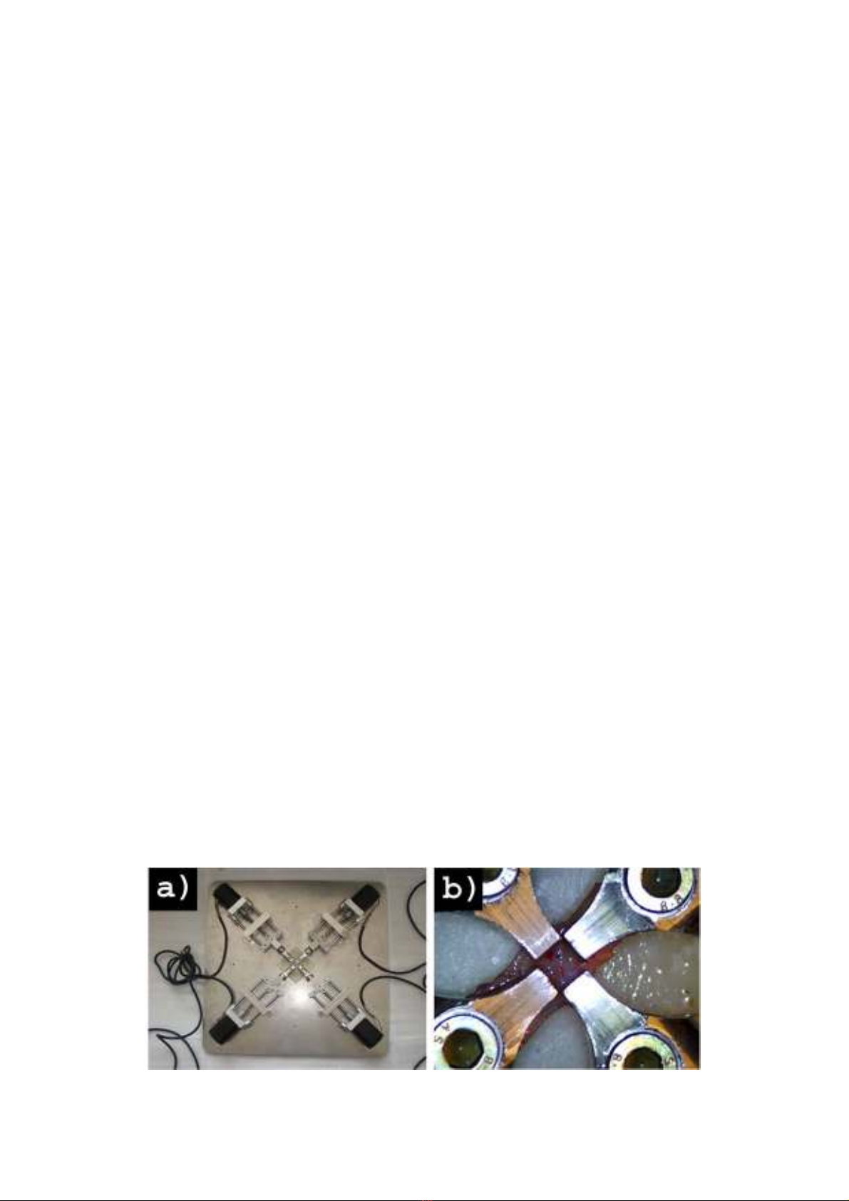
2.1 Developing a dedicated biaxial tensile test device
Regarding the dimensions of samples and the range of applied forces, a biaxial tensile test device was
designed and developed. This device is able to perform a quasi-static test with proper force
sensitivity. The clamps of this device are able to directly hold samples with dimensions of ≥ 5×5
mm2. The device can keep the tissue wet during the test so that its properties do not change due to
evaporation. Tensile forces are measured by two 2-channels 16 bit with 5 to 8 sample/s ADC
(Analogue to Digital Converter) and load cell conditioners, for UMAA 2 kgf load cell (Dacell Co.,
Ltd, Korea Corporation, Korea) attached to the aluminum clamps with a precision of 16 bits. The

46
required tensile forces in this device are applied by four micro stepper motors (made by Autonics
Corporation, Gyeonggi-do, Korea) with a resolution of 0.36 degrees and with a torque of 1.2 kg.cm.
For visual measurement of the tissue deformation, a USB digital microscope camera (with zoom:
300X, frequency: 30 Hz and resolution: 480×640) was used in the test device. Four drivers were used
to start the stepper motors (Autonics model MD5-H14). Data sent by our controller are transferred to
a computer and saved there. For synchronizing the results, the data of load cells and cameras were
both written simultaneously with a frequency of 5 Hz in a Python programmed code and saved. After
completion, the device was calibrated with standard balance weights. Moreover, we tested
successfully a silicon sample with known properties to validate our device measurements.
2.2 Strain Energy Density Function
Most experimental data are generally analyzed using the strain energy density function. It is a scalar
measure of the energy stored in the material as a result of deformation. If there is a one-to-one
relationship between strain and stress, then the theory of elasticity states that there exists a strain
energy density function W, from which the stresses can be computed from the strains as follows
(Humphrey et al., 1990b; Sun et al., 2003; Monson et al., 2008):
(1)
1
2.
(2)
where Sij, Eij, Fij, Iij are the components of the second Piola-Kirchhoff stress tensor ( s ), the Green-
Lagrange strain tensor ( E ), the finite strain deformation tensor ( F ) and the identity unit tensor (I),
respectively.
The most common strain energy density functions used to determine the mechanical properties of an
artery are the functions of the Finger tensor (B) or more explicitly in terms of its invariants (I3, I2, I1)
(Sun et al., 2003). The Finger tensor B gives us the relative local change in area within the sample
and is defined as:
.
. (3)
The finite strain deformation tensor (F) is defined as F= ∂x/∂x', where x' and x are the initial and
current configurations, respectively. The invariants of this tensor are given by:
trB, (4)
=1/2 , (5)
. (6)
For an incompressible material I3=1. Tr and det refer to transpose of a matrix and determinant of a
matrix, respectively.
2.2.1 Hyperelastic models
2.2.1.1 Mooney-Rivlin model
Mooney-Rivlin model (Mooney, 1940; Rivlin, 1948) used in this work includes 5 parameters. This
model was used before in many studies for blood vessels (Ally et al., 2004). The strain energy
function used was also employed before by Humphrey et al., (1990b), to describe the non-elastic
pseudolinear behavior of myocardium (Humphrey et al., 1990b; Monson et al., 2008).
،=1
1
3
1
3
3
, (7)
M. Shafigh et al. / Engineering Solid Mechanics 1 (2013)
47
where W is the strain energy function, I1 and I2 are invariants of right Cauchy deformation tensor, and
C1-C5 are empiric constants to be fitted.
2.2.1.2 Other models developed to describe the behavior of arteries
Several researchers have developed many mathematical expressions that describe the stress-strain
relationship for mechanical tests, but the most common are those based on polynomial or exponential
functions. A three dimensional strain energy function which is appropriate for the analysis of thick-
walled tubes or a two dimensional strain energy function can be used for modeling the mechanical
properties of arteries. Some of these functions are described below:
Ogden (Keshaw, 2001) proposed an isotropic, hyperelastic constitutive equation that has six material
parameters (three dimensional: µ1, µ2,µ3, and three non-dimensional: α1, α2,α3), which is a function of
the principal stretch ratios, λl,λ2 and λ3. Another three dimensional strain energy function was
developed by Humphrey et al. (1990b) and Monson et al. (2008). For doing that, they based their
development on the extension of the two dimensional strain energy function proposed by Fung et al.,
(1971). Until now, the architecture of the arterial wall has not been considered in the formulation of
the constitutive equations that are conventionally used to model the mechanical behavior of the
arterial wall. That is why several researchers (Holzapfel et al., 1996; Holzapfel et al., 2000) have
formulated constitutive models which incorporate some histological information. Hence, the material
parameters involved may be associated with the histological structure of the arterial walls.
2.3. Test method
Thirteen samples were obtained from 23 middle cerebral arteries (MCA) extracted according to a
special protocol from 20 human cadavers whose death was not due to injuries or diseases of cerebral
blood vessels. These samples were subjected to the test by our biaxial tensile device within 12 hours
after their resection. Before resecting the samples, the families of the deceased were asked to sign a
consensus form. To reach a uniform stress distribution, special clamps were designed such that could
grip tissues with dimensions as small as 5×5 mm2 directly. These lightweight clamps were then
attached to the load cells. In order to prevent damages of the vessels, brain of the deceased were first
removed completely. Then all cerebral vessels were separated from soft tissue by an expert
neurosurgeon. To standardize the tests, 10mm proximal segment of MCA was resected for all
specimens. To maintain the physiological conditions and freshness of samples, all the tests were done
on the day of acquiring samples. Specimens were stored in physiological saline 0.9%. Thickness of
specimens was measured with a vernier caliper. Then the specimens were cut to 5×5 mm2with a
special cutter. During the test, the samples were stored in 0.9% physiological saline heated by a
heater to 37°C. The specimens, after attachment to the four clamps, were stretched simultaneously in
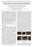
two dimensions (four directions) by the movement of four stepper motors (Fig. 1).
Fig. 1. Biaxial tensile test device (a) and cerebral vessel specimen under loading (b)