JST: Engineering and Technology for Sustainable Development
Volume 35, Issue 2, April 2025, 009-017
9
Flavonoids with Their Anti-Melanogenic Activity
from Glycyrrhiza glabra L. in Vietnam
Nguyen Thi Viet Thanh*, Hoang Tran Nhu, Tran Minh Anh
Hanoi University of Science and Technology, Ha Noi, Vietnam
Corresponding author email: thanh.nguyenthiviet@hust.edu.vn
Abstract
Glycyrrhiza glabra L. has a long-standing history in traditional medicine across both Eastern and Western
areas. The plant's phytochemials, such as glycyrrhizin, glycyrrhetic acid, and various flavonoids, offer
significant therapeutic potential. Further research could explore its application in modern drug development
for the treatment of systemic and non-systemic diseases. In the course of study on the chemical composition
of Glycyrrhiza glabra L. in Vietnam, this paper described the extraction and structure evaluation of four
compounds, including glabridin (1); 4'-O-methylglabridin (2); glabrol (3); kanzonol Y (4) as well as the melanin
inhibitory activity of these compounds. The stems of this plant were collected, identified, dried and extracted
in different polarity solvents. These substances were isolated from the ethyl acetate extract on the basis of
column chromatography combined with thin layer chromatography. Their structures were identified based on
spectroscopic evaluation and comparison of corresponding authentic compounds.
Keywords: Glycyrrhiza glabra, glabridin, flavonoid, anti-melanogenic.
1. Introduction
1
Glycyrrhiza glabra L. (G. glabra), commonly
known as licorice root, is a perennial shrub from the
Fabaceae family. The plant is native to China but is
now widely cultivated in various regions of Vietnam,
such as Tuyen Quang, Ha Giang, Dien Bien, and Son
La. In Europe and Asia, it has been used as both a
natural sweetener and a pharmaceutical [1]. According
to previous phytochemical reports, G. glabra contains
numerous bioactive compounds, including triterpenes,
saponins, flavonoids, polysaccharides, and
glycyrrhizin [2, 3]. Glycyrrhizin, a triterpenoid
glycoside, is responsible for the sweet taste of licorice
root. It consists of calcium, potassium, and magnesium
salts of glycyrrhizic acid [4]. In addition, many
saponins, such as oleanane triterpenoid saponin, have
also been isolated. Flavonoids, including flavanones,
flavonols, flavones, isoflavones, isoflavones, and
chalcones, are abundant and contribute to the yellow
color of the plant [2, 4].
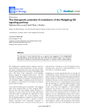
Fig. 1. Some chemical constituents from Glycyrrhiza glabra L.
1
ISSN 2734-9381
https://doi.org/10.51316/jst.181.etsd.2025.35.2.2
Received: Jan 17, 2025; revised: Feb 12, 2025
accepted: Feb 20, 2025
JST: Engineering and Technology for Sustainable Development
Volume 35, Issue 2, April 2025, 009-017
10
The root of G. glabra possesses diverse
biological activities, including antioxidant,
anti-inflammatory, expectorant, detoxifying, and
hepatoprotective properties. It is used in the treatment
of various conditions, such as peptic ulcers, bronchitis,
eczema, herpes, and immune-related diseases.
G. glabra also shows potential in cancer prevention,
antioxidation, antibacterial action, and hormonal
regulation. Extracts from the root have estrogenic and
anti-estrogenic activities and can reduce serum
testosterone levels in women. It is beneficial in
managing autoimmune conditions and immune
deficiencies such as AIDS [5, 6].
Melanin protects the skin against harmful stimuli,
while its overproduction leads to skin
hyperpigmentation, which can result in diseases such
as lentigines, melasma, freckles, and skin cancer.
Currently, the roots of G. glabra are commercially
used as a natural treatment for skin whitening. Several
compounds and extracts from G. glabra have been
proven to inhibit melanogenesis [7]. However, limited
research on chemical properties and melanin inhibition
has been conducted in Vietnam. In this study, glabridin
(1), 4'-O-methylglabridin (2), glabrol (3), and
kanzonol Y (4), along with their melanin synthesis
inhibitory activity, were investigated for the first time
from G. glabra in Vietnam.
2. Materials and Methods
2.1. Plant Materials
Roots of G.glabra (Fig. 2) were collected in Kim
Phu, Yen Son, Tuyen Quang province in September
2023. This plant was identified as Glycyrrhiza glabra
L., belongs to genus Glycyrrhiza, family Fabaceae.
Fig. 2. Dry root of Glycyrrhiza glabra L.
2.2. General Experimental Procedures
The method of analysis and separation of the
extracted residues was thin layer and column
chromatography. The isolated compounds were
identified by modern analytical methods such as 1D,
2D-NMR, and HR-MS.
Preparative HPLC was carried out using an
AGILENT 1200 HPLC system, HPLC column YMC
J'sphere ODS-H80 (4 µm, 20 × 250 mm) of brand
YMC. Column chromatography was performed using
either silica gel (Kieselgel 60, 70-230 mesh, and
230- 400 mesh, Merck) or RP-18 resin (150 µm, Fuji
Silysia Chemical Ltd.) as the reversed-phase. Thin
layer chromatography (TLC) was performed using
60 F254 (0.25 mm, Merck) and RP-18 F254S
(0.25 mm, Merck) plates.
NMR spectra (including 1H-NMR - Proton
nuclear magnetic resonance, 13C-NMR carbon-13
nuclear magnetic resonance, HMBC - Heteronuclear
Multiple Bond Correlation, HSQC - Heteronuclear
Single Quantum Coherence) were recorded on an
Agilent 600 NMR spectrometer (600 MHz for
1H-NMR and 150 MHz for 13C-NMR). Chemical shift
(δ) was reported in parts per million (ppm) and
abbreviations such as s (single), d (double), t (triplet),
q (quadruplet), m (multiple), and br. (extensive) were
used to report data. HR-MS mass spectra were
recorded on an Agilent 1200 series LC-MSD Ion Trap.
The melting point was measured on a Cole-Parmer
Instrument electrothermal melting point instrument
with serial number R216000334. Plant samples were
extracted with methanol using Vevor Ultrasonic model
JPS-100A.
2.3. Extraction and Isolation
The cleaned G. glabra roots were chopped, dried,
and crushed. The resulting dry powder (12.0 kg) was
extracted with methanol at room temperature (e times
× 15 L, each extraction for 1 hour). The extracts were
filtered and then distilled under reduced pressure to
recover the solvent, yielding 400.0 g of methanol
extract. The extract residue was dissolved in 2 liters of
distilled water and then extracted with ethyl acetate
(EtOAc), resulting in both EtOAc and aqueous
extracts. These two extracts were concentrated under
reduced pressure at 40-50 °C to recover the solvents.
This process yielded an ethyl acetate residue
(E, 158.78 g) and an aqueous residue (W).
The ethyl acetate extract (E, 158.78 g) of
G. glabra powder was subjected to column
chromatography, using silica gel as the stationary
phase. The elution solvent system consisted of a
dichloromethane/methanol (100/0, 50/1, 25/1, 10/1,
5/1, 2.5/1, v/v). The resulting fractions were
evaporated under reduced pressure, yielding six
fractions: E1, E2, E3, E4, E5, and E6.
Fraction E1 (23.71 g) was separated using silica
gel as the adsorbent and eluted with a gradient solvent
system of n-hexane/ethyl acetate (from 100/0 to 1/1,
v/v), yielding seven fractions: E1A, E1B, E1C, E1D,
E1E, E1F, and E1G. Fraction E1D (3.57 g) was
impregnated with silica gel, evaporated under reduced
pressure until the powder became loose and dry, and
then subjected to column chromatography using silica
JST: Engineering and Technology for Sustainable Development
Volume 35, Issue 2, April 2025, 009-017
11
gel as the stationary phase with
dichloromethane/acetone (100/1, 50/1, v/v) elution
solvent system. Fractions were collected in 20 mL test
tubes, and the elution was monitored by TLC using
appropriate solvent systems and silica gel plates.
Fractions displaying similar spots on TLC were
combined into larger fractions, and the solvent was
evaporated under reduced pressure to obtain eight
subfractions (E1D1 to E1D8).
Fraction E1D5 (0.69 g) was further purified by
column chromatography with silica gel as the
stationary phase, using a solvent system of
n-hexane/acetone/methanol (4/1/0.1, v/v/v), yielding
three fractions: E1D5’, E1D5’’, and E1D5’’’. Fraction
E1D5’’’ (0.36 g) was further purified by column
chromatography with silica gel as the stationary phase,
using a solvent system of n-hexane/acetone/methanol
(7/1/0.1, v/v/v), resulting in the isolation of compound
2 (70.08 mg).
Fraction E2 (33.83 g) was further separated by
silica gel column chromatography using a gradient
solvent system, yielding six fractions: E2A to E2F.
Fraction E2B (3.79 g) was separated using silica gel as
the stationary phase and dichloromethane:acetone
(15/1, v/v) as the eluent, yielding three fractions:
E2B1, E2B2, and E2B3. Fraction E2B2 was further
purified by column chromatography using
n-hexane/acetone/methanol (3/1/0.1, v/v/v), resulting
in compound 1 (100.05 mg).
Fraction E2B1 (0.96 g) was subjected to column
chromatography using silica gel as the stationary phase
and n-hexane/dichloromethane/methanol (1/1/0.2,
v/v/v) as the eluent system. The eluates were collected
in 20 mL test tubes, and the progress of the elution was
monitored by TLC on both normal- and reverse-phase
plates with appropriate solvent systems. Tubes with
similar TLC profiles were pooled into larger fractions,
which were then evaporated under reduced pressure to
yield six main fractions, E2B1A to E2B1F. Fraction
E2B1B was then subjected to reverse-phase YMC
column chromatography with acetone/water (2/1, v/v)
as the eluent, resulting in four fractions: E2B1B’,
E2B1B”, E2B1B”’, and E2B1B””. Finally, fraction
E2B1B” was purified by high-performance liquid
chromatography (HPLC) using an acetonitrile 65% in
water as the solvent system with a retention time of
45.25 minutes, yielding compound 3 (12.62 mg), while
fraction E2B1B”’ was purified by HPLC using an
acetonitrile 65% in water as the solvent system with a
retention time of 48.54 minutes, yielding compound 4
(22.17 mg).
2.4. Anti-Melanogenic Assay
The melanin synthesis inhibitory activity of
compounds 1 and 2 was evaluated based on the
melanin content in cultured cells. Prior to cellular
melanin measurement, B16.F10 cells were cultured in
6-well plates at a concentration of 1 × 10⁵ cells/mL and
stabilized overnight. The cells were then incubated
with different concentrations of the sample in the
presence of α-MSH (10 nM) for 48 hours. Kojic acid
(HiMedia) was used as a reference control. After
48 hours of incubation at 37 °C and 5% CO2, the cells
were harvested and washed with phosphate buffered
saline. The cell residue was then dissolved in 1N
NaOH solution containing 10% dimethyl sulfoxide
(DMSO) and incubated at 80 °C for 1 hour. The
absorbance of the extracted melanin was then
measured at wavelengths 405/450 nm [8].
Fig. 3. Diagram of isolation of compounds 1-4 from G.glabra
JST: Engineering and Technology for Sustainable Development
Volume 35, Issue 2, April 2025, 009-017
12
Fig. 4. The chemical structures of compounds 1-4
Glabrindin (1): HR-MS:
m/z= 325.1421[M+H]+, calcd. for [C20H21O4]+ =
325.1434.
1H-NMR (600 MHz, CDCl3): δH (ppm) 7.29
(s, -OH ); 7.26 (s, -OH); 6.90 (1H, d, J = 8.4 Hz, H-6');
6.80 (1H, d, J = 8.4 Hz, H-5); 6.64 (1H, d, J = 9.6 Hz,
H-4''); 6.41 (1H, d, J = 2.4 Hz, H-3'); 6.37 (1H, dd,
J = 8.4, 2.4 Hz, H-5'); 6.35 (1H, d, J = 8.4 Hz, H-6);
5.54 (1H, d, J = 9,6 Hz, H-3''); 4.36 (1H, ddd, J = 10.2,
3.6, 1.8 Hz, H-2eq); 4.00 (1H, dd, J = 10.2 Hz, 10.2,
H-2ax); 3.50 (1H, m, H-3ax); 2.96 (1H, dd, J = 15.6,
11.4 Hz, H-4ax), 2.82 (1H, ddd, J = 15.6, 4.8, 1.8 Hz,
H-4eq); 1.42 (3H, s, H-5'') and 1.40 (3H, s, H-6'')
13C-NMR (150 MHz, CDCl3): δC (ppm) 155.8
(C-4'); 155.2 (C-2'); 151.7 (C-7); 149.8 (C-9); 129.2
(C-5); 128.9 (C-3''); 128.1 (C-6'); 119.5 (C-1'); 117.0
(C-4''); 114.7 (C-10); 109.9 (C-8); 108.6 (C-6); 107.5
(C-5'); 103.2 (C-3'); 75.6 (C-2''); 70.1 (C-2); 30.9
(C-3); 30.6 (C-4); 27.7 (C-5''); 27.5 (C-6'').
4’-O-methylglabridin (2): HR-MS:
m/z = 339.1580 [M+H]+, calcd. for [C21H23O4]+,
M = 339.1591.
1H-NMR (600 MHz; CDCl3): 𝛿H (ppm) 6.99 (1H,
d, J = 8.4 Hz, H-6); 6.81 (1H, d, J = 8.4 Hz, H-6'); 6.64
(1H, d, J = 10.2 Hz, H-4''); 6.45 (1H, dd, J = 8.4, 2.4
Hz, H-5'); 6.37 (1H, d, J = 8.4 Hz, H-5); 6.34 (1H, d,
J = 2.4 Hz, H-3'); 5.76 (1H, s, -OH); 5.55 (1H, d,
J = 10.2 Hz, H-3''); 4.37 (1H, ddd, J = 10.2, 3.6, 1.8
Hz, H-2eq); 4.01 (1H, dd, J = 10.2, 10.2 Hz, H-2ax);
3.73 (3H, s, -OCH3); 3.49 (1H, m, H-3ax); 2.96 (1H,
dd, J = 15.6, 11.4 Hz, H-4ax H-4a); 2.84 (1H, ddd,
J = 15.6, 4.8, 1.8 Hz, H-4eq); 1.42 (3H, s, H-5''); 1.41
(3H, s, H-6'').
13C-NMR (150MHz, CDCl3): 𝛿C (ppm) 159.3
(C-4'); 154.6 (C-2'); 151.8 (C-7); 149.8 (C-9); 129.3
(C-5); 129.0 (C-3''); 128.2 (C-6'); 120.1 (C-4''); 117.0
(C-1'); 114.6 (C-10); 110.0 (C-8); 108.7 (C-6); 105.9
(C-5'); 102.2 (C-3'); 75.7 (C-2''); 70.1 (C-2); 31.7
(C-4), 30.6 (C-3); 27.8 (C-5''); 27.5 (C-4'') and 55.4
(-OCH3).
Glabrol (3): HR-MS: m/z = 391.1905 [M]+,
calcd. for [C25H26O4]+, M = 391.1904.
1H- NMR (600 MHz; MeOD): 𝛿H (ppm) 7.59
(1H, d, J = 8.4 Hz, H-5); 7.20 (1H, d, J = 2.4 Hz,
H-2'); 7.12 (1H, d, J = 8.4, 2.4 Hz, H-6'); 6.80 (1H, d,
J = 8.4 Hz, H-5'); 6.52 (1H, d, J = 8.4 Hz, H-6); 5.33
(1H, m, H-2); 5.29 (1H, m, H-2''); 5.20 (1H, m, H-2''');
3.33 (2H, m, H-1''); 3.30 (2H, m, H-1'''); 2.97 (1H, m,
H-3a); 2,70 (1H, m, H-3b); 1.73 (3H x2, s, H-4''',
H-5''') and 1,62 (3H x2, s, H-4'', H-5'').
13C-NMR (150 MHz; MeOD): δC (ppm) 194.3
(C-4); 163.9 (C-7); 163.0 (C-9); 156.3 (C-4'); 133.1
(C-3'''); 132.1 (C-3''); 131.4 (C-1'); 129.4 (C-3'); 128.9
(C-2'); 126.7 (C-5); 125.9 (C-6'); 123.8 (C-2''); 123.3
JST: Engineering and Technology for Sustainable Development
Volume 35, Issue 2, April 2025, 009-017
13
(C-2'''); 117.1 (C-8); 115.6 (C-5'); 115.1 (C-10); 110.7
(C-6); 80.8 (C-2); 44.7 (C-3); 29.2 (C-1'''); 26.0 (C-4'',
C4'''); 23.1 (C-1''); 18.0 (C-5''') and 17.9 (C-5'').
Kanzonol Y (4): Amorphous powder, HR-MS:
m/z = 411.2153 [M+H]+, calcd. for [C25H31O5]+,
M = 411.2166
1H-NMR (600 MHz, MeOD): δH (ppm) 7.36 (1H,
s, H-6'); 6.87 (1H, dd, J = 8.4, 2.4 Hz, H-6); 6.79 (1H,
d, J = 2.4 Hz, H-2); 6.67 (1H, d, J = 8.4 Hz, H-5); 6.30
(1H, s, H-3'); 5.28 (1H, m, H-8'); 5.21 (1H, m, H-8);
5.10 (1H, dd, J= 7.2, 5.4, H-
); 3.22 (2H, d, J = 7.2
Hz, H2-7); 3.18 (2H, d, J = 7.2 Hz, H2-7'); 2.98 (1H,
dd, J1 = 13.8, 5.4 Hz, H-β); 2.84 (1H, dd, J1 = 13.8, 7.2
Hz, H-β); 1.78 (3H, s, H3-11'); 1.72 (3H, s, H3-10'),
1.71 (3H, s, H3-11) and 1.67 (3H, s, H3-10).
13C-NMR (150 MHz, MeOD): δC (ppm) 205.0
(C=O); 165.1 (C-2'), 165.0 (C-4'); 154.8 (C-4); 133.9
(C-9); 132.8 (C-9'); 131. 7 (C-6'), 131.5 (C-2); 129.1
(C-1); 129.0 (C-3); 128.6 (C-6); 124. 0 (C-8); 123.4
(C-8'); 122.1 (C-5'), 111.8 (C-1'); 115.7 (C-5); 103.2
(C-3'); 74.5 (C-
); 42.9 (C-β); 29.1 (C-7); 28.4 (C-7');
26.0 (C-11); 25.9 (C-11'); 17.9 (C-10) and 17.8
(C-10').
3. Results and Discussions
Four compounds were isolated from ethyl acetate
extract of G.glabra. The chemical structures of the
isolated compounds were determined based on modern
spectroscopic methods such as one- and two-
dimensional nuclear magnetic resonance and mass
spectroscopy.
Compound 1 was isolated as an amorphous
powder. 1H-NMR spectrum of 1 showed
oxymethylene group signals at δH 4.36 (1H, ddd,
J = 10.2, 3.6, 1.8 Hz, H-2eq); 4.00 (1H, dd, J = 10.2,
10.2 Hz, H-2ax), one proton signal of methylene group
at 3.50 (1H, m, H-3ax); 2 proton signals of methylene
group at δH 2.96 (1H, dd, J = 15.6, 11.4 Hz, H-4ax),
2.82 (1H, ddd, J = 15.6, 4.8, 1.8 Hz, H-4eq). These data
revealed an structure of an isoflavan [9]. Additionally,
the spectrum presented signals of ortho-coupled
aromatic protons and ABX spin coupling systems: [δH
6.80 (1H, d, J = 8.4 Hz, H-5), 6.35 (1H, d, J = 8.4 Hz,
H-6) and δH 6.90 (1H, d, J = 8.4 Hz, H-6'), 6.37 (1H,
dd, J = 8.4, 2.4 Hz, H-5'), 6.41 (1H, d, J = 2.4 Hz,
H-3')], characteristic of two aromatic rings.
Furthermore, 1H-NMR spectrum showed the
proton signals assignable to one γ,γ-dimethylallyl δH
6.64 (1H, d, J = 9.6 Hz, H-4''), 5.54 (1H, d, J = 9.6 Hz,
H-3''), 1.42 (3H, s, H-5''), 1.40 (3H, s, H-6'').
Based on the above spectral data, compound 1
was suggested as an isoflavan skeleton, conjugating
with a γ,γ-dimethylallyl group.
The 13C-NMR spectrum combined with the
HSQC spectrum showed the appearances of 20 carbon
signals: 12 sp2 aromatic carbon signals including:
quaternary carbons bearing oxygen signals δC 155.8
(C-4'), 155.2 (C-2'), 151.7 (C-7), 149.8 (C-9); one
oxymethylen at δC 70.1 (C-2); 1 CH group at δC 30.9
(C-3) suggesting a CO bond in the pyran ring.
Combined with the 1H-NMR spectrum, it could be
confirmed that this compound is composed of an
isoflavan framework. In addition, 3 carbon signals of
CH group were dedermined as: 1 double bond C=C at
δC 128.9 (C-3''), 117.0 (C-4''), one quaternary carbon
signal attached to oxygen element at δC 75.6 (C-2'');
2 carbon signals of dimethylallyl group at δC 27.7
(C-5''), 27.5 (C-6'').
HMBC spectrum allowed the confirmation of the
position of groups in the molecule. The correlations
between H-2 (δH 4.3, 4.0)/H-3 (δH 3.5)/H-4 (δH 2.96,
2.82)/H-5' (δH 6.37)/H-6' (δH 6.90) with C-1' and the
correlations between H-6' (δH 6.90)/H-3 (δH 3.5)/H-4
(δH 2.96, 2.82) with C-3 (δC 30.9) indicated the linkage
between the pyran ring and the aromatic ring at C-3,
C-1'. The interactions between H-4'' (δH 6.64) with
C-7 (δC 151.7), C-9 (δC 149.8), C-8 (δC 109.9), C-2''
(δC 75.6), between H-3'' (δH 5.54) with C-8 (δC 109.9),
C-2” (δC 75.6) established the pyran ring position at
C-7 and C-8; The interaction of protons in the pyran
ring with other carbons were implied based on the
interaction between H-2 (δH 4.3, 4.0) with C-9
(δC 149.8)/C-3 (δC 30.9)/C-4 (δC 30.6); between H-4
(δH 2.96, 2.82) with C-9 (δC 149.9)/C-10 (δC 114.7)/
C-2 (δC 70.1)/C-3 (δC 30.9). These data were closely
resembling those of glabrindin suggesting that 1
was glabridin (Table 1)
Moreover, the HRMS spectrum of compound 1
showed an ion peak m/z= 325.1421 [M+H]+, which
suggested the molecular formula of C20H20O4. From
the above evidence, compound 1 was identified as
glabridin.
Compound 2 was obtained as yellow solid. The
NMR spectra of compound 2 was similar to NMR
spectrum of 1 exception for the presence of proton
signals of -OCH3 at 3.73 ppm (3H, s) in 1H-NMR and
the signal of -OCH3 (55.4 ppm) presented in the
13C-NMR spectra. These data suggested that
compound 2 was the oxmethyl derivative of glabridin.
HSQC and HMBC spectra confirmed the structure of
2. Especially the interaction of C-4' (δC 155.8) and
-OCH3 (δH 3.73) revealed the position of -OCH3 at
C-4'. Moreover, the molecular formula of 2 was
C20H20O4 as determined from its HR-MS (found
m/z= 325.1421 [M+H]+, calcd. for [C20H21O4]+ =
325.1434.
In addition, The NMR data of 2 were
perfectly equivalent to those of 4'-O-methylglabridin
(Table 1). From the above data, combined with the
reference documents, it can be confirmed that
compound 2 is 4'-O-methylglabridin.