Mixed lineage leukemia histone methylases play critical roles in estrogen-mediated regulation of HOXC13 Khairul I. Ansari, Sahba Kasiri, Imran Hussain and Subhrangsu S. Mandal
Department of Chemistry and Biochemistry, The University of Texas at Arlington, TX, USA
Keywords estrogen; estrogen receptor; HOXC13 gene regulation; mixed lineage leukemia; nuclear receptor
Correspondence S. S. Mandal, Department of Chemistry and Biochemistry, The University of Texas at Arlington, Arlington, TX 76019, USA Fax: +1 817 272 3808 Tel: +1 817 272 3804 E-mail: smandal@uta.edu
(Received 28 July 2009, revised 12 October 2009, accepted 20 October 2009)
doi:10.1111/j.1742-4658.2009.07453.x
HOXC13, a homeobox-containing gene, is involved in hair development and human leukemia. The regulatory mechanism that drives HOXC13 expression is mostly unknown. Our studies have demonstrated that HOXC13 is transcriptionally activated by the steroid hormone estrogen (17b-estradiol; E2). The HOXC13 promoter contains several estrogen- response elements (EREs), including ERE1 and ERE2, which are close to the transcription start site, and are associated with E2-mediated activation of HOXC13. Knockdown of the estrogen receptors (ERs) ERa and ERb suppressed E2-mediated activation of HOXC13. Similarly, knockdown of mixed lineage leukemia histone methylase (MLL)3 suppressed E2-induced activation of HOXC13. MLLs (MLL1–MLL4) were bound to the HOXC13 promoter in an E2-dependent manner. Knockdown of either ERa or ERb affected the E2-dependent binding of MLLs (MLL1–MLL4) into HOXC13 EREs, suggesting critical roles of ERs in recruiting MLLs in the HOXC13 promoter. Overall, our studies have demonstrated that HOXC13 is transcriptionally regulated by E2 and MLLs, which, in coordi- nation with ERa and ERb, play critical roles in this process. Although MLLs are known to regulate HOX genes, the roles of MLLs in hormone- mediated regulation of HOX genes are unknown. Herein, we have demon- strated that MLLs are critical players in E2-dependent regulation of the HOX gene.
Introduction
are key players
roles
plays critical in embryogenesis and organo- genesis. The nature of a body structure depends on the specific combination of HOX gene products, and the expression of specific HOX genes varies at different stages of development. Therefore, proper regulation for and maintenance of HOX genes are essential normal physiological functions and growth.
Homeobox-containing in genes embryogenesis and development [1,2]. Misregulation of homeobox genes is associated with tumorigenesis. More than 200 homeobox-containing genes have been identified in vertebrates, and they have been classified into two major groups, class I and II. Class I homeo- box-containing genes share a high degree of identity (more than 80%) and are called HOX genes. In humans, there are 39 different HOX genes, clustered into four different groups, called HOXA, HOXB, HOXC, and HOXD, located on chromosomes 7, 17, 12, and 2, respectively [1,2]. Each of these HOX genes
HOXC13 is a critical gene involved in the regulation of the hair keratin gene cluster and alopecia [3–5]. Transgenic mice overexpressing HOXC13 in differenti- ating keratinocytes of hair follicles develop alopecia, accompanied by a progressive pathological skin condi-
Abbreviations ChIP, chromatin immunoprecipitation; DEPC, diethyl pyrocarbonate; E2, estrogen (17b-estradiol); ER, estrogen receptor; ERE, estrogen- response element; H3K4, histone H3 lysine 4; HMT, histone methyltransferase; MLL, mixed lineage leukemia histone methylase; NR, nuclear receptor; RNAPII, RNA polymerase II.
FEBS Journal 276 (2009) 7400–7411 ª 2009 The Authors Journal compilation ª 2009 FEBS
7400
K. I. Ansari et al.
Estrogen-mediated HOXC13 activation involving MLL
Although MLLs are recognized as major regulators of HOX genes during embryogenesis, they are not impli- cated in steroid hormone-mediated HOX gene regula- tion. Herein, we have investigated the roles of the MLL family of HMTs in E2-mediated regulation of HOXC13. Our results show that HOXC13 is transcrip- tionally regulated by E2, and that MLLs, in coordina- tion with ERs, regulate E2-induced activation of HOXC13.
Results
HOXC13 is transcriptionally regulated by E2
tion that resembles ichthyosis [4,5]. HOXC13 mutant mice lack external hair, suggesting a critical role for the gene in hair development [5]. HOXC13 has also been found to be a fusion partner of NUP98 in adult acute myeloid leukemia [6,7]. This protein also binds to the ETS family transcription factor PU.1 and affects the differentiation of murine erythroleukemia [8]. Although HOX13 is critical player in hair development and disease, little is known about its own regulation. Steroid hormones are critical players in sexual differen- tiation. Steroid hormones such as estrogen (17b-estra- diol; E2) and androgens are also linked with hair in hair patterning follicle growth and differences between males and females [9,10]. However, the molec- ular mechanism of the roles of steroid hormones in hair development is poorly understood. Herein, we have investigated whether HOXC13, a critical player in hair follicle development, is regulated by steroid hormones.
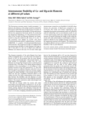
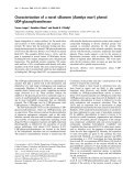
ERs are major players in E2-mediated regulation of E2-responsive genes [41,42]. In general, upon binding to E2, ERs are activated. The activated ERs bind to E2 response elements (EREs) present in the promoter of E2-responsive genes, leading to their transcriptional activation [43]. In this work, before examining the E2-mediated regulation of HOXC13, we analyzed its promoter for the presence of any EREs. Our results demonstrated that the HOXC13 promoter contains six putative EREs (ERE1 ⁄ 2 sites) within )1 to )3000 bp upstream of the transcription start site (Fig. 1). All of the EREs show 100% homology with ERE1 ⁄ 2 sites (GGTCA or TGACC) but not with the consensus full ERE sequence (GGTCAnnnTGACC). The presence of these EREs in close proximity to the transcription start site indicated that HOXC13 might be potentially regu- lated by E2 via the involvement of ERs.
Mixed lineage leukemia histone methylases (MLLs) are human histone H3 lysine 4 (H3K4)-specific histone methyltransferases (HMTs) that play critical roles in gene activation. MLLs are key players in HOX gene regulation [11–22]. MLLs are also well known to be rearranged in acute lymphoblastic and myeloid leuke- mias [12,15]. In humans, there are several MLL families of proteins, such as MLL1, MLL2, MLL3, and MLL4. Each of them possesses H3K4-specific HMT activity and exists as a multiprotein complex with several com- mon protein subunits [12,23,24]. Recently, we demon- strated that human CpG-binding protein interacts with MLL1, MLL2, and hSet1, and regulates the expression of MLL target HOX genes [11]. Studies from our labo- ratory (and others) have demonstrated that MLLs are important players in cell cycle regulation and stress responses [25–33]. Knockdown of MLL1 resulted in cell cycle arrest at the G2 ⁄ M phase [34].
Recent
studies have demonstrated that
several MLLs (MLL2, MLL3, and MLL4) act as coregulators for E2-mediated activation of E2-sensitive genes [12,35–38]. MLL2 interacts with E2 receptor (ER) in an E2-dependent manner, and regulates the activation of cathepsin D [35,38]. MLL3 and MLL4 regulate the E2-sensitive gene encoding liver X-receptor [36,39,40].
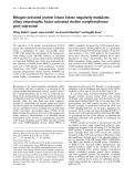
In order to examine whether HOXC13 is regulated by E2, we treated a steroidogenic human cell line (JAR cells, of choriocarcinoma placental origin, cul- tured in phenol-red free medium containing activated charcoal-treated fetal bovine serum) with different concentrations (1–1000 nm) of E2 for 8 h. The RNA was isolated from the E2-treated cells and analyzed by RT-PCR, using HOXC13-specific primers (Fig. 2; Table 1). Interestingly, our results demonstrated that HOXC13 was overexpressed upon exposure to E2 in a concentration-dependent manner (Fig. 2A,B). In com- parison with the control, HOXC13 expression was four-fold to five-fold higher in the presence of 100 and
+1 +1
–3000
–234 –234 ERE1 ERE1
–1260 ERE2 ERE2
–1788 ERE3 ERE3
–2000 –2288 –2152 ERE6 ERE6 ERE4 ERE4 ERE5 ERE5
Fig. 1. Schematic diagram showing different EREs located in the HOXC13 promoter. All of the EREs analyzed in the HOXC13 promoter are ERE1 ⁄ 2 sites (GGTCA or TGACC).
FEBS Journal 276 (2009) 7400–7411 ª 2009 The Authors Journal compilation ª 2009 FEBS
7401
K. I. Ansari et al.
Estrogen-mediated HOXC13 activation involving MLL
E2 (nM)
A
0 0.1 1.0 10 100 1000
strated that HOXC13 activation was maximum after 6–8 h of E2 treatment (Fig. 2C,D; with 100 nm E2, lanes 4 and 5).
β-actin
HOXC13
1 2 3 4 5 6
ERs play a critical role in E2-induced HOXC13 expression
0.8
B
0.6
) n i t c a
n o i s s e r p x e
0.4
0.2
o t e v i t a l e r (
3 1 C X O H
0
0
0 1
1 1 . 0
0 0 1
0 0 0 1
[E2] (nM)
E2 (100 nM)
C
Time (h)
0 2 4 6 8 12 16 24
β
-actin
HOXC13
1 2 3 4 5 6 7 8
0.8
D
0.6
0.4
0.2
) n i t c a o t e v i t a l e r (
n o i s s e r p x e 3 1 C X O H
0
0
2
4
6
8 12 16 24
Time (h)
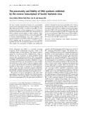
In order to examine the potential role of ERs in E2-induced activation of HOXC13, we knocked down ERa and ERb separately, using specific antisense oli- gonucleotides, in JAR cells and exposed the cells to 100 nm E2 for an additional 8 h. A scramble antisense oligonucleotide (with no homology to ERs) was used as a negative control. Our results demonstrated that application of ERa or ERb antisense oligonucleotide knocked down the respective genes efficiently, at both the mRNA and the protein level (Fig. 3A,B, lanes 4–6, and data not shown; the quantifications are shown in the respective bottom panels). After confirming effec- tive knockdown, we analyzed the RNA from these ER knockdown and E2-treated cells for the expression lev- els of HOXC13 using RT-PCR. As seen in Fig. 3A,B, HOXC13 expression was increased upon exposure to E2 (compare lanes 1 and 2), and application of scram- ble antisense oligonucleotide did not have any signifi- cant effect on E2-mediated activation of HOXC13. Interestingly, upon knockdown of either ERa or ERb, the E2-dependent activation of HOXC13 was sup- pressed almost to the basal level (Fig. 3 A,B, compare lanes 5 and 6 with lanes 1 and 2; quantifications are shown in the respective bottom panels). These results demonstrated that both ERa and ERb are essential for E2-mediated transcriptional activation of HOXC13.
MLLs play critical roles in E2-induced HOXC13 expression
Fig. 2. Effect of E2 on HOXC13 gene expression. (A, B) JAR cells were initially grown in phenol red-free medium, and treated with different concentrations (0–1000 nM) of E2 for 8 h. The total RNA was isolated and analyzed by RT-PCR, using primers specific for HOXC13. b-Actin was used as control. Quantification of RT-PCR products is shown in (B). (C, D) JAR cells were treated with 100 nM E2 for different time periods (0–24 h). The total RNA was isolated and analyzed by RT-PCR, using primers specific for HOXC13. b-Actin was used as control. The RT-PCR products were quantified, and the relative expression of HOXC13 (relative to actin) is shown in (D). Each of these experiments was repeated three times, and values were assumed to be significantly different at P £ 0.05.
1000 nm E2 (Fig. 2A,B; compare lane 1 with lanes 5 and 6). As 100 nm was most effective, we analyzed HOXC13 expression using an E2 concentration range closer to 100 nm (20, 50, 100 and 250 nm), and found that 100 nm was the optimal concentration for the E2-mediated induction of HOXC13 (data not shown). The stimulation of HOXC13 expression upon exposure to E2 demonstrated that HOXC13 is transcriptionally regulated by E2. Time-dependence experiments demon-
As MLLs are well known as master regulators of HOX genes, and several MLLs are implicated in E2 signaling, we examined whether any of the MLLs are involved in E2-dependent stimulation of HOXC13 expression. We knocked down different MLL genes (MLL1, MLL2, MLL3, and MLL4) separately by using specific phosphorothioate antisense oligonucleo- tides, and then exposed the cells to E2 (100 nm for 8 h). Before performing E2-related experiments, we examined the efficacies of different MLL (MLL1– MLL4)-specific antisense oligonucleotides and their most effective doses. The specific MLL knockdowns were confirmed by analyzing their respective gene expression at both the mRNA and protein levels (data not shown). On the basis of these initial experiments, we applied the specific concentration of each of the
FEBS Journal 276 (2009) 7400–7411 ª 2009 The Authors Journal compilation ª 2009 FEBS
7402
K. I. Ansari et al.
Estrogen-mediated HOXC13 activation involving MLL
Table 1. Primers used for RT-PCR, ChIP and antisense oligonucleotide experiments.
Primers
Forward primer (5¢- to 3¢)
Reverse primer (5¢- to 3¢)
AGCACTGTGTTGGCGTACAG CGATTTGCTGACCACCTTCT GGAGCAAGAGGTTCAGCATC GCACAATGCTGTCAGGAGAA ACAAGCCATAGGAGGTGGTG AGTCTGCATCCCCGTATTTG CAGGTCTCCTGGGGTTCC TCTGCTTTACCTCGCTGGAT CGCGGGTAGTAGAAGTGGAA AGCCTTTGGGAGTAGGAACC
CTCTTCCAGCCTTCCTTCCT GGAAGTCTCCCTTCCCAGAC GAGGACCCCGGATTAAACAT GTGCAGCAGAAGATGGTGAA AAGCAAACGGACTCAGAGGA GTCTATGCGCAGTGGAGACA GCGTCTCCCTGTCCCTTTA TTGCCGAGTATATTCCATTGC TTTCAGGCCCTTTGTTTCTC TGCCCTCATATAAACCTGGAA CATGGTCATGGTCAGa GAATGTCATAGCTGAa TGCCAGTCGTTCCTCTCCACa ACTCTGCCACTTCCCGCTCAa CCATCTGTTCCTTCCACTCCCa CCTTCTCTTCTCCCTCCTTGTa CGTTTGTCCCTCCAGCATCTa
b-Actin HOXC13-ORF MLL1 MLL2 MLL3 MLL4 HOXC13-ERE1 HOXC13-ERE2 HOXC13-ERE3 HOXC13-ERE4 ERa antisense ERb antisense MLL1 antisense MLL2 antisense MLL3 antisense MLL4 antisense Scramble antisense
a Phosphorothioate antisense oligonucleotide.
irrespective of
the E2-mediated
activation
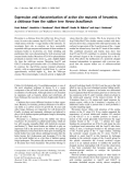
MLL antisense oligonucleotides that showed the most effective knockdown of the respective gene and then exposed the cells to E2 (100 nm for 8 h) in an MLL knockdown environment. In parallel, we also applied a scramble antisense oligonucleotide (no homology with any of the MLLs) as a negative control. As seen in Fig. 4A, upon application of MLL1 antisense oligonu- cleotide followed by exposure to E2, MLL1 was effi- scramble antisense ciently knocked down, whereas oligonucleotide had no significant effect on the level of MLL1 mRNA. Interestingly, upon downregulation of MLL1, E2-mediated upregulation of HOXC13 was lane 3). Similar results slightly decreased (Fig. 4A, were observed for MLL2 and MLL4 downregulation (Fig. 4B,D). The knockdown of MLL3 almost abol- ished of HOXC13 (Fig. 4C). These results demonstrated that the MLL family of HMTs, especially MLL3, play critical roles in the E2-mediated regulation of HOXC13.
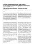
against ERs and MLLs. ChIP experiments were also performed in parallel with the use of antibody against actin as a nonspecific negative control. In brief, JAR cells were treated with 100 nm E2 for 6 h, and control and E2-treated cells were then subjected to ChIP anal- ysis. The immunoprecipitated DNA fragments were PCR amplified using primers specific for ERE1, ERE2, ERE3 and ERE4 of the HOXC13 promoter. As seen in Fig. 5A, no significant binding of actin was observed in any of the the EREs, absence and presence of E2. Binding of ERa and ERb was the increased in both ERE1 and ERE2 of HOXC13 promoter (Fig. 5A, lanes 1–4). The levels of E2-induced binding of ERa and ERb were higher in ERE2 than in ERE1. ERE3 and ERE4 were not sensi- tive to ER binding as a function of E2, probably because of their distance from the transcription start site, although some amount of constitutive binding was observed in both regions.
E2-induced recruitment of ERs and MLLs in the HOXC13 promoter
As the HOXC13 promoter contains several ERE1 ⁄ 2 regions within the first 3000 nucleotides upstream of the transcription start site, we analyzed the involve- ment of some of these EREs (ERE1–ERE4, located at )234, )1260, )1788 and )2000 bp upstream) by ana- lyzing the in vivo binding of ERs and MLLs. We ana- lyzed the in vivo binding of the different factors in the absence and presence of E2, using chromatin immuno- [34], using antibodies precipitation (ChIP) assays
The binding profiles of different MLLs were interest- ing. First, although some amount of binding of MLL1 was observed in ERE3, no significant E2-dependent binding of any of the MLLs was observed in ERE3 and ERE4 (Fig. 5A, lanes 5–8). Significant amounts of constitutive binding of MLL1, MLL3 and MLL4 were observed in ERE1, even in the absence of E2 (Fig. 5A, lane 1). However, MLL2 binding to ERE1 was enhanced upon addition of E2 (Fig. 5A, lanes 1 and 2). Interestingly, binding of all of the MLLs (MLL1– MLL4) was greatly enhanced upon addition of E2 in ERE2 (Fig. 5A, lanes 3 and 4). These results demon- strated that ERE1 ()234 bp) and ERE2 ()1260 bp),
FEBS Journal 276 (2009) 7400–7411 ª 2009 The Authors Journal compilation ª 2009 FEBS
7403
K. I. Ansari et al.
Estrogen-mediated HOXC13 activation involving MLL
Scramble ERα
A
Antisense (µg)
6
3 6 9 – + + + + +
E2 (100 nM)
ERE4, which are located far upstream ()1788 bp or further), were not sensitive to E2-dependent binding of any of the MLLs ⁄ ERs, indicating no significant roles of these EREs in HOXC13 activation (Fig. 5A).
β-actin
ERα
HOXC13
1 2 3 4 5 6
0.8
ERα HOXC13
0.6
0.4
0.2
n o i s s e r p x E
) n i t c a o t e v i t a l e r (
0.0
1
2
3
4
5
6
Scramble ERβ
B
6 3 6 9 + – + + + +
Antisense (µg) E2 (100 nM)
β-actin
ERβ
HOXC13
1 2 3 4 5 6
To further confirm the E2-dependent binding of ERs and MLLs to the HOXC13 promoter, we analyzed their binding profiles in a time-dependent manner in ERE1 and ERE2 (Fig. 5B). In agreement with the above findings, binding of ERa and ERb was increased in both ERE1 and ERE2 in the presence of E2. Interestingly, the kinetics of E2-dependent binding of ERa and ERb to both ERE1 and ERE2 are differ- ent. The binding of ERa is very low in the absence of E2, and is significantly enhanced in the presence of E2 n both ERE1 and ERE2. However, in the case of ERb, some constitutive binding was observed in ERE2 even in the absence of E2, and this binding was increased in the presence of E2 (Fig. 5A,B; compare 0 h and 6–8 h time points). These differences in the kinetic profiles of binding of ERa and ERb suggest that they have distinct modes of action in regulating target gene activation. It is important to mention that, although it is poorly understood, the difference in the kinetics of binding of ERa and ERb to the target gene promoters has been previously observed by other laboratories [44].
ERβ HOXC13
n o i s s e r p x E
) n i t c a o t e v i t a l e r (
1.0 0.8 0.6 0.4 0.2 0.0
1
2
3
4
5
6
Fig. 3. Effect of depletion of ERa and ERb on E2-induced expres- sion of HOXC13. JAR cells were grown up to 60% confluency prior to treatment with different concentrations of ERa-specific and ERb-specific phosphorothioate oligonucleotides by using ifect transfection (MoleculA). Control cells were treated with a scramble antisense oligonucleotide with no homology with the ERa and ERb genes. The antisense oligonucleotide-transfected cells were incu- bated for 24 h and then treated with E2 (100 nM) for an additional 8 h. Cells were harvested and subjected to RNA preparation. The mRNA was subjected to RT-PCR analysis by using primers specific for HOXC13 along with ERa and ERb. b-Actin was used as control. The RT-PCR products were analyzed in agarose gel. Quantification of transcript accumulation on the basis of RT-PCR products (average of three replicates) is shown beneath the respective gel image. Bars indicate standard errors. Values were assumed to be significantly different at P £ 0.05. The results of experiments involv- ing ERa and ERb are shown in (A) and (B), respectively.
which are close to the transcription start site, are mostly responsible for E2-dependent binding of ERs and MLLs and hence the regulation of HOXC13. ERE2 appeared to have more critical roles (sensitivity to E2) than the other EREs examined. ERE3 and
E2-dependent binding of MLLs (MLL1–MLL4) was primarily observed in ERE2 (Fig. 5B). Again, as seen above, MLL2 binding was observed in ERE1 as a func- left panel). The E2-dependent tion of E2 (Fig. 5B, increase in binding of MLLs to the EREs were observed at as early 30 min post-E2 exposure, and increased with time, reaching a maximum at (cid:2) 6 h (Fig. 5B). The binding of MLL3 to ERE2 appeared to be most prominent, although E2-induced binding of other MLLs (MLL1, MLL2, and MLL3) was also significant (Fig. 5B). In addition, we also analyzed the status of RNA polymerase II (RNAPII) and H3K4- trimethylation level in ERE1 and ERE2. Our results demonstrated that in both ERE1 and ERE2, the levels of RNAPII and H3K4-trimethylation were increased in the presence of E2 (Fig. 5B). These results demon- strated that both ERE1 and ERE2 (especially ERE2) coordinate the binding of ER and MLL coregulators as well as RNAPII, and regulate the E2-mediated tran- scriptional activation of HOXC13. It is important to note that although ERE2 is located far upstream (1260 bp away from the transcription start site), we still observed significant transcription-dependent increases in RNAPII binding to these EREs. These observations suggest that there is probably a looping of the large promoter regions so that far upstream cis-elements could be placed closer to the promoter proximal sites
FEBS Journal 276 (2009) 7400–7411 ª 2009 The Authors Journal compilation ª 2009 FEBS
7404
K. I. Ansari et al.
Estrogen-mediated HOXC13 activation involving MLL
B
A
L L 2 L L 2
L L 1
Antisense
Scra m ble
Scra m ble Scra m ble
+
M M +
M +
–
+
Antisense Antisense E2 (100 nM) – β-actin β-actin
E2 (100 nM) β-actin
MLL1
MLL2 MLL2
HOXC13
HOXC13 HOXC13
1 2 1 2
3 3
1 2
3
1.2
1.2
MLL1 HOXC13
MLL2 HOXC13
0.8
0.8
0.4
0.4
n o i s s e r p x E
n o i s s e r p x E
) n i t c a o t e v i t a l e r (
) n i t c a o t e v i t a l e r (
0.0
0.0
1
2
3
1
2
3
C
D
L L 3 L L 3
L L 4 L L 4
Antisense Antisense
Antisense Antisense
Scra m ble Scra m ble
Scra m ble Scra m ble
M M +
+
+
M M +
E2 (100 nM) – β-actin β-actin
E2 (100 nM) – β-actin β-actin
MLL4 MLL4
MLL3 MLL3
HOXC13 HOXC13
HOXC13 HOXC13
1 2 1 2
3 3
1 2 1 2
3 3
1.2
0.8
MLL4 HOXC13
MLL3 HOXC13
0.6
) n i t c a
) n i t c a
0.8
0.4
0.4
n o i s s e r p x E
n o i s s e r p x E
0.2
o t e v i t a l e r (
o t e v i t a l e r (
0.0
0.0
1
2
3
1
2
3
Fig. 4. Effect of depletion of MLL1, MLL2, MLL3 and MLL4 on E2-induced expression of HOXC13. JAR cells were grown up to 60% confluency, and then separately trans- fected with phosphorothioate oligonucleo- tides specific for MLL1 (A), MLL2 (B), MLL3 (C) and MLL4 (D) by using ifect transfection reagent. Control cells were treated with a scramble antisense oligonucleotide with no homology with the MLL1, MLL2, MLL3 or MLL4 gene. The antisense oligonucleotide- treated cells were incubated for 24 h, and then treated with E2 (100 nM) for 8 h and subjected to RNA preparation. The mRNA was analyzed by RT-PCR, using primers specific for HOXC13 along with respective MLLs (MLL1–MLL4). b-Actin was used as loading control. The RT-PCR products were analyzed in agarose gel. Quantification of transcript accumulation based on RT-PCR product (average of three replicates) is shown at the bottom of the respective gel. Bars indicate standard errors. Values were assumed to be significantly different at P £ 0.05.
and coordinate with RNAPII and other transcription factors during transcription initiation [45,46].
to initiate efficient
positioned to coordinate with transcription factors and coactivators transcription. The other possibility is that, in addition to ERE1 ⁄ 2 sites, other neighboring promoter elements coordinate with it, and that this ultimately drives the assembly of the MLLs and other coregulator complexes around the specific ERE.
Recruitment of MLLs to the HOXC13 EREs is mediated via ERs
genes
via
ERs are well known to bind directly to the EREs of their DNA-binding the E2-responsive domains. MLLs (MLL1–MLL4) also have DNA- binding domains that might be involved in direct
In addition, binding of some MLLs to certain EREs even prior to the addition of E2 suggests that this binding might be linked to the basal transcriptional regulation of the gene. Furthermore, we also observed that the recruitment of MLL2 is induced by E2 at both ERE1 and ERE2. However, the recruitment of other MLLs (i.e. MLL1, MLL3, and MLL4) at ERE1 is not induced by E2 (Fig. 5). These differences in recruit- ment profiles can be attributed to different possibili- ties. One of the possibilities is that, even if there is an ERE, it may not be responsive (not participating in the activation) all of the time, probably because of the presence of other EREs that are more appropriately
FEBS Journal 276 (2009) 7400–7411 ª 2009 The Authors Journal compilation ª 2009 FEBS
7405
K. I. Ansari et al.
Estrogen-mediated HOXC13 activation involving MLL
ERE1 ERE2 ERE3 ERE4
A
E2 (100 nM)
– + – + – + – +
Input
ERα
ERβ
MLL1
MLL2
MLL3
MLL4
β-actin
1 2 3 4 5 6 7 8
ERE1 ERE2
B
Time (h) 0 ¼ ½ 1 2 4 6 8 0 ¼ ½ 1 2 4 6 8
Input ERα
ERβ
MLL1
MLL2
MLL3
MLL4 H3K4-tri Met RNAPII
1 2 3 4 5 6 7 8 1 2 3 4 5 6 7 8
Fig. 5. E2-dependent recruitment of ERa, ERb and MLLs (MLL1–MLL4) in ERE1, ERE2, ERE3 and ERE4 of the HOXC13 promoter. (A) E2-treated (100 nM for 6 h) and untreated JAR cells were subjected toChIP assay, using antibodies against ERa, ERb MLL1, MLL2, MLL3, and MLL4. b-Actin antibody was used as control IgG. The immunoprecipitated DNA fragments were PCR amplified using primers specific for ERE1, ERE2, ERE3 and ERE4 of the HOXC13 promoter. (B) Dynamics of recruit- ment of ERa, ERb and MLLs (MLL1–MLL4), H3K4-trimethyl and RNAPII into ERE1 and ERE2 of the HOXC13 promoter under E2 treatment using ChIP assays. JAR cells were treated with 100 nM E2 for different time periods (0–8 h), and then subjected to ChIP assay using different antibodies. Immunoprecipitated DNA fragments were PCR amplified using primers specific for ERE1 and ERE2 of the HOXC13 promoter.
transcription of
(direct or
binding of promoters. This binding may be critical for regulation of basal the target genes. On the other hand, MLLs might be recruited to the HOXC13 promoter via protein–protein interac- tions indirect) with ERs. Amino acid sequence analysis demonstrated that MLL1–MLL4 have LXXLL domains [also called nuclear receptor (NR) boxes], which are known to be involved in E2-dependent interactions with ERs [12]. MLL1 has only one LXXLL domain, whereas MLL2, MLL3 and MLL4 have multiple LXXLL domains [12]. In fact, MLL2, MLL3 and MLL4 have recently been shown to interact with ERs, and are involved in the E2-mediated activation of E2-responsive genes [12,35–38]. In the present study, we examined whether all of the MLLs that are involved in the E2-mediated activation of HOXC13 directly bind to the EREs, or whether they are recruited to EREs via interactions
with ERs in an E2-dependent manner. To examine this, we knocked down both ERa and ERb sepa- rately, exposed the cell to 100 nm E2 for 6 h, and analyzed the status of the binding of all the MLLs to ERE1 and ERE2 of the HOXC13 promoter (Fig. 6). As expected, our results demonstrated that binding of each of the MLLs (MLL1–MLL4) was increased in ERE2 of the HOXC13 promoter in the presence of E2 in the cells that were treated with scramble anti- sense oligonucleotide (Fig. 6, lanes 5 and 6). How- ever, knockdown of either ERa or ERb significantly the recruitment of decreased (or even abolished) MLLs, especially into ERE2 (Fig. 6, lanes 3 and 4, results demonstrated that and 7 and 8). These E2-induced binding of each of the MLLs to the HOXC13 promoter was mediated via interaction (direct or indirect via other MLL-interacting proteins) with ERa and ERb.
FEBS Journal 276 (2009) 7400–7411 ª 2009 The Authors Journal compilation ª 2009 FEBS
7406
K. I. Ansari et al.
Estrogen-mediated HOXC13 activation involving MLL
A
ERE1 ERE2 ble ble
Antisense
α
α
β
β
o n e o n e
R R
R R
R R
S cra m S cra m
S cra m S cra m
o n e o n e N N –
ble ble R R N N E E E E + + –
+
E E E E + + +
E2 (100 nM)
Input Input
MLL1 MLL1
MLL2 MLL2
MLL3 MLL3
MLL4 MLL4
1 2 3 4 5 6 7 8
Anti-MLL3 IP Beads
Nuclear extract
B E2 (100 nM) – + – + – +
MLL3
ERα
ERβ
Fig. 6. (A) Roles of ERa and ERb in E2-dependent recruitment of MLLs (MLL1– MLL4) into ERE1 and ERE2 of the HOXC13 promoter. JAR cells were grown up to 60% confluence, transfected with ERa and ERb antisense oligonucleotides for 24 h, and exposed to E2 (100 nM) for an additional 6 h. Cells were harvested and subjected to ChIP assay using antibodies against MLL1, MLL2, MLL3, and MLL4. The immunopre- cipitated DNA fragments were PCR ampli- fied using primers specific for ERE1 and ERE2 of the HOXC13 promoter. (B) Interac- tion of MLL3 with ERs. JAR cells were treated with 100 nM E2 for 6 h before being harvested for preparation of nuclear extract. The extracts were immunoprecipitated by using MLL3 antibody. The immuno- precipitated MLL3 complexes were then analyzed by western blot, using ERa and ERb antibodies. Immunoprecipitation with protein G agarose beads was used as negative control.
they are necessary for functional differentiation [47]. In general, HOX gene products act as transcription factors that regulate critical genes that are necessary for cell differentiation and development [1,2]. Despite their critical and well-characterized functions, the regu- latory mechanisms that drive HOX gene expression are mostly unknown. Although the mechanism is unclear, several hormones have recently been shown to regulate HOX gene expression, and the endocrine regulation of HOX genes appears to allow the generation of struc- tural and functional diversity in both developing and adult tissues [47].
The physical interactions of MLLs with ERs were further confirmed by using coimmunoprecipitation experiments. As MLL3 showed the most potent activ- ity in E2-dependent HOXC13 regulation, we analyze the interaction of MLL3 with ERa and ERb sepa- rately. In brief, JAR cells were treated with 100 nm E2 for 6 h. Nuclear extracts were prepared from these E2-treated and untreated cells, and were incubated with MLL3 antibody (bound to protein G agarose beads) overnight at 4 (cid:2)C. Proteins bound to the MLL3-attached and control beads were analyzed by western blotting using antibodies specific for ERa, ERb, and MLL3. Our results demonstrated that the interactions of both ERa and ERb with MLL3 were increased in the presence of E2 (Fig. 5B). The direct physical interaction between MLL2 and ERa, MLL3 and ERa and MLL4 and ERa have been previously shown by other laboratories. Thus, our results, in agreement with other reported data, demonstrated that MLLs are recruited to the HOXC13 promoter via interactions (direct or indirect) with ERs.
oligonucleotide-mediated
knockdown
Discussion
HOX genes play major role in embryonic development, where they determine the anteroposterior body axis [1]. HOX genes are also expressed in adult tissues, where
HOXC13 is a homeobox-containing gene that plays critical roles in hair development. Hair follicle develop- ment, male-specific and female-specific hair patterning and sexual differentiation are strongly dependent on steroid hormones such as E2, progesterone, and andro- gens [3–5,10]. Herein, we have demonstrated that the HOXC13 gene is transcriptionally regulated by E2. ERa and ERb are two major players in E2-dependent gene activation [41]. Our studies demonstrated that antisense of either ERa or ERb downregulated the E2-mediated activation of HOXC13, indicating their critical roles in the process. ER-mediated regulation of E2-sensitive genes is a complicated process [43]. In the presence of E2, ERs are activated and bind to the EREs of
FEBS Journal 276 (2009) 7400–7411 ª 2009 The Authors Journal compilation ª 2009 FEBS
7407
K. I. Ansari et al.
Estrogen-mediated HOXC13 activation involving MLL
information
relative
about
terms of activity of the enzyme, although it provides important binding efficiency. This might explain the difference in MLL binding profile (ChIP data) versus their activity in knockdown experiments.
Our
studies demonstrated that,
E2-responsive genes, eventually resulting in transcrip- tion activation [41]. In addition to ERs, E2-mediated gene activation requires various other coregulators and coactivators that result in chromatin modification and remodeling [40,48]. Our results described herein dem- onstrated that MLLs and ERs play crucial roles in the E2-mediated regulation of HOXC13. Knockdown of MLLs (especially MLL3) suppressed the E2-mediated activation of HOXC13.
genes
sequence
[41]. Our
the potential
gene
activation
in E2-mediated
In general, ERs, along with various coregulators, are recruited to EREs present in the promoters of E2-responsive analysis demonstrated that the HOXC13 promoter contains at least six EREs within )3000 bp upstream of the tran- scription start site. In vivo binding analysis (ChIP) demonstrated that, in the presence of E2, ERs bind primarily to ERE1 ()234 bp) and ERE2 ()1260 bp), which are closer to the transcription start site. These results suggest that ERE1 and ERE2 of the HOXC13 promoter are primarily responsible for E2-mediated gene activation.
ChIP analysis
also demonstrated that MLLs (MLL1–MLL4) were bound to the responsible EREs in an E2-dependent manner. Knockdown of ERa and ERb downregulated the recruitment of MLLs into the HOXC13 EREs, demonstrating important roles of ER in recruiting MLLs into the HOXC13 promoter. Furthermore, our coimmunoprecipitation experiments demonstrated that MLL3 interacts with both ERa and ERb in an E2-dependent manner. Consistent with our observations, MLL2, MLL3 and MLL4 have previ- ously been shown to interact with ERa in an E2-dependent manner [12,35–38].
Importantly,
functions
enzymatic
It
(knockdown experiments)
there are so many MLLs (MLL1– MLL5) with similar (H3K4- specific HMT activity), and they are probably involved in regulating different target genes. Because of the differences in promoter cis-elements and their organi- zation, different genes require different activators and coactivators. On the basis of our knockdown experi- ments, MLL3 is the most important MLL coactivator for HOXC13 expression. However, we observed that other MLLs (MLL1, MLL2, and MLL4) are also involved in HOXC13 regulation, although with weaker effects than MLL3. As MLL1, MLL2 and MLL4 are involved in E2-mediated HOXC13 expression, we expected (as observed; Fig. 5) them to bind to HOXC13 EREs as a function of E2. However, irrespective of the relative importance of the MLLs (MLL1–MLL4), ChIP analysis (Fig. 5) showed the MLLs efficient E2-dependent binding of all in ERE2. It should be noted that the ChIP assay does not provide a truly quantitative measurement in
in addition to MLL2–MLL4, MLL1 is also recruited to ERE2 of the HOXC13 promoter in an E2-dependent manner. Amino acid sequence analysis demonstrated that each MLL (MLL1–MLL4) contains one or more LXXLL domains (NR boxes), which are known to interact with nuclear receptors (NRs) and mediate ligand- dependent gene activation [12]. MLL1 contains one NR box, whereas MLL2–MLL4 contain several (three to four) NR boxes, indicating that each of the MLLs to interact with ERs and be has [12]. involved Although further studies are needed to understand the detailed roles of different MLLs and their coordi- nation with ERs, our studies have demonstrated that MLL1–MLL4 are involved in E2-mediated HOXC13 regulation. Furthermore, the E2-dependent increase in histone H3K4-trimethylation level suggested that some of the MLLs might be critical in regulating his- tone H3K4-methylation in the HOXC13 promoter, which is crucial for gene activation. Although MLLs are well known as major regulators of HOX genes, their roles in the endocrine regulation of HOX genes are unknown. Our results have demonstrated that MLLs play critical roles in the E2-dependent regula- tion of HOX gene expression. Steroid hormones have been linked with hair growth, sex differentiation and difference in hair patterning between males and females. Our studies provide a molecular link between steroid hormones and the regulation of HOXC13 that may have implications for our understanding of the mechanism of sex-specific hair development. In addi- tion, our results have demonstrated that HOXC13 expression is induced by the steroid hormone E2 in JAR cells, which have a placental origin. Although, at this time, the role of HOX genes in placental func- tion is not clear, this particular organ is critical in is well embryogenesis and fetal development. known that the placenta produces several steroid hor- mones that are circulated maternally and to the fetus, and play critical roles in pregnancy and fetal growth [49]. Significant amounts of these hormones remain in the placental tissue, and may regulate placental genes, development, and function. On the basis of our observations, we hypothesize that E2-mediated expres- sion of HOXC13, and possibly various other HOX genes, may have crucial roles in placental function, and this aspect needs to be further investigated.
FEBS Journal 276 (2009) 7400–7411 ª 2009 The Authors Journal compilation ª 2009 FEBS
7408
K. I. Ansari et al.
Estrogen-mediated HOXC13 activation involving MLL
RT-PCR and western blot analysis
Experimental procedures
Cell culture, E2 treatment, and antisense oligonucleotide experiments
The first cDNA was synthesized in a 25 lL reaction volume containing 500 ng of RNA, 2.4 lm oligo(dT) (Promega, Madison, WI, USA), 100 units of Moloney murine leukemia virus reverse transcriptase, 1· first-strand buffer (Promega), 100 lm each of dATP, dGTP, dCTP, and dTTP (Invitrogen, Carlsbad, CA, USA), 1 mm dithiothreitol, and 20 units of RNaseOut (Invitrogen). The cDNA was diluted to 100 lL, and 5 lL of the diluted cDNA was used for PCR performed with the gene-specific primer pairs described in Table 1. The PCR program consisted of 30 cycles of 94 (cid:2)C for 30 s, 60 (cid:2)C for 30 s, and 72 (cid:2)C for 45 s, with a final extension at 72 (cid:2)C for 5 min. Each of the experiments was repeated three times. The normality of the data was analyzed by using t-tests, and ANOVAs were performed at a 5% level of significance. Human choriocarcinoma placenta (JAR) cells obtained from the ATCC were maintained in DMEM (Sigma, St Louis, MO, USA) supplemented with 10% fetal bovine serum, 2 mm l-glutamine and penicillin ⁄ streptomycin (100 units and 0.1 mgÆmL)1, respectively) in a humidified CO2 incu- bator, as described previously [11,50,51]. Prior to E2 treat- ment, JAR cells were grown in phenol red-free DMEM-F12 (Sigma), containing 10% activated charcoal-stripped fetal bovine serum for at least three generations. The final round of the cells were grown up to 70% confluency and treated with different concentrations (0–1000 nm) of E2 for varying time periods. The cells were then harvested and subjected to either RNA and protein extraction or ChIP assay.
ChIP assays
For western blot analysis, 25 lg of protein extract was subjected to SDS ⁄ PAGE and transferred to nitrocellulose membranes. The membranes were then probed with anti- bodies against MLL1 (Bethyl laboratory), MLL2 (Bethyl laboratory), MLL3 (Abgent, San Diego, CA, USA), MLL4 (Sigma), ERa (Santa Cruz Biotechnology, Santa Cruz, CA, USA), ERb (Santa Cruz), and b-actin (Sigma), and devel- oped using the alkaline phosphatase method.
Preparation of RNA and protein extract
For treatment of JAR cells with antisense oligonucleo- tides, cells were grown up to 60% confluency in 60 mm cul- ture plates and transfected with varying amounts (3–9 lg) of different antisense oligonucleotides. Briefly, cocktails of different amounts of antisense oligonucleotide and transfec- tion reagents (ifect, MoleculA) were made in the presence of 300 lL of culture medium (without supplements) by incubating for 30 min, as instructed by the manufacturer. Cells were washed twice with supplement-free culture med- ium, and finally submerged in 1.7 mL of medium (without supplements). The antisense oligonucleotide ⁄ transfection reagent cocktail was applied to the cells and incubated for 7 h before the addition of 2 mL of culture medium with all supplements and 20% activated charcoal-stripped fetal bovine serum. The cells were then incubated for an addi- tional 24 h before being treating with E2.
Coimmunoprecipitation of MLL–ER complexes
ChIP assays were performed by using an EZ Chip chroma- tin immunoprecipitation kit (Upstate, Billerica, MA, USA), as described previously [34]. In brief, cells were fixed in 4% formaldehyde, lysed, and sonicated to shear the chromatin. The fragmented chromatins were precleaned with protein G agarose and subjected to overnight immunoprecipitation with antibodies specific for ERa, ERb, MLL1, MLL2, MLL3, and MLL4. Immunoprecipitated chromatins were washed and deproteinized, and DNA fragments were purified by phenol ⁄ chloroform extraction followed by precipitation overnight at )80 (cid:2)C. The purified DNA frag- ments were then used as templates in PCR amplification of four EREs of the HOXC13 promoter, using the primer pairs listed in Table 1.
The cells harvested from culture plates were collected by centrifugation at 500 g for 5 min at 4 (cid:2)C. The cells were then resuspended in diethyl pyrocarbonate (DEPC)-treated buffer A (20 mm Tris ⁄ HCl, pH 7.9, 1.5 mm MCl2, 10 mm KCl, 0.5 mm dithiothreitol, 0.2 mm phenylmethanesulfonyl fluoride) for 10 min on ice, and centrifuged at 3500 g for 5 min. The supernatant was subjected to phenol ⁄ chloro- form extraction, followed by LiCl precipitation of cytoplas- mic mRNA by incubating for 1 h at )80 (cid:2)C. The mRNA was washed with DEPC-treated 70% ethanol, air dried, and resuspended in DEPC-treated water [29].
FEBS Journal 276 (2009) 7400–7411 ª 2009 The Authors Journal compilation ª 2009 FEBS
7409
In order to confirm physical interaction of MLLs with ERa and ERb, we performed coimmunoprecipitation from JAR cells in the absence and presence of E2. In brief, cells were treated with 100 nm E2 for 6 h, and harvested for prepara- tion of nuclear extract. E2-treated and untreated nuclear extracts were incubated overnight at 4 (cid:2)C with MLL3 anti- bodies bound to the protein G agarose beads. The beads separated, and washed with buffer C (20 mm were Tris ⁄ HCl, pH 7.9, 5 mm MgCl2, 420 mm KCl, 0.5 mm dith- iothreitol, 0.2 mm phenylmethanesulfonyl) in the presence For preparation of protein extract, cells were incubated with whole cell extract buffer (50 mm Tris ⁄ HCI, pH 8.0, 150 mm NaCl, 5 mm EDTA, 0.05% NP-40, 0.2 mm phen- ylmethanesulfonyl fluoride, 1· protease inhibitors) for 20 min on ice, and centrifuged at 10 000 g for 10 min. The supernatant containing the whole cell protein extract was stored at )80 (cid:2)C until further analysis.
K. I. Ansari et al.
Estrogen-mediated HOXC13 activation involving MLL
10 Ohnemus U, Uenalan M, Inzunza J, Gustafsson JA & Paus R (2006) The hair follicle as an estrogen target and source. Endocr Rev 27, 677–706. 11 Ansari KI, Mishra BP & Mandal SS (2008) Human
Acknowledgements
of 0.1% NP-40. The affinity-bound proteins were eluted from the beads using 0.2 m glycine (pH 2.9), and analyzed by western blot, using specific bodies, for the presence of ERa, ERb, and MLL3. Western blots were developed using ECL-Plus (GE Healthcare, Piscataway, NJ, USA), and detected with a phosphorimager (Storm840). CpG binding protein interacts with MLL1, MLL2 and hSet1 and regulates Hox gene expression. Biochim Biophys Acta 1779, 66–73.
12 Ansari KI, Mishra BP & Mandal SS (2009) MLL histone methylases in gene expression, hormone signaling and cell cycle. Front Biosci 14, 3483–3495. 13 Bannister AJ & Kouzarides T (2004) Histone
We thank S. Mandal, B. P. Mishra and other labora- tory members for helpful discussions. This work was supported in part by ARP (00365-0009-2006) and the American Heart Association (0765160Y).
References
methylation: recognizing the methyl mark. Methods Enzymol 376, 269–288. 14 Fischle W, Wang Y & Allis CD (2003) Binary switches
and modification cassettes in histone biology and beyond. Nature 425, 475–479. 15 Hess JL (2004) MLL: a histone methyltransferase
disrupted in leukemia. Trends Mol Med 10, 500–507. 16 Jenuwein T & Allis CD (2001) Translating the histone 1 Lappin TR, Grier DG, Thompson A & Halliday HL (2006) HOX genes: seductive science, mysterious mechanisms. Ulster Med J 75, 23–31. code. Science 293, 1074–1080. 2 Lawrence HJ, Sauvageau G, Humphries RK & 17 Klose RJ & Zhang Y (2007) Regulation of histone
methylation by demethylimination and demethylation. Nat Rev Mol Cell Biol 8, 307–318. Largman C (1996) The role of HOX homeobox genes in normal and leukemic hematopoiesis. Stem Cells 14, 281–291. 3 Jave-Suarez LF & Schweizer J (2006) The HOXC13-
18 Krogan NJ, Dover J, Khorrami S, Greenblatt JF, Schneider J, Johnston M & Shilatifard A (2002) COMPASS, a histone H3 (lysine 4) methyltransferase required for telomeric silencing of gene expression. J Biol Chem 277, 10753–10755. controlled expression of early hair keratin genes in the human hair follicle does not involve TALE proteins MEIS and PREP as cofactors. Arch Dermatol Res 297, 372–376. 19 Peterson CL & Laniel MA (2004) Histones and histone modifications. Curr Biol 14, R546–551. 4 Awgulewitsch A (2003) Hox in hair growth and development. Naturwissenschaften 90, 193–211. 5 Godwin AR & Capecchi MR (1999) Hair defects in
20 Schneider R, Bannister AJ & Kouzarides T (2002) Unsafe SETs: histone lysine methyltransferases and cancer. Trends Biochem Sci 27, 396–402. Hoxc13 mutant mice. J Investig Dermatol Symp Proc 4, 244–247. 21 Schneider R, Bannister AJ, Myers FA, Thorne AW, 6 Kobzev YN, Martinez-Climent J, Lee S, Chen J &
Crane-Robinson C & Kouzarides T (2004) Histone H3 lysine 4 methylation patterns in higher eukaryotic genes. Nat Cell Biol 6, 73–77.
Rowley JD (2004) Analysis of translocations that involve the NUP98 gene in patients with 11p15 chromosomal rearrangements. Genes Chromosomes Cancer 41, 339– 352.
22 Sims RJ III & Reinberg D (2006) Histone H3 Lys 4 methylation: caught in a bind? Genes Dev 20, 2779– 2786.
23 Crawford BD & Hess JL (2006) MLL core components give the green light to histone methylation. ACS Chem Biol 1, 495–498. 24 Dou Y, Milne TA, Ruthenburg AJ, Lee S, Lee JW, 7 La Starza R, Trubia M, Crescenzi B, Matteucci C, Negrini M, Martelli MF, Pelicci PG & Mecucci C (2003) Human homeobox gene HOXC13 is the partner of NUP98 in adult acute myeloid leukemia with t(11;12)(p15;q13). Genes Chromosomes Cancer 36, 420–423. 8 Yamada T, Shimizu T, Suzuki M, Kihara-Negishi F,
Verdine GL, Allis CD & Roeder RG (2006) Regulation of MLL1 H3K4 methyltransferase activity by its core components. Nat Struct Mol Biol 13, 713–719.
25 Deng LW, Chiu I & Strominger JL (2004) MLL 5 pro- tein forms intranuclear foci, and overexpression inhibits cell cycle progression. Proc Natl Acad Sci USA 101, 757–762. Nanashima N, Sakurai T, Fan Y, Akita M, Oikawa T & Tsuchida S (2008) Interaction between the homeo- domain protein HOXC13 and ETS family transcription factor PU.1 and its implication in the differentiation of murine erythroleukemia cells. Exp Cell Res 314, 847– 858. 26 Hsieh JJ, Cheng EH & Korsmeyer SJ (2003) Taspase1:
FEBS Journal 276 (2009) 7400–7411 ª 2009 The Authors Journal compilation ª 2009 FEBS
7410
a threonine aspartase required for cleavage of MLL and proper HOX gene expression. Cell 115, 293–303. 9 Drummond AE (2006) The role of steroids in follicular growth. Reprod Biol Endocrinol 4, 16, doi:10.1186/1477- 7827-4-16.
K. I. Ansari et al.
Estrogen-mediated HOXC13 activation involving MLL
27 Hsieh JJD, Ernst P, Erdjument-Bromage H, Tempst P & Korsmeyer SJ (2003) Proteolytic cleavage of MLL generates a complex of N- and C-terminal fragments that confers protein stability and subnuclear localiza- tion. Mol Cell Biol 23, 186–194. 28 Takeda S, Chen DY, Westergard TD, Fisher JK, 39 Lee J, Kim DH, Lee S, Yang QH, Lee DK, Lee SK, Roeder RG & Lee JW (2009) A tumor suppressive coactivator complex of p53 containing ASC-2 and histone H3-lysine-4 methyltransferase MLL3 or its paralogue MLL4. Proc Natl Acad Sci USA 106, 8513– 8518. 40 Lee S, Lee J, Lee SK & Lee JW (2008) Activating
Rubens JA, Sasagawa S, Kan JT, Korsmeyer SJ, Cheng EH & Hsieh JJ (2006) Proteolysis of MLL family proteins is essential for taspase1-orchestrated cell cycle progression. Genes Dev 20, 2397–2409. 29 Ansari KI, Hussain I, Das HK & Mandal SS (2009) signal cointegrator-2 is an essential adaptor to recruit histone H3 lysine 4 methyltransferases MLL3 and MLL4 to the liver X receptors. Mol Endocrinol 22, 1312–1319. 41 Nilsson S, Makela S, Treuter E, Tujague M, Thomsen
Overexpression of human histone methylase MLL1 upon exposure to a food contaminant mycotoxin, deoxynivalenol. FEBS J 276, 3299–3307. J, Andersson G, Enmark E, Pettersson K, Warner M & Gustafsson JA (2001) Mechanisms of estrogen action. Physiol Rev 81, 1535–1565. 42 Lalmansingh AS & Uht RM (2008) Estradiol regulates
30 Libura J, Ward M, Solecka J & Richardson C (2008) Etoposide-initiated MLL rearrangements detected at high frequency in human primitive hematopoietic stem cells with in vitro and in vivo long-term repopulating potential. Eur J Haematol 81, 185–195. 31 Moneypenny CG, Shao J, Song Y & Gallagher EP
corticotropin-releasing hormone gene (crh) expression in a rapid and phasic manner that parallels estrogen receptor-alpha and -beta recruitment to a 3¢,5¢-cyclic adenosine 5¢-monophosphate regulatory region of the proximal crh promoter. Endocrinology 149, 346–357. (2006) MLL rearrangements are induced by low doses of etoposide in human fetal hematopoietic stem cells. Carcinogenesis 27, 874–881. 32 Chantrain CF, Sauvage D, Brichard B, Dupont S,
43 Nilsson S & Gustafsson JA (2002) Estrogen receptor action. Crit Rev Eukaryot Gene Expr 12, 237–257. 44 Miller WJ, Suzuki S, Miller LK, Handa R & Uht RM (2004) Estrogen receptor (ER)beta isoforms rather than ERalpha regulate corticotropin-releasing hormone pro- moter activity through an alternate pathway. J Neurosci 24, 10628–10635. Poirel HA, Ameye G, De Weer A, Vandenberghe P, Detaille T, Anslot C et al. (2009) Neonatal acute myeloid leukemia in an infant whose mother was exposed to diethylstilboestrol in utero. Pediatr Blood Cancer 53, 220–222. 33 Schnyder S, Du NT, Le HB, Singh S, Loredo GA &
45 Ansari A & Hampsey M (2005) A role for the CPF 3¢-end processing machinery in RNAP II-dependent gene looping. Genes Dev 19, 2969–2978. Vaughan AT (2009) Estrogen treatment induces MLL aberrations in human lymphoblastoid cells. Leuk Res 33, 1400–1404.
46 Singh BN & Hampsey M (2007) A transcription-inde- pendent role for TFIIB in gene looping. Mol Cell 27, 806–816. 47 Daftary GS & Taylor HS (2006) Endocrine regulation of HOX genes. Endocr Rev 27, 331–355. 34 Mishra BP, Ansari KI & Mandal SS (2009) Dynamic association of MLL1, H3K4 trimethylation with chromatin and Hox gene expression during the cell cycle. FEBS J 276, 1629–1640. 48 Dowhan DH, Hong EP, Auboeuf D, Dennis AP, 35 Dreijerink KM, Mulder KW, Winkler GS, Hoppener
JW, Lips CJ & Timmers HT (2006) Menin links estro- gen receptor activation to histone H3K4 trimethylation. Cancer Res 66, 4929–4935. Wilson MM, Berget SM & O’Malley BW (2005) Steroid hormone receptor coactivation and alternative RNA splicing by U2AF65-related proteins CAPERalpha and CAPERbeta. Mol Cell 17, 429–439. 49 Strauss JF III, Martinez F & Kiriakidou M (1996)
Placental steroid hormone synthesis: unique features and unanswered questions. Biol Reprod 54, 303–311. 36 Lee J, Saha PK, Yang QH, Lee S, Park JY, Suh Y, Lee SK, Chan L, Roeder RG & Lee JW (2008) Targeted inactivation of MLL3 histone H3-Lys-4 methyltransfer- ase activity in the mouse reveals vital roles for MLL3 in adipogenesis. Proc Natl Acad Sci USA 105, 19229– 19234. 37 Lee S, Kim DH, Goo YH, Lee YC, Lee SK & Lee JW
50 Woldemariam GA & Mandal SS (2008) Iron(III)-salen damages DNA and induces apoptosis in human cell via mitochondrial pathway. J Inorg Biochem 102, 740–747. (2009) Crucial roles for interactions between MLL3 ⁄ 4 and INI1 in nuclear receptor transactivation. Mol Endo- crinol 23, 610–619.
FEBS Journal 276 (2009) 7400–7411 ª 2009 The Authors Journal compilation ª 2009 FEBS
7411
51 Ansari KI, Grant JD, Woldemariam GA, Kasiri S & Mandal SS (2009) Iron(III)-salen complexes with less DNA cleavage activity exhibit more efficient apoptosis in MCF7 cells. Org Biomol Chem 7, 926–932. 38 Mo R, Rao SM & Zhu YJ (2006) Identification of the MLL2 complex as a coactivator for estrogen receptor alpha. J Biol Chem 281, 15714–15720.