Regulatory feedback loop between NF-jB and MCP-1-induced protein 1 RNase Lukasz Skalniak, Danuta Mizgalska, Adrian Zarebski, Paulina Wyrzykowska, Aleksander Koj and Jolanta Jura
Department of Cell Biochemistry, Faculty of Biochemistry, Biophysics and Biotechnology, Jagiellonian University, Krakow, Poland
Keywords inflammation; MCPIP; NF-kappa B; transcription start site; transcriptional regulation
Correspondence J. Jura, Department of Cell Biochemistry, Faculty of Biochemistry, Biophysics and Biotechnology, 7 Gronostajowa St, 30-387 Krakow, Poland Fax: +48 12 664 6902 Tel: +48 12 664 6359 E-mail: jolanta.jura@uj.edu.pl Website: http://biotka.mol.uj.edu.pl/
(Received 23 June 2009, revised 6 August 2009, accepted 10 August 2009)
doi:10.1111/j.1742-4658.2009.07273.x
involve disturbances in the functioning of A novel gene ZC3H12A, encoding MCP-1-induced protein 1 (MCPIP), was recently identified in human peripheral blood monocytes treated with monocyte chemotactic protein 1 (MCP-1) and in human monocyte-derived macrophages experiments stimulated with interleukin (IL)-1b. These revealed that the gene undergoes rapid and potent transcription induction upon stimulation with proinflammatory molecules, such as MCP-1, IL-1b, tumour necrosis factor a and lipopolysaccharide. Here we show that the induction of ZC3H12A by IL-1b is predominantly NF-jB-dependent because inhibition of this signalling pathway results in the impairment of ZC3H12A transcription activation. Our results indicate the presence of an IL-1b-responding region within the second intron of the ZC3H12A gene, which contains four functional NF-jB-binding sites. Therefore, we propose this transcription enhancer transduces a ZC3H12A transcription- that inducing signal after IL-1b stimulation. Recent reports suggest that MCPIP acts as a negative regulator of inflammatory processes because it is engaged in the degradation of transcripts coding for certain proinflammatory cytokines. Our observations provide evidence for a novel negative feed- back loop in the activation of NF-jB and point to potential significance of MCPIP in the treatment of various pathological states, such as diabetes or cancer that the NF-jB system.
Introduction
Abbreviations IL, interleukin; IjB, inhibitor of jB; LPS, lipopolysaccharide; MCP-1, monocyte chemotactic protein 1; MCPIP, MCP-1-induced protein 1; RLM-RACE, RNA ligase-mediated rapid amplification of cDNA ends; TNFa, tumour necrosis factor a; TSS, transcription start site.
FEBS Journal 276 (2009) 5892–5905 ª 2009 The Authors Journal compilation ª 2009 FEBS
5892
The binding of cytokines to receptors localized at the surface of a target cell activates a cascade of biochemi- cal events, often resulting in an alteration of the cell’s transcriptome profile. Recently, microarray analysis of human peripheral blood monocytes treated with mono- cyte chemotactic protein 1 (MCP-1) [1] and human monocyte-derived macrophages treated with interleu- kin (IL)-1b [2], led to the identification of a novel gene, termed ZC3H12A. The gene, which encodes a protein named MCP-1-induced protein 1 (MCPIP), undergoes rapid and potent transcription induction upon stimulation of cells with MCP-1 or IL-1b. Fur- ther studies showed that MCPIP plays an important role in both physiological and pathological processes related to inflammation. The protein was postulated to act as an executor of MCP-1 action in chronic inflam- mation-related ischaemic heart disease [1,3] and MCP- 1-induced angiogenesis [4]. MCPIP was also proved to be a negative regulator of macrophage activation. Preliminary studies revealed that this regulation, at
L. Skalniak et al.
Interplay between NF-kappaB and MCPIP RNase
least partially, occurs via interference with the NF-jB signalling pathway [5].
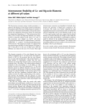
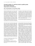
(IjBa(S ⁄ A)] [7]. The cells were stimulated with IL-1b for 4 h and real-time PCR analysis of MCPIP-coding transcript level was carried out. The control HepG2- mock cell line showed an almost 14-fold increase in line the transcript level, whereas in the HepG2 cell expressing the mutant form of the IjB inhibitor only a fivefold increase was observed (Fig. 1). These results clearly indicate that IL-1b-induced ZC3H12A gene expression is predominantly NF-jB dependent.
Determination of ZC3H12A gene transcription start site
In this study, we show that
the
18
16
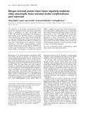
Although it has been proven that the ZC3H12A gene is composed of six exons, the first of which contains exclusively a noncoding sequence, localization of the gene’s transcription start site (TSS) is still not clear. Four sequences in the Entrez Gene database (http:// www.ncbi.nlm.nih.gov/sites/entrez?db=gene) match the ID of the MCPIP transcript, each having a distinct 5¢-UTR length (Fig. 2A). To identify the authentic TSS of the ZC3H12A gene we performed the 5¢ RNA ligase-mediated rapid amplification of cDNA ends (RLM-RACE) procedure. Analysis of RACE products by sequencing revealed the presence of two groups of (Fig. 2C). sequences with slightly different 5¢-ends Both sequences were registered to the NCBI database under a single accession number FJ695517. Because the first of the detected TSSs was represented by the majority of the sequenced clones (60%), we postulated
-
14
HepG2-mock HepG2-IκBα (S /A)
-
12
10
8
Although the contribution of MCPIP to inflamma- tion is indisputable, the mode of action of this protein seems to be ambiguous. As the protein contains a sin- gle CCCH-type zinc finger, a putative nuclear localiza- tion signal and two proline-rich potential activation domains, it was initially classified as a transcription factor [1,4]. Nevertheless, the data of Matsushita et al. [6] and our data (in preparation) suggest that MCPIP exhibits RNase properties, mediated by the PilT N-ter- minus domain. The experiments indicate that MCPIP may be an important factor regulating the half-life of transcripts coding for such proteins as IL-1b and IL-6. the transcriptional induction of the ZC3H12A gene after IL-1b stimula- tion is predominantly NF-jB-dependent. We prove that this induction is mediated by a transcriptional enhancer localized within the second intron of the ZC3H12A gene. The enhancer contains four functional jB sites (NF-jB-binding sites), revealed by computa- tional analysis and verified by chromatin immuno- precipitation, mutagenesis studies and gel-retardation assay (EMSA). Using EMSA, we also show that over- expression of MCPIP impedes formation of NF-jB ⁄ DNA complexes following IL-1b stimulation, suggesting an inhibitory effect on NF-jB activation. The effect was confirmed by studying the influence of MCPIP on the induction of NF-jB-dependent genes, namely IjBa and TNFa after IL-1b stimulation. The results presented here, together with previously reported findings on the significance of MCPIP in the inhibition of NF-jB activation [5], provide evidence of a novel negative feedback loop in the activation of this transcription factor, and indicate the potential signifi- cance of MCPIP in the treatment of NF-jB-related diseases.
Results
**
6
A N R m P I P C M
4
l o r t n o c s v e g n a h c d l o F
2
0
Participation of the NF-jB pathway in activation of ZC3H12A gene
–
+
IL-1β
Fig. 1. Contribution of NF-jB pathway to induction of ZC3H12A transcription after IL-1b stimulation. In the experiment, the level of MCPIP transcript was measured by real-time PCR in control HepG2 cells (HepG2-mock) and cells deprived of NF-jB activation ability [HepG2-IjBa(S ⁄ A)]. Cells were serum-starved for 24 h and stimu- lated for 4 h with 60 UÆmL)1 IL-1b. Graphs present fold increase of MCPIP transcript level normalized to unstimulated cells. The results are means ± SD of three experiments. Student’s t-test was used for statistics: **P < 0.01.
FEBS Journal 276 (2009) 5892–5905 ª 2009 The Authors Journal compilation ª 2009 FEBS
5893
The degradation of inhibitors of jB (IjBs) is a crucial step in the activation of the NF-jB transcription factor signalling pathway and is followed by translocation of the transcription factor to the nucleus. To investigate the role of this pathway in the induction of transcrip- tion of the ZC3H12A gene after exposure of cells to IL-1b, we used a HepG2 cell line deprived of NF-jB activation ability [cells stably transduced with the retroviral vector pCFG5-IEG2 containing a domi- nant negative mutant form of the NF-jB inhibitor
L. Skalniak et al.
Interplay between NF-kappaB and MCPIP RNase
A
B
C
D
Fig. 2. Analysis of ZC3H12A transcription start site localization. (A) Set of the ZC3H12A gene transcription start sites represented by entries in Entrez Gene database. (B) Visualization of 5¢-ends of sequences extracted from Expressed Sequence Tags database revealed by BLAST search with the ZC3H12A gene query. The size of the arrows corresponds to the number of entries in dbEST matching the particular 5¢-end. (C) The result of 5¢ RLM-RACE experiment. Numbers denote the percent of sequenced clones corresponding to each sequence. (D) Visuali- zation of ZC3H12A minimal promoter with determined transcription start sites and putative promoter elements of the gene. Inr ) initiator, +1 indicates the major TSS.
tool [11]. Human Rel-family protein (c-Rel, p50 and p65) potential binding sites were detected with the TF score cut-off of 85%. The result of the computational analysis is presented in Fig. 3.
it to be the strongest TSS of the ZC3H12A gene in HepG2 cells and designated the corresponding nucleo- tide as +1 in the gene sequence (Fig. 2D). The second group of sequences obtained in the experiment cover the remaining 40% of the clones and start three nucle- otides downstream at the +4 position.
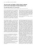
The prediction procedure revealed the accumulation of potential NF-jB-binding sites within the second intron of the ZC3H12A gene. Thus, after initial experi- mental verification of the activity of the ZC3H12A promoter region, we focused on this intronic sequence while designing further luciferase deletion mutants (see below).
ZC3H12A minimal promoter
Two potential elements of the classical-type poly- merase II promoter: TATA box and Initiator element (Inr) were found in the nearest neighbourhood of the described TSSs (Fig. 2D). In addition, the two TSSs of ZC3H12A are the most represented in the Expressed Sequence Tags database as 5¢-ends of sequences under- going transcription and corresponding to MCPIP- coding transcript (Fig. 2B). This additional proof increases the reliability of indicated TSSs.
Computational analysis of potential jB sites within the ZC3H12A gene and in its neighbourhood
are elements
after
FEBS Journal 276 (2009) 5892–5905 ª 2009 The Authors Journal compilation ª 2009 FEBS
5894
Transcription regulatory commonly located within a short sequence upstream of the mini- mal promoter. Nevertheless, many groups have reported the presence of regulatory elements more dis- tant either upstream or downstream of the regulated gene, as well as within the gene [8–10]. In order to localize potential regulatory elements in the human ZC3H12A locus containing NF-jB-binding sites, com- putational transcription factor binding-site prediction analysis was performed. The sequence ranging from )2.5 kb and reaching the end of the ZC3H12A gene (+9796 bp) was analysed using the consite online Computational analysis identified no significant poten- tial NF-jB-binding site within the region directly upstream of the ZC3H12A gene and suggested the existence of only two potential c-Rel recognition sites within the first intron of the gene (Fig. 3). To verify the importance of those two sites and to investigate the ZC3H12A promoter potential toward gene activa- tion in response to IL-1b stimulation, initial deletion mutants were constructed (Fig. 3, constructs F2-R2, F3-R2 and F4-R2). Luciferase activity assay revealed that the observed ability of the ZC3H12A promoter- containing vectors to mediate transcription induction is significantly below the observed increase of the MCPIP transcript IL-1b stimulation level (2.5- versus >14-fold increase in the fourth hour of stimulation; Fig. 3). It is rather comparable with the induction of luciferase gene transcription driven by an together empty vector (Fig. 3). These observations,
L. Skalniak et al.
Interplay between NF-kappaB and MCPIP RNase
c-REL p50 p50
c-REL
c-REL p65
c-REL c-REL p50 p65 p65 c-REL
c-REL p50 c-REL p65 c-REL
c-REL
c-REL c-REL p65
c-REL
c-REL
p50
p50 c-REL p65
c-REL
p50 p50 p65
Fig. 3. Localization of IL-1b-responding element in the second intron of the ZC3H12A gene, as revealed by luciferase activity assay. The upper part of the figure represents computational prediction of putative NF-jB-binding sites within the ZC3H12A gene. The analysis was performed starting from 2.5 kb upstream from the transcription start site. The sequence was analysed with CONSITE software towards Homo sapiens Rel family members: c-Rel, p50 and p65 with the TF score cut-off of 85%. For luciferase activity assay HepG2 cells were transfected with 25 fmol of pGL4.17[luc2 ⁄ Neo] plasmid containing investigated fragments of ZC3H12A cloned adjacent to luc2 reporter gene, and 5 fmol of pEF1 ⁄ Myc–His ⁄ Gal vector coding for b-galactosidase as an internal transfection control. Following 48 h incubation, cells were stimulated for 8 h with 60 UÆmL)1 of IL-1b and chemiluminescence-based luciferase activity assay was performed. Each data point rep- resents the mean ± SD of three independent experiments, each performed in duplicates, and is presented as fold stimulation (normalized firstly to b-galactosidase and then to basal construct activity). Student’s t-test was used for statistics: **P < 0.01. luc2, reporter gene; E1, E2, E3, E4, E5, E6, ZC3H12A exons; —, intron ⁄ intergenic sequence; h, noncoding exonic region; , coding region; EnhA, sequence bearing multiple potential NF-jB-binding sites localized in second intron of the ZC3H12A gene.
with the absence of potential jB sites within the near- est 5¢ neighbourhood of the first exon, suggest that IL-1b-dependent transcription activation of the gene requires other regulatory elements. fragments of second intron of
is
Further experiments revealed that the shortest exam- ined sequence which maintains full activity of the ZC3H12A promoter located between nucleotide )124 and +18 (Fig. 3, construct F14-R9). Therefore, this construct was used to generate further constructs for experiments testing the postulated transcription enhancer (see below).
Localization of transcription enhancer in the second intron of the ZC3H12A gene
FEBS Journal 276 (2009) 5892–5905 ª 2009 The Authors Journal compilation ª 2009 FEBS
5895
described computational previously, As analysis revealed the presence of potential NF-jB-binding sites in the second intron of the ZC3H12A gene. For this reason, we focused on this region while preparing fur- ther reporter gene constructs. A luciferase activity assay showed that only two constructs containing the longest the the ZC3H12A gene are able to significantly increase the transcript level after IL-1b stimulation. This indicates the presence of IL-1b-responding elements within a sequence located between nucleotides +2626 and +2950 (Fig. 3), which was earlier predicted to contain multiple potential jB sites. Taking into account this prediction, a shorter sequence from this region was extracted (+2791 to +3088) and termed EnhA. For further experiments, this sequence was then fused with a minimal promoter of the ZC3H12A gene to create an A ⁄ F14-R9 construct, the activity of which was com- parable with F2-R7 and F2-R8 constructs (Fig. 3). The
L. Skalniak et al.
Interplay between NF-kappaB and MCPIP RNase
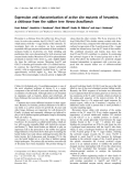
Fig. 4. Verification of four putative NF-jB-binding sites, localized in the second intron of the ZC3H12A gene in the EnhA sequence. (A) The effect of point mutagenesis of jB sites on transcription activation properties of the A ⁄ F14-R9 construct, as measured by luciferase activity assay. HepG2 cells were transfected with 25 fmol of pGL4.17[luc2 ⁄ Neo] plasmid or the plasmid containing fragments of ZC3H12A regula- tory elements cloned adjacent to luc2 luciferase-coding reporter gene, as indicated on the figure, and 5 fmol of pEF1 ⁄ Myc–His ⁄ Gal vector coding b-galactosidase as an internal transfection control. Following 48 h of incubation, cells were stimulated with 60 UÆmL)1 of IL-1b for 8 h and chemiluminescence-based luciferase activity assay was performed. Each data point represents the mean ± SEM of three experi- ments, each performed in duplicate, and is presented as fold stimulation (normalized first to b-galactosidase and then to basal construct activity). Student’s t-test was used for statistics: *P < 0.05. luc2, reporter gene; N1, N2, N3, N4, putative jB sites; E1, E2, E3, E4, E5, E6, ZC3H12A exons; —, intron ⁄ intergenic sequence; h, noncoding exonic region; , coding region, red cross indicates the mutation of the jB site. (B) Chromatin immunoprecipitation experiment indicating the presence of NF-jB-binding sites within the EnhA sequence. PCR was performed on 10· (1 : 10) or 100· (1 : 100) diluted, sonicated DNA isolated from control and stimulated cells (Input) and on the same DNA samples subjected to immunoprecipitation with antibody against p-65 NF-jB subunit (ChIP, a-p65). Unspecific antibodies served as an iso- type control (IgG) of the chromatin immunoprecipitation experiment. Primers used in PCR were designed to amplify the EnhA sequence in the ZC3H12A gene (MCPIP) or the IjBa gene promoter region containing known NF-jB-binding sites as a control (IjBa). Primers are listed in Table 1. (C) Verification of the relevance of four NF-jB-binding sites from the EnhA sequence by EMSA. Nuclear extracts isolated from IL-1b-stimulated ⁄ unstimulated HepG2 cells were incubated with radiolabelled oligonucleotides corresponding to N1, N2, N3 and N4 jB sites (wt) or mutant forms of these oligonucleotides deprived of NF-jB recognition sequences (mut). A radiolabelled oligonucleotide bearing the NF-jB-binding sequence from the HIV enhancer was used as a positive control (lanes 9 and 10). Lanes 1, 3 and 9, extracts from unstimu- lated Hep-G2 cells; lanes 2, 4-8 and 10, extracts from Hep-G2 stimulated for 40 min with 60 UÆmL)1 IL-1b; lane 5, the effect of addition of a 100-fold excess of cold probe; lanes 6–8, the effect of addition of antibody against p-65 (lane 6), p-50 (lane 7) or control antibody (lane 8).
presence of NF-jB-binding sites within the EnhA region was confirmed by chromatin immunoprecipita- tion assay, in which an antibody against the human p65 NF-jB subunit was used (Fig. 4B).
EnhA enhancer contains four NF-jB-binding sites stimulated with cells and
For each probe bearing an original sequence of the examined NF-jB-binding site, the gel-shift band was observed after stimulation with IL-1b (Fig. 4C, lanes 2). The band corresponding to the NF-jB ⁄ probe complex was present neither in lane 1 (original probes, control cells), nor in lanes 3 and 4 (mutated probes, control IL-1b, cells respectively), indicating that the shift is stimulation- dependent and all mutations abolish the NF-jB-bind- ing capability of the examined binding sites. Addition of a 100-fold excess of a cold original probe as a bind- ing competitor also abolished the formation of observed complexes (lanes 5).
To increase the sensitivity of NF-jB-binding site pre- diction within the EnhA sequence, computational anal- ysis was repeated using the consite online tool and a TF score cut-off of 78%. The analysis revealed the presence of four potential binding sites for NF-jB subunits. Each of these sites was given a separate label: N1, N2, N3 and N4, in accordance with the decreasing TF score. Then, point mutagenesis was carried out on the A ⁄ F14-R9 construct in order to create four mutant forms of this construct, deprived of consecutive sites. The primers used in the point mutagenesis procedure are listed in Table 1.
Interestingly,
that at least
FEBS Journal 276 (2009) 5892–5905 ª 2009 The Authors Journal compilation ª 2009 FEBS
5896
To further examine the composition of the com- plexes observed in lane 2 (Fig. 4C), a supershift assay was performed with antibodies against human p65 and p50 NF-jB subunits. A characteristic super- shift band was observed for all examined probes when anti-p65 was added (Fig. 4C, lanes 6), suggest- ing the presence of this subunit in the NF-jB ⁄ probe complexes. the addition of anti-p50 IgG (lanes 7) resulted in a weakening of the signal corresponding to the shifted band in comparison with the signal observed for control antibody (lanes 8). The described effect of antibody addition may result from competition between antibody and DNA probe in binding to the p50 NF-jB subunit. In our hands, this is a common phenomenon observed for supershift assays with probes known to bind NF-jB lane 2 and 3). The obtained complexes (see Fig. 5, some NF-jB com- suggests result plexes bound to sites N1, N3 and N4 contain p50 subunits. The luciferase activity assay indicated the importance of each of the predicted binding sites after IL-1b stimu- lation (Fig. 4A) suggesting their potential role in acti- vation of ZC3H12A gene transcription. In addition, the mutagenesis of all four jB sites together reduced the transcription-activating potential of the A ⁄ F14-R9 con- struct to the base level observed for the F14-R9 con- struct. To verify the ability of sites N1–N4 to bind activated NF-jB complexes, EMSA was performed. Radiolabelled probes corresponding to the original sequences of nominated NF-jB-binding sites and to the sequences carrying identical mutations as mutant forms of the A ⁄ F14-R9 construct were used (Fig. 4C). The probes used in this experiment are listed in Table 2.
L. Skalniak et al.
Interplay between NF-kappaB and MCPIP RNase
A
B
C
FEBS Journal 276 (2009) 5892–5905 ª 2009 The Authors Journal compilation ª 2009 FEBS
5897
L. Skalniak et al.
Interplay between NF-kappaB and MCPIP RNase
MCPIP exerts an inhibitory effect on NF-jB activation
was examined using real-time PCR. Overexpression of MCPIP resulted in a lowering of the amount of both TNFa- and IjBa-coding transcripts (Fig. 6A), whereas silencing of the gene increased the transcript level of those proteins after IL-1b treatment (Fig. 6B). Overex- pression of MCPIP was confirmed by western blotting and the silencing of MCPIP was verified by real-time PCR (Fig. 6A and B, respectively, left-hand panel).
Discussion
the ZC3H12A gene was previously Expression of shown to be induced by inflammation-related factors, such as MCP-1, TNFa, LPS and IL-1b [1,5]. More- over, the importance of the ZC3H12A gene product, namely MCPIP, has been postulated in the immune system. The protein was identified as being able to reduce macrophage activation upon LPS recognition in the murine macrophage cell line Raw264.7 [5]. The authors concluded that MCPIP may be involved in the regulation of macrophage activation and implicated in the pathogenesis of inflammatory diseases. It was shown recently that MCPIP overexpression abolishes the p65-induced transcription from NF-jB- activated promoters [5]. To further test the influence of MCPIP on the NF-jB signalling pathway, EMSA was performed. HepG2 cells were transfected either with the construct coding for MCPIP, or with an empty vector, and stimulated for 40 min with 60 UÆmL)1 of IL-1b. Alternatively, cells were treated with siRNA specific for MCPIP and stimulated in the same manner. Following stimulation, nuclear extracts were isolated and the formation of NF-jB ⁄ DNA complexes was examined using a radiolabelled probe named NF-jBor (Table 2) bearing a well-known NF-jB- binding DNA sequence from the human immuno- deficiency virus enhancer [12] (Fig. 5A). Overexpres- sion of MCPIP was confirmed by western blotting (Fig. 5B) and siRNA-mediated inhibition of MCPIP gene expression was verified by real-time PCR (Fig. 5C).
In this study, we identified two major transcription start sites of the ZC3H12A gene. Computational anal- ysis of the direct neighbourhood of those TSSs indi- cated the presence of two motifs with high similarity to well-characterized cis-acting elements of the RNA polymerase II core promoter: the TATA box and the it seems that the ZC3H12A Inr element [13]. Thus, promoter belongs to the TATA-containing group of RNA polymerase II core promoters.
Stimulation of cells with IL-1b resulted in activation of NF-jB in control cells (Fig. 5, lanes 1–3, 5 and 9). To ensure that the observed shift is a consequence of binding of the NF-jB transcription factor, the involve- ment of the p65 NF-jB subunit in formed complexes was confirmed by the addition of a p65-specific anti- body, resulting in the appearance of a supershift band (lane 1). Moreover, addition of an anti-p50 IgG weakened the NF-jB gel-shift signal, suggesting the presence of complexes containing this subunit (lane 2). Control anti-STAT1 IgG had no influence on the observed gel band shift (lane 3).
Overexpression of MCPIP significantly reduced the signal of activated NF-jB bound to the radioactive probe (lane 7 versus 5). Conversely, MCPIP silencing augmented the activation of NF-jB (lane 13 versus 11 and 9). Both results suggest the presence of an inhibitory effect of the MCPIP protein on NF-jB activation.
MCPIP inhibits the induction of NF-jB dependent genes
Large-scale mapping of human minimal promoter topology revealed that promoters of genes displaying tissue-specific expression often contain a TATA box element within the minimal promoter region. Previous studies have shown that TATA box-associated pro- moters are involved in the tight regulation of genes, allowing diversification of expression in different cell types under various conditions [14]. These data seem to be in accordance with our recent observation that the level of MCPIP mRNA varies between different tissues. However, the functionality of the predicted TATA box needs to be verified experimentally. The existence of two or more TSSs within the Inr element is not an unusual situation, because transcription initi- ation sites are currently known to be described rather as short sequences than a single nucleotide [15].
FEBS Journal 276 (2009) 5892–5905 ª 2009 The Authors Journal compilation ª 2009 FEBS
5898
It was shown that in cells overexpressing MCPIP and stimulated with lipopolysaccharide (LPS) activation of NF-jB-dependent genes is reduced [5]. To test this in our model, HepG2 cells were transfected with a vector encoding MCPIP or treated with siRNA specific for MCPIP. Cells were then stimulated with IL-1b and the expression of transcripts coding for tumour necrosis factor a (TNFa) and IjBa, well known NF-jB targets, Interestingly, none of the TSSs revealed in the RACE experiment match any of the TSSs postulated previ- ously. However, this does not imply that those sites are irrelevant, because their existence may be a part of the mechanism altering the expression of the ZC3H12A gene in other tissues [14,16]. Additional transcription
L. Skalniak et al.
Interplay between NF-kappaB and MCPIP RNase
Table 2. Oligonucleotides used as molecular probes for EMSA. Font styles provide the following information: bold, nucleotides within NF-jB binding sites that were subjected to mutagenesis.
Probe name
Primers alignment
NF-jBor
5¢ AGCTTCAGAGGGGACTTTCCGAGAGG 3¢
Table 1. Primers used in experimental approaches. All sequences are given in the 5¢ fi 3¢ direction. Font styles provide the follow- ing information: bold, ZC3H12A start codon mutation and nucleo- tides within NF-jB binding sites that were subjected to mutagenesis; italic, restriction enzyme recognition site. RLM-RACE, RNA ligase-mediated rapid amplification of cDNA ends; ChIP, chro- matin immunoprecipitation.
3¢ AGTCTCCCCTGAAAGGCTCTCCTCGA 5¢
GSN1or
5¢ AGGAGGGGAATTCCAGC 3¢
Primer
Sequence
3¢ TCCCCTTAAGGTCGGAG 5¢
Real-time PCR primers
GSN1mu
5¢ AGGAGCTCAATTCGAGC 3¢
3¢ TCGAGTTAAGCTCGGAG 5¢
GSN2or
5¢ GGTGGGGAAATTCACC 3¢
3¢ CCCCTTTAAGTGGAGG 5¢
GSN2mu
5¢ GGTGGCCAAAGGTACC 3¢
3¢ CCGGTTTCCATGGAGG 5¢
GSN3or
5¢ GGCTCGGGGGTTTCTG 3¢
3¢ AGCCCCCAAAGACTGG 5¢
GGCAGCGACCTGAGACCAGTG GGTGTGTGATGGGCACGTCGG AACCTGCAGCAGACTCCACTCC ACACGTGTGGCCATTGTAGTTGG CAGGCGGTGCTTGTTCCTCAG GGGCTACAGGCTTGTCACTCG GACATCACCAAGGGTGTGCAG TCAGCACACTGGCATAGAGGC
ZFFq ZFRq hqIkBaF hqIkBaR hTNFf hTNFr EF2F EF2R
GSN3mu
5¢ GGCTCGGAGGCATATG 3¢
3¢ AGCCTCCGTATACTGG 5¢
GSN4or
5¢ GGAAGGGAATTTTTT 3¢
3¢ TCCCTTAAAAAAAGG 5¢
GSN4mu
5¢ GGAAGGAGATCTTTT 3¢
Primers used in the RLM-RACE procedure CGACTGGAGCACGAGGACACTGA AGCCCAGCTTCCGGAAGAAGTCC GGACACTGACATGGACTGAAGGAGTA CCCAGATCTGCCACTGATAGCTCAGACTCCTG
raceF1 raceR1 raceF2 raceR2
3¢ TCCTCTAGAAAAAGG 5¢
Primers used for constructs preparation
CCG CTCGAG CTCCAGCGTGTGGGCTCTGTG CCG CTCGAG CCGTCCGCACCTCGGTCAGTG CCG CTCGAG AGCAGGAAGGGGCGAGGCAGC CCC AGATCT CCCTGTGGAGAGAAGCCTGTCC CCG CTCGAG AGGCAGCCCCGCCCCCGGG CCC AGATCT GCCACTGATAGCTCAGACTCCTG CATTCCTGTGCTGGGGGAT GCTACATGAGGCTGGACACT CCC AAGCTT TTGCATATGATGGGGGGGCTAGC CCC GGATCC AAGCTT GCTGGGAGGGAGAGGACAGGG CCC AAGCTT TCCATGGGGCCGAGTCCTGGG CCG GGTACC TGAGAAGCAGAGCCACGCACCC CCG CTCGAG CCAGCTAGGCTGCTCCTGCCC
F2 F3 F4 F11 F14 R2 R4 R6 R7 R8 R9 EnhAF1 EnhAR1
Point mutagenesis
and both computational
GGTAGTGGCAG GAGCTC AATTCGAGCCTCAAACTTCC GGAAGTTTGAGGCTCGAATT GAGCTC CTGCCACTACC CATATGCAAATCATGGCCAAA GGTACC TCTGTTTCC GGAAACAGA GGTACC TTTGGCCATGATTTGCATATG CGCACCCACCTCGGAGG CATATG AGCTGGGGTAGTGGC GCCACTACCCCAGCT CATATG CCTCCGAGGTGGGTGCG CTGTTTCCTTCTAAGG AGATCT TTTTCCACTCGCCAGGC GCCTGGCGAGTGGAAAA AGATCT CCTTAGAAGGAAACAG
CCGGGTACCTGAGAAGCAGAGCCACGCACCC CCCAAGCTTAGAAGGAAACAGAGGTGAATTTCCC GACGACCCCAATTCAAATCG TCAGGCTCGGGGAATTTCC
MuN1F MuN1R MuN2F MuN2R MuN3F MuN3R MuN4F MuN4R ChIP ChipF ChipR ChipIkBf ChipIkBr
gene activation provide proof of the NF-jB-depen- dency of the transcriptional induction of this gene. experimental Using approaches, we identified a transcription enhancer con- taining four NF-jB-binding sites located within the second intron of the ZC3H12A gene. We postulate that this enhancer, termed EnhA, mediates the activa- tion of ZC3H12A upon IL-1b stimulation. However, taking into consideration that other proinflammatory molecules may act through distinct signal transduction pathways, the question of whether the gene response to those stimulants also requires EnhA activation warrants future investigation.
the ZC3H12A gene was
start sites could then be engaged in altered transcrip- tion initiation in some other cell types.
In this study, we also show that MCPIP exerts an inhibitory effect on the activation of the transcrip- tion factor NF-jB. This inhibitory effect results in decreased induction of transcript levels for NF-jB- dependent genes, namely IjBa and TNFa. The observations are in agreement with recently published data showing NF-jB-mediated transcription inhibi- tion in the case of MCPIP overexpression [5]. These results argue that the MCPIP protein acts as a novel negative regulator for the NF-jB signalling pathway. shown to be Because induced predominantly in a NF-jB-dependent man- ner, this observation places the MCPIP in the nega- tive regulatory loop of the cell’s response to the inflammatory state.
FEBS Journal 276 (2009) 5892–5905 ª 2009 The Authors Journal compilation ª 2009 FEBS
5899
It is well known that at least three species of mole- cules activating ZC3H12A expression (TNFa, LPS and IL-1b) are potent inducers of the NF-jB signalling pathway [17]. Our studies on IL-1b-induced ZC3H12A NF-jB is a transcription factor known to play a central and crucial role in regulating such complex
L. Skalniak et al.
Interplay between NF-kappaB and MCPIP RNase
A
B
C
Fig. 5. MCPIP inhibits NF-jB activation after IL-1b stimulation. (A) EMSA presenting the effect of MCPIP overexpression and silencing on NF-jB activation. For the experiment HepG2 cells were cultured on six-well plates and transfected with a MCPIP-containing construct (lanes 6 and 7) or an empty vector (lanes 1–5). For MCPIP silencing cells were treated with 50 nM MCPIP siRNA (lanes 12 and 13) or scram- bled siRNA (lanes 10 and 11) and plated on a 12-well plate. In addition, untreated cells were analysed (lanes 8 and 9). After 48 h cells were stimulated for 40 min with 60 UÆmL)1 of human recombinant IL-1b (lanes 1–3, 5, 7, 9, 11 and 13). Unstimulated cells were used as a control (lanes 4, 6, 8, 10 and 12). Nuclear extracts were incubated with radiolabelled oligonucleotides bearing the NF-jB-binding DNA sequence from the HIV enhancer. Lane 1, 2 and 3 represent the effect of addition of antibody against p-65, p-50 and control anti-STAT1 IgG, respectively. Overexpression of MCPIP was verified by western blotting (B) and MCPIP silencing was verified by real-time PCR (C). Student’s t-test was used for statistics: **P < 0.01.
interesting object to study and a putative target for future therapeutic approaches.
Materials and methods
Human recombinant IL-1b was purchased from Promokine (Heidelberg, Germany). Restriction endonucleases, i.e. KpnI, XhoI, HindIII, SacI, KpnI, NdeI and BglII, were obtained from New England Biolabs (Ipswich, MA, USA). Rabbit polyclonal IgG antibodies specific for human p65 (cat. sc-109), p50 (cat. sc-114) and unspecific IgG were from Santa Cruz Biotechnology (Santa Cruz, CA, USA). [32P]dCTP[aP] was purchased from Hartmann Analytic GmbH (Braunschweig, Germany).
Materials and reagents
Human hepatocellular carcinoma-derived cells (HepG2 line from ATCC) were cultured in Dulbecco’s Modified Eagle’s
processes as the immune response, differentiation and tumorigenesis [18,19]. The diversity of biological roles fulfilled by NF-jB is achieved throughout a sophisti- cated regulatory network, providing discriminatory effects in signal transduction pathways initiated by diverse stimulatory factors. Much effort has been made recently to uncover those networks, because these incontestably increase our understanding of many physiological and pathological processes, enabling the improvement of many NF-jB-associated disease treat- ments [20]. This study, along with other recent studies cited in this article, determines the MCPIP protein as a novel cog in the NF-jB-regulating machine. Despite the reliability of both the observed NF-jB influence on ZC3H12A transcription and the negative effect of MCPIP on NF-jB activation, the exact mode and stage of MCPIP action still need to be determined. Altogether, observations Cell lines
FEBS Journal 276 (2009) 5892–5905 ª 2009 The Authors Journal compilation ª 2009 FEBS
5900
concerning the mutual its regulatory effect between MCPIP and NF-jB, implication in the inflammatory state and the ambi- guous nature of the protein’s action, make MCPIP an
L. Skalniak et al.
Interplay between NF-kappaB and MCPIP RNase
A
B
Fig. 6. Effect of MCPIP overexpression (A) and silencing (B) on induction of IjBa and TNFa transcript level after stimulation with IL-1b. HepG2 cells were cultured on 12-well plates and transfected with MCPIP-containing construct or an empty vector as indicated (A). For MCPIP silencing cells were treated with 50 nM MCPIP siRNA or scrambled siRNA as indicated (B) and plated on 12-well plate. After 48 h cells were stimulated for 3 h with 60 UÆmL)1 of human recombinant IL-1b and transcript level for IjBa (A and B, central panel) and TNFa (A and B, right-hand panel) was examined by real-time PCR. Overexpression of MCPIP was verified by western blotting (A, left-hand panel) and MCPIP silencing was verified by real-time PCR (B, left-hand panel). Student’s t-test was used for statistics: *P < 0.05, **P < 0.01, ***P < 0.001.
with 5% (v ⁄ v) fetal bovine serum and 1 mgÆmL)1 Zeocin (Invitrogen, Carlsbad, CA, USA).
Medium (Sigma, St Louis, MO, USA) supplemented with 5% (v ⁄ v) fetal bovine serum (Gibco, Carlsbad, CA, USA). Cell cultures were maintained at 37 (cid:2)C in a humidified atmo- sphere of 5% CO2 and passaged every 4–5 days. For experi- ments, cells were seeded on poly-l-lysine (Sigma) coated 12- or 24-well plates (BD Falcon, San Jose, CA, USA).
siRNA for MCPIP (5¢-CCCUGUUGAUACACAUU- GUTT), as well as scrambled siRNA were obtained from Ambion. Transfection was performed with siPORT NeoFx agent (Ambion, Austin, TX, USA) according to the manu- facturer’s protocol. Shortly, for single transfection 3 lL of siPORT NeoFx and siRNA to a final concentration of 50 nm were diluted separately in 50 lL of OPTI-MEM,
HepG2 cells stably transduced with a retroviral vector encoding green fluorescent protein alone (HepG2-mock), or encoding both green fluorescent protein and the dominant negative form of the IjBa inhibitor [HepG2-IjBa(S ⁄ A)], were kindly provided by S. Ludwig (Heinrich-Heine Uni- versity, Du¨ sseldorf, Germany) [7]. The transduced cells were cultivated in Dulbecco’s modified Eagle’s medium
FEBS Journal 276 (2009) 5892–5905 ª 2009 The Authors Journal compilation ª 2009 FEBS
5901
MCPIP silencing
L. Skalniak et al.
Interplay between NF-kappaB and MCPIP RNase
after 10 min of incubation they were combined and left for another 10 min to allow complex formation. Next, the mix- ture was dispensed on 12-well plates and overlaid with 8 · 104 HepG2 cells. The effect of gene silencing was assessed 48 h post transfection.
(Sigma)
fluoride Inhibitor Cocktail
For RNA isolation, cells were seeded on poly-l-lysine coated 12-well plates, serum-starved overnight in Opti-Mem serum-free medium (Gibco) and stimulated for 4 h with 60 UÆmL)1 of IL-1b. Cells were washed twice with NaCl ⁄ Pi and total RNA was isolated using the modified Chomczyn- ski–Sacchi method, as described previously [2]. RNA concentration was measured with a ND-1000 spectropho- tometer (NanoDrop, Wilmington, DE, USA) and RNA integrity was verified on a 1% denaturating agarose gel.
Nuclear extracts were prepared by a mini-extraction proce- dure. After a brief wash with cold NaCl ⁄ Pi, cells were collected with a rubber policeman and centrifuged for 5 min at 400 RCF. Pelleted cells were resuspended in 200 lL of buffer containing 10 mm Hepes pH 7.9, 10 mm KCl, 0.1 mm EDTA, 0.1 mm EGTA, 1 mm Na3VO4, 1 mm dithiothreitol, 0.2 mm phenylmethanesulonyl and Complete Protease (Roche, Basel, Switzerland). After incubation on ice for 10 min, 15 lL of 10% Nonidet NP-40 was added. Nuclei were collected by centrifugation at 500 g for 3 min at 4 (cid:2)C and resuspended in 50 lL of extraction buffer (20 mm Hepes pH 7.9, 400 mm NaCl, 1 mm EDTA, 1 mm EGTA, 1 mm Na3VO4, 1 mm dithiothreitol and Complete Protease Inhibitor cocktail). Samples were incubated for 15 min on ice and mixed by vor- texing every 3 min. Following incubation, the nuclei were centrifuged for 5 min at 14 000 g. Protein concentration was determined by BCA assay (Sigma). For storage, glycerol was added to a final concentration of 10% and nuclear extracts were frozen in liquid nitrogen and placed at )80 (cid:2)C. For the NF-jB-directed EMSA, double-stranded probes were pre- pared by equimolar mixing of primer pairs as indicated in Table 2, denaturation at 98 (cid:2)C for 5 min and refolding at room temperature. One picomole of double-stranded probes were labelled with 3.33 pmol of [32P]dCTP[aP] and Klenow polymerase (Fermentas, Burlington, Canada) for 1 h at 37(cid:2)C in the presence of 1 nmol dATP, dGTP and dTTP mix. Radioactive probes were purified with QIAquick PCR Purification Kit (Qiagen, Du¨ sseldorf, Germany) and sus- pended in 50 lL of Tris buffer.
For the real-time PCR experiment, 1 lg of total RNA was reverse-transcribed using oligo(dT) 15 primer (Promega, Madison, WI, USA) and M-MLV reverse transcriptase (Promega). Following synthesis, cDNA was diluted 5· and real-time PCR was carried out using Rotor-Gene 3000 (Corbett, Cambridge, UK) system and Sybr Green-based master mix (Finnzymes, Espoo, Finland). After an initial denaturation step for 10 min at 95 (cid:2)C, conditions for cycling were: 40 cycles of 20 s at 95 (cid:2)C, 20 s at 62 (cid:2)C and 30 s at 72 (cid:2)C. The fluorescence signal was measured right after the extension step at 72 (cid:2)C. At the end of the PCR cycling, a melting curve was generated to verify specificity of the PCR product. For the normalization of each sample, the amount of eukaryotic translation elongation factor 2 cDNA was measured (primers EF2F and EF2R). All samples were run in triplicates. The primers used in real-time PCR are listed in Table 1.
Real-time PCR
To investigate the human ZC3H12A transcription start site localization, RLM-RACE was carried out with the GeneR- acer Kit (Invitrogen). Total RNA from HepG2 cells was isolated using the modified Chomczynski–Sacchi method and processed according to the GeneRacer manual. The raceR1 primer was used for the reverse transcription step (Table 1). The first PCR was carried out with the use of raceF1 and raceR1 primers. For the nested PCR, the prod- uct of the first PCR was used and the specific product was amplified with raceF2 and raceR2 primers (Table 1).
Five micrograms of nuclear extracts from control cells and cells stimulated with IL-1b were incubated for 10 min at room temperature with 1 ng of poly(dI-dC) (Sigma) in binding buffer (10 mm Hepes, pH 7.9, 100 mm NaCl, 0.5 mm EDTA, 10% v ⁄ v glycerol and 0.2 mm dithiothrei- tol). Next, 1 lL of purified labelled probe was added and samples were incubated for 45 min at room temperature. For the supershift assay, after the initial 15 min of incuba- tion with radiolabelled probe, the anti-p65, anti-p50 or con- trol IgG was added as indicated in Fig. 4C and samples were incubated at room temperature for additional 30 min. Following incubation, the samples were loaded on 5% (w ⁄ v) non-denaturating polyacrylamide gel. Electrophoresis was run at 160 V for 2.5 h in 0.5 · TBE. Gels were trans- ferred to 3 mm Chr chromatography paper (Whatman, Maidstone, UK) and dried under vacuum. The bands were visualized by 48 h exposure to a phosphoimage screen and read using molecular imager fx and quantity one soft- ware (BioRad, Hercules, CA, USA).
Mapping the transcription start site
EMSA
All DNA sequences were extracted from the Entrez Nucleotide database, National Center for Biotechnology
HepG2, HepG2-mock and HepG2-IjBa(S ⁄ A) cell lines were cultured on 60 mm cell culture dishes. After overnight serum starvation, cells were stimulated for 40 min with 60 UÆmL)1 IL-1b. Unstimulated cells served as an experimental control.
FEBS Journal 276 (2009) 5892–5905 ª 2009 The Authors Journal compilation ª 2009 FEBS
5902
Computational analysis
L. Skalniak et al.
Interplay between NF-kappaB and MCPIP RNase
each
constructs
25 fmol 2.5 fmol
transfection, and
Information (http://www.ncbi.nlm.nih.gov). To identify potential NF-jB-binding sites within the ZC3H12A gene and its promoter region, DNA sequence ranging from )2500 to +9796 (3¢-end of the gene) was analysed with ConSite regulatory elements prediction software (http:// www.phylofoot.org/consite) [11]. The sequence was analy- sed towards Homo sapiens Rel family members: c-Rel, p65 and p50 with the TF score cut-off of 85%.
of Mem medium. For pGL4.17[luc2 ⁄ Neo]-based of pEF1 ⁄ Myc-His ⁄ Gal vector were used. After 12 h incuba- tion, fresh Opti-Mem medium was applied and cells were incubated for additional 36 h. Cells were stimulated for 8 h with 60 UÆmL)1 IL-1b and luciferase activity assay was per- formed using chemiluminescence-based Dual-Light System (Applied Biosystems, Foster City, CA, USA) according to the manufacturer’s protocol. Unstimulated cells served as a basal promoter activity control. Chemiluminescence was measured with MiniLuminat LB 96P (EG&G Berthold, Bad Wildbad, Germany). The luciferase activity of each construct was normalized to b-galactosidase activity as an integral transfection control. Data points were presented as fold stimulation of promoter activity (normalized to basal promoter).
Constructs
To investigate regulatory elements controlling the human ZC3H12A gene, a large panel of luciferase reporter con- structs was prepared. ZC3H12A gene and promoter frag- ments were PCR-amplified using Advantage(cid:3) 2 PCR Enzyme System (BD Biosciences Clontech, Mountain View, CA, USA) and cloned into the pGL4.17[luc2 ⁄ Neo] plasmid (Promega) with T4 DNA ligase (New England Biolabs). All constructs were verified by restriction digest and sequenc- ing. All primers utilized in the preparation of the constructs are listed in Table 1 and were designed basing on two Entrez Nucleotide NCBI entries: AL449284 and AL034379. The nucleotide corresponding to the TSS determined in the RLM-RACE experiment (see below) was marked as ‘+1’ and the numeration presented here is related to this nucleotide.
All point mutants presented here, except for the ATG mutants described before, were prepared based on the A ⁄ F14-R9 construct. For mutagenesis, the QuikChange XL Site-Directed Mutagenesis Kit (Stratagene, Cedar Creek, TX, USA) was used according to the manufacturer’s proto- col. Mutations were designed in a way to introduce novel restriction enzyme recognition sites (for sites N1, N2, N3 and N4 respectively SacI, KpnI, NdeI and BglII). Primers used in this procedure (listed in Table 1) were HPLC-puri- fied. All mutations were confirmed by restriction cleavage and sequencing.
Point mutagenesis
the ZC3H12A gene was
Constructs F2-R2, F3-R2 and F4-R2 (Fig. 3), containing the first exon of the ZC3H12A gene, first intron and first three codons of the ZC3H12A coding sequence, were pre- pared with F2, F3, F4 and R2 primers and XhoI ⁄ BglII (F2-R4, F2-R6, restriction enzymes. Further constructs F2-R7 and F2-R8, Fig. 3) were prepared in a two-step manner. First, the F2-R2 promoter region was cloned into the pGL4.17[luc2 ⁄ Neo] vector using F2 and R2 primers and XhoI ⁄ BglII restriction enzymes, and then the rest of the DNA fragment was added by subsequent cloning with BglII and HindIII (primers F11 and R4, R6, R7 or R8). Additional constructs were prepared with the F14 and R9 primers and cloned with XhoI and HindIII enzymes (con- struct F14-R9 containing the ZC3H12A minimal promoter: nucleotides from )124 to +18, Fig. 3) or by initial cloning with XhoI and HindIII and subsequent cloning with KpnI and XhoI with the use of primers EnhAF1 and EnhAR1 (i.e. A ⁄ F14-R9). In all prepared constructs, the first ATG codon of subjected to mutagenesis (ATG fi ATC) to allow the translation of the luciferase reporter gene. The pEF1 ⁄ Myc–His ⁄ Gal plasmid encoding b-galactosidase was used as an internal transfection control for the luciferase activity assays.
Chromatin immunoprecipitation
HepG2 cells were plated onto poly-l-lysine-coated 24-wells plates. Transfection was performed with Lipofectamine 2000 (Invitrogen) after overnight serum starvation in Opti-
HepG2 cells were cultured on a 100 mm cell culture dish and, where indicated, stimulated for 30 min with IL-1b (60 UÆmL)1) after overnight serum starvation. Proteins were cross-linked to DNA by incubation with 1% formaldehyde for 10 min at 37 (cid:2)C. The cells were washed with 0.125 m gly- cine in NaCl ⁄ Pi and collected with a rubber policeman in 1 mL of NaCl ⁄ Pi containing Complete Protease Inhibitor cocktail and 1 mm phenylmethanesulonyl fluoride. Cells were pelleted and washed once with a buffer containing 10 mm Hepes, pH 6.5, 0.5 mm EGTA, 10 mm EDTA and 0.25% Triton X-100 and then with a buffer containing 10 mm Hepes, pH 6.5, 0.5 mm EGTA, 1 mm EDTA and 200 mm NaCl. Cells were lysed with lysis buffer (50 mm Tris ⁄ HCl, pH 8.0, 1% SDS and 10 mm EDTA). Lysates were sonicated to produce DNA fragments between 300 and 600 bp. After centrifugation, the samples were diluted 1 to 10 with IP buffer (16.7 mm Tris ⁄ HCl, pH 8.0, 0.01% SDS, 1.1% Triton X-100, 1.2 mm EDTA, 16.7 mm NaCl and Complete Protease Inhibitor Cocktail) and immunoprecipi- tated overnight at 4 (cid:2)C using the anti-p65 IgG or the control unspecific IgG. Following immunoprecipitation, samples were incubated for 1 h with 20 lg of salmon testes DNA
FEBS Journal 276 (2009) 5892–5905 ª 2009 The Authors Journal compilation ª 2009 FEBS
5903
Luciferase activity assay
L. Skalniak et al.
Interplay between NF-kappaB and MCPIP RNase
References
1 Zhou L, Azfer A, Niu J, Graham S, Choudhury M,
Adamski FM, Younce C, Binkley PF & Kolattukudy PE (2006) Monocyte chemoattractant protein-1 induces a novel transcription factor that causes cardiac myocyte apoptosis and ventricular dysfunction. Circ Res 98, 1177–1185.
2 Jura J, Wegrzyn P, Korostyn´ ski M, Guzik K, Oczko- Wojciechowska M, Jarzab M, Kowalska M, Piechota M, Przewocki R & Koj A (2008) Identification of inter- leukin-1 and interleukin-6-responsive genes in human monocyte-derived macrophages using microarrays. Biochim Biophys Acta 1779, 383–389.
3 Bidzhekov K, Zernecke A & Weber C (2006) MCP-1 induces a novel transcription factor with proapoptotic activity. Circ Res 98, 1107–1109.
4 Niu J, Azfer A, Zhelyabovska O, Fatma S &
(Sigma) and 200 lg of BSA and then 20 lL of protein G dynal beads (Dynal Biotech, Oslo, Norway) was added to each sample. After a 3 h incubation, the beads were washed with the following buffers: TSE I (20 mm Tris, pH 8.0, 2 mm EDTA, 150 mm NaCl, 1% Triton X-100 and 0.1% SDS), TSE II (20 mm Tris, pH 8.0, 2 mm EDTA, 500 mm NaCl, 1% Triton X-100 and 0.1% SDS), Buffer III (10 mm Tris, pH 8.0, 0.25 m LiCl, 1 mm EDTA, 1% NP-40 and 1% sodium deoxycholate) and TE (10 mm Tris, pH 8.0 and 1 mm EDTA). DNA was eluted with elution buffer (0.1 m NaHCO3 and 1% SDS). NaCl (200 mm) was added to the eluted samples and to the input samples and cross-linking was reversed by overnight incubation at 65 (cid:2)C. After diges- tion with 20 lg of proteinase K for 1 h at 45 (cid:2)C, DNA was purified using a Qiagen PCR cleanup kit. Primers ChipF and ChipR (Table 1) were used in the PCR to amplify the human ZC3H12A second intron fragment, ranging from +2791 to +2982, and containing postulated NF-jB-binding sites. As a positive control a PCR was car- ried out with primers specific to the IjBa promoter region (Table 1, primers ChipIkBf and ChipIkBr).
Kolattukudy PE (2008) Monocyte chemotactic protein (MCP)-1 promotes angiogenesis via a novel transcription factor, MCP-1-induced protein (MCPIP). J Biol Chem 283, 14542–14551.
5 Liang J, Wang J, Azfer A, Song W, Tromp G, Kol-
attukudy PE & Fu M (2008) A novel CCCH-zinc finger protein family regulates proinflammatory activation of macrophages. J Biol Chem 283, 6337–6346.
6 Matsushita K, Takeuchi O, Standley DM, Kumagai Y, Kawagoe T, Miyake T, Satoh T, Kato H, Tsujimura T, Nakamura H et al. (2009) ZC3H12A is an RNase essential for controlling immune responses by regulating mRNA decay. Nature 458, 1185–1190.
7 Yang XP, Albrecht U, Zakowski V, Sobota RM, Ha¨ us- singer D, Heinrich PC, Ludwig S, Bode JG & Schaper F (2004) Dual function of interleukin-1beta for the regulation of interleukin-6-induced suppressor of cyto- kine signaling 3 expression. J Biol Chem 279, 45279– 45289.
Cytoplasmic extracts (20 lg) were separated on SDS ⁄ PAGE 10% polyacrylamide gel. Following electrotransfer to poly(vinylidene difluoride) membrane (Millipore, Billerica, MA, USA) and blocking in 2% BSA (BioShop, Burlington, Canada) dissolved in Tris-buffered saline containing 0.1% Nonidet, membranes were incubated with primary antibody at 4 (cid:2)C overnight. After addition of secondary antibodies, chemiluminescence detection was performed using SuperSig- nal (Thermo Scientific, Waltham, MA, USA). Membranes were exposed to Kodak Medical X-ray Film (Kodak, New York, NY, USA) and developed. The following antibodies were used: rabbit anti-MCPIP (self-made, final concentration 1 lgÆmL)1); mouse anti-actin (1 : 5000; Sigma); peroxidase- conjugated anti-rabbit (1 : 40 000; Sigma) and peroxidase- conjugated anti-mouse (1 : 10 000, Sigma).
Western blotting
Acknowledgements
8 Cleynen I, Brants JR, Peeters K, Deckers R, Debiec- Rychter M, Sciot R, Van de Ven WJ & Petit MM (2007) HMGA2 regulates transcription of the Imp2 gene via an intronic regulatory element in cooperation with nuclear factor-kappaB. Mol Cancer Res 5, 363–372. 9 St Clair DK, Porntadavity S, Xu Y & Kiningham K (2002) Transcription regulation of human manganese superoxide dismutase gene. Methods Enzymol 349, 306–312.
10 Smith AN, Barth ML, McDowell TL, Moulin DS, Nuthall HN, Hollingsworth MA & Harris A (1996) A regulatory element in intron 1 of the cystic fibrosis transmembrane conductance regulator gene. J Biol Chem 271, 9947–9954.
11 Sandelin A, Wasserman WW & Lenhard B (2004)
ConSite: web-based prediction of regulatory elements using cross-species comparison. Nucleic Acids Res 32, W249–W252.
FEBS Journal 276 (2009) 5892–5905 ª 2009 The Authors Journal compilation ª 2009 FEBS
5904
We thank Prof. Stephan Ludwig (Heinrich-Heine Uni- versity, Du¨ sseldorf, Germany) for delivering HepG2- IjBa(S ⁄ A) and HepG2-mock cell lines. We would like to thank Dr J. Miedzobrodzki (Department of Micro- biology, Faculty of Biochemistry, Biophysics and Biotechnology) for providing the luminescence micro- plate reader used in the luciferase activity studies. The work was supported by the European Community’s FP6: MTKD-CT-2006-042586 and LSHM-CT-2006- 036903 and Polish Ministry of Scientific Research and Information Technology: 63 ⁄ 6PR-UE ⁄ 2007 ⁄ 7 and 339 ⁄ 6PR-UE ⁄ 2007 ⁄ 7.
L. Skalniak et al.
Interplay between NF-kappaB and MCPIP RNase
12 Franza BR Jr, Josephs SF, Gilman MZ, Ryan W &
Clarkson B (1987) Characterization of cellular proteins recognizing the HIV enhancer using a microscale DNA- affinity precipitation assay. Nature 330, 391–395.
16 Kimura K, Wakamatsu A, Suzuki Y, Ota T, Nishikawa T, Yamashita R, Yamamoto J, Sekine M, Tsuritani K, Wakaguri H et al. (2006) Diversification of transcrip- tional modulation: large-scale identification and charac- terization of putative alternative promoters of human genes. Genome Res 16, 55–65.
17 Mu¨ ller JM, Ziegler-Heitbrock HW & Baeuerle PA
13 Juven-Gershon T, Hsu JY, Theisen JW & Kadonaga JT (2008) The RNA polymerase II core promoter – the gateway to transcription. Curr Opin Cell Biol 20, 253– 259.
(1993) Nuclear factor kappa B, a mediator of lipopoly- saccharide effects. Immunobiology 187, 233–256. 18 Hayden MS & Ghosh S (2008) Shared principles in
NF-kappaB signaling. Cell 132, 344–362.
19 Karin M (2006) Nuclear factor-kappaB in cancer devel-
14 Mu¨ ller F, Deme´ ny MA & Tora L (2007) New problems in RNA polymerase II transcription initiation: matching the diversity of core promoters with a variety of promoter recognition factors. J Biol Chem 282, 14685–14689.
opment and progression. Nature 441, 431–436. 20 Courtois G & Gilmore TD (2006) Mutations in the
NF-kappaB signaling pathway: implications for human disease. Oncogene 25, 6831–6843.
15 Suzuki Y, Taira H, Tsunoda T, Mizushima-Sugano J, Sese J, Hata H, Ota T, Isogai T, Tanaka T, Morishita S et al. (2001) Diverse transcriptional initiation revealed by fine, large-scale mapping of mRNA start sites. EMBO Rep 2, 388–393.
FEBS Journal 276 (2009) 5892–5905 ª 2009 The Authors Journal compilation ª 2009 FEBS
5905