Journal of Pharmaceutical Research and Drug Information, 2024, 15: 2-9
Journal homepage: jprdi.vn/JP
Journal of Pharmaceutical Research and Drug Information
An official journal of Hanoi University of Pharmacy
*Corresponding author: Duc-Vinh Pham; e-mail address: vinhpd@hup.edu.vn 2
https://doi.org/10.59882/1859-364X/120
Research Article
Synergistic effect of Erythrina variegata L. and tamoxifen against the
growth of breast cancer cells
Van Thi-Hong Trana, Thi Van Anh Hoangb, Viet Hung Laic, Thi Hong Khanh Dod, Duc-Vinh
Phamb,*
aFaculty of Pharmacology and Biochemistry, National Institute of Medicinal Materials, 3B Quang Trung,
Hanoi, Vietnam
bFaculty of Pharmacology - Clinical Pharmacy, Hanoi University of Pharmacy, 13-15 Le Thanh Tong, Hanoi,
Vietnam
cCenter of Medicinal Material Resources, National Institute of Medicinal Materials, 3B Quang Trung, Hanoi,
Vietnam
dFaculty of Pharmacology, University of Medicine and Pharmacy Vietnam National University, 144 Xuan
Thuy, Hanoi, Vietnam
A R T I C L E I N F O
A B S T R A C T
Article history
Received 04 Nov 2023
Revised 25 Jan 2024
Accepted 8 Feb 2024
Breast cancer is the most common malignancy and the second
leading cause of death in women. While tamoxifen (TAM) remains
to be a first-line agent for prevention and treatment of estrogen-
receptor (ER) positive breast tumors, TAM resistance represents one
of the biggest clinical challenges in breast cancer therapy. Therefore,
there has been growing interest in the identification of novel agents
including those of plant origin that enhance the TAM sensitivity of
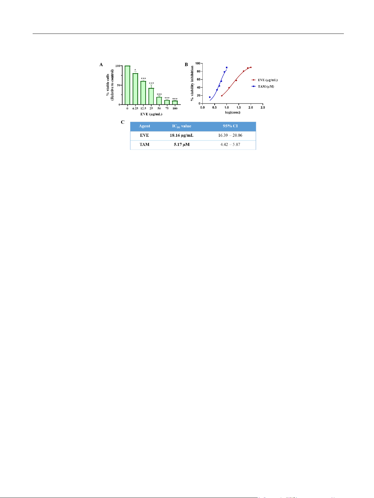
breast cancer cells. In the present study, we aimed to evaluate the
synergistic effect of Erythrina variegata extract (EVE) and TAM on
the suppression of breast cancer cell growth using MCF-7 cell model
and conventional bioassays such as MTT and colony formation
assay, cell cycle analysis, and immunocytochemistry. Interestingly,
MTT assay in combination with Chou-Talalay analysis demonstrated
the potent synergism between EVE and TAM against MCF-7 cell
survival. Additionally, co-treatment of MCF-7 cells with EVE and
TAM more significantly suppressed colony formation, cell cycle
progression, and the nuclear expression of proliferative marker Ki67
compared to TAM alone. These findings imply that EVE potentiates
the anti-proliferative effect of TAM in MCF-7 breast cancer cells and
may become a promising agent in the treatment of ER positive breast
cancer.
Keywords
Breast cancer
Erythrina variegata
Hormon receptor
MCF-7 cells
Tamoxifen
Van Thi-Hong Tran et al.
3
Introduction
Breast cancer (BC) is the most common type of
cancer and the second leading cause of cancer-
associated deaths in women [1]. Due to the genetic
heterogeneity of breast tumor cells, therapeutic agents
will be indicated based on the expression of molecular
markers. Of various types of BC cells, hormone
receptor (HR) positive tumors are the main
pathological variant accounting for 70% of all BC
cases and anti-estrogen therapies including selective
estrogen receptor modulators (SERMs), selective
estrogen receptor degrader (SERDs), or aromatase
inhibitors serve as first-line adjuvant therapeutic
agents for this variant [2]. Despite recent introduction
of newer drug classes, tamoxifen, classified as a first-
generation SERM, remains the drug of choice for the
prevention and treatment of BC, especially in
premenopausal women, since it reduces the mortality
and recurrence by 30 and 50%, respectively [3].
Unfortunately, approximately 30% of BC patients will
develop the resistance to tamoxifen either at the
beginning of treatment (de novo resistance) or during
the treatment (acquired resistance) that may lead to BC
relapse, development of secondary tumors, and higher
risks of treatment failure and mortality [4]. Given the
importance of tamoxifen in the therapeutic regimen of
BC, discovering the agents which enhance the
sensitivity or reduce the resistance of BC cells to
tamoxifen has attracted a great deal of attention in
recent years.
Over the last decades, numerous studies have
focused on seeking novel anti-breast cancer agents of
herbal origin which led to the introduction of more
than 70 plant species with the inhibitory activity of BC
cell growth [5]. Nevertheless, little has been made to
figure out the potential of these plants in the
combination therapy with tamoxifen. In fact, even
though many plant extracts exhibited the arrest of BC
cell growth, their effects on the anti-breast cancer
effect of tamoxifen were not always positive. Indeed,
while certain plants (e.g., Viscum album and Trifolium
pratense) were demonstrated the synergistic effect
with tamoxifen on suppression of cancer cell growth
[6, 7], many other species such as Angelica sinensis,
Paeonia lactiflora, Rehmannia glutinosa, Astragalus
mongholicus, and Glycyrrhiza glabra antagonized the
anti-breast cancer effect of tamoxifen, possibly due to
presence of estrogen-like compounds in their chemical
composition [8]. These surprising findings indicate
that careful considerations will be required in the
utilization of medicinal plants as supplements to
conventional endocrine therapy even when their anti-
cancer effects were previously reported.
Erythrina variegata. L is a Vietnamese native
plant traditionally used to treat insomnia,
inflammation-related disorders, and some types of
tumors. A previous study by Nguyen et al. also
reported that several compounds isolated from this
plant showed good in vitro inhibition on α-glucosidase
enzyme [9]. Notably, several studies have shown that
E. variegata possesses anti-breast cancer effects. For
example, Teti Herlina et al. reported the cytotoxic
activity of the methanol extract from E. variegata in
T47D cells with an IC50 value of 40.5 µg/mL [10].
Likewise, this plant extract exhibited to inhibit MCF-
7 and MDA-MB-231 cells via induction of apoptosis
[11]. However, there has been not any report on anti-
cancer effect of E. variegate collected from Vietnam.
Also, the synergistic effect of E. variegata and
tamoxifen on the arrest of BC cell growth has not been
investigated. Therefore, the present study aimed to
examine if E. variegata extract (EVE) enhances the
sensitivity of ER-positive BC cells to tamoxifen.
Materials and methods
Cell culture and reagents
MCF-7 breast cancer cell line of ATCC origin was
kindly provided by Prof. Suresh Awale from the
University of Toyama (Japan) and Prof. Pil-Hoon Park
from Yeungnam University (Korea), and routinely
cultured in DMEM media supplemented with 10%
FBS and 1% penicillin/streptomycin. Cells were
maintained in a humidified incubator at 37oC with 5%
CO2. All reagents for cell culture were purchased from
Thermo Scientific (Waltham, MA, USA). Apparatuses
for cell culture (tissue culture 96-well plates, 10-mm
dishes, 35-mm dishes, and 8-well slides) were
acquired from Corning Incorporated (Somerville,
Massachusetts, USA).
Collection and preparation of plant extract
Leaves of E. variegata were collected at the
Research Center of Medicinal Plants, Hanoi in August
2022. The botanical identification was performed by
Mr. Lai Viet Hung (Center of Medicinal Material
Resources). Voucher specimens (No. Nimm-19205)
were deposited at the Herbarium of Medicinal Material
Resources Centre (NIMM), Vietnam.
The dried plant sample (1 kg) was coarsely ground
and extracted thrice with 70% aqueous ethanol under
reflux for 3h. The extracts were then combined,
filtered, and the solvent was evaporated completely
under reduced pressure to give a total ethanol extract
Van Thi-Hong Tran et al.
4
of E. variegata (EVE). The extraction yield was
5.58%.
Cell viability assay
Cell viability was determined using MTT (3-(4,5-
Dimethylthiazol-2-yl)-2,5-Diphenyltetrazolium
Bromide) assay [12]. In brief, cells were seeded into a
96-well plate at the density of 104 cells/well. After
overnight incubation, cells were treated with
tamoxifen (Sigma-Aldrich, USA) with or without
EVE at increasing concentrations for 48h. Cells were
then incubated with MTT (Sigma-Aldrich, USA)
solution at the final concentration of 0.5mg/mL for 2h.
Subsequently, media were removed, and the resultant
formazan crystals were dissolved in 200µL dimethyl
sulfoxide (Sigma-Aldrich, USA). Finally, the
absorbance was measured at 570nm using a microplate
reader (SpectraMax iD3, Molecular Devices, USA).
For evaluation of synergistic effects, IC50 values
(concentrations that reduce the cell viability by 50%)
for tamoxifen and EVE were first estimated. Then, the
effect of tamoxifen and EVE (alone or combination)
on the cell viability was examined at ~ 0.25×IC50,
0.5×IC50, 1.0×IC50, 1.5×IC50, and 2.0×IC50 for each
agent (combination ratio was fixed at 1:1 based on IC50
values).
Colony formation assay
Colony formation assay was performed as
previously described [13]. Cells were seeded into 35-
mm dishes at a low density (2×10³ cells/dish). On the
next day, cells were treated with tamoxifen and/or
EVE for 16 h. After that, treatment media were
replaced with fresh media to allow cells to grow.
Media were then changed every 3 days. On the 14th
day, cells were fixed with 70% ice-cold ethanol,
followed by staining with crystal violet (Sigma-
Aldrich, USA) solution (0.5%) for 30 min. Cells were
properly washed with PBS and the images were
acquired using a light microscope (Zeiss, Germany).
The number of cell colonies (were defined as clusters
of at least 50 cells) counted using Image J software.
Cell cycle analysis
Cell proliferation was examined using cell cycle
analysis as previously described [14, 15]. Cells were
seeded into 60-mm dishes at the density of 3×10⁵
cells/dish. After treatments as indicated, cells were
collected by trypsinization and washed with PBS. The
single cells were fixed with 70% ice-cold ethanol in
PBS for 2 h at 4oC. After removing the fixing solution,
cells were incubated with 50 µg/mL propidium iodide
solution containing RNase I (550U/mL) (Abcam,
Cambridge, MA, USA) for 20 min at room
temperature. The nuclear incorporation of propidium
iodide was analysed using a flow cytometry (BD
FACS Calibur, BD Biosciences, San Jose, CA, USA).
The proportion of cells in each phase of cell cycle was
estimated using Flowjo 7.6 software.
Immunocytochemistry
Immunocytochemistry (ICC) was used to
determine the nuclear level of Ki67, a proliferative
marker [14]. Cells were seeded into an 8-well glass
chamber slides at the density of 4×10⁴ cells/well. After
treatment with tamoxifen and EVE, cells were fixed
with 4% paraformaldehyde for 20 min, followed by
permeabilization with 0.2% Triton X100 in PBS for 10
min and blocking with 3% bovine serum albumin for
45 min. Then, cells were sequentially incubated with a
primary antibody against Ki67 (1:200) (Cell Signaling
Technology Inc., Beverly, MA, USA) for 16 h at 4oC
and an Alexa-488 conjugated secondary antibody
(1:500) (Abcam) for 90 min at room temperature. The
slides were covered with a coverslip using a mounting
reagent containing DAPI (Thermo Scientific). The
images were acquired using a fluorescent microscope
(Nikon, Tokyo, Japan). The proportion of Ki67-
positive cells was calculated based on Image J
software.
Statistical analysis
Data acquired from at least three independent
experiments are expressed as mean ± standard error
(SE). Statistical analyses were performed using
GraphPad Prism 8.3.0 (San Diego, CA, USA). In
particular, IC50 values were estimated using a non-
linear regression model. Statistical differences among
groups were determined by one-way ANOVA in
combination with post-hoc Turkey’s test or t-test (if
only 2 groups were presented). In addition, Chou-
Talalay method along with CompuSyn software were
employed to specifically estimate combination index
(CI) and Dose-reduction index (DRI).
Results and discussions
Results
EVE suppresses the viability of MCF-7 breast cancer
cells
It has been reported that E. variegata suppressed
BC cell growth [9, 10]. Hence, we first confirmed the
effect of EVE on MCF-7 cell viability using MTT
assay. As expected, EVE decreased the MCF-7 cell
survival in a concentration-dependent manner (Fig. 1A
and 1B). The non-linear regression analysis suggested
a sigmoidal dose-response model for EVE which was
essentially similar to TAM. The IC50 of EVE was
Van Thi-Hong Tran et al.
5
18.16 µg/mL (95% CI: 16.39 20.06), demonstrating
a relatively potent anti-breast cancer activity of EVE.
Figure 1. Effect of EVE on the cell viability of MCF-7 breast cancer cells. (A) MCF-7 cells were treated with EVE at
indicated concentrations for 48 h. Cell viability was examined by MTT assay. (B) The concentration-response curves of
EVE and TAM were established based on a non-regression regression analysis by Graphpad software. (C) The estimated
IC50 values of EVE and TAM were presented along with their 95% confident intervals (CI). * p < 0.05, *** p < 0.001
compared to the control cells; TAM: Tamoxifen; EVE: crude ethanol extract of E. vaiegata.
Chou-Talalay analysis of synergism between TAM and
EVE
Based on the estimated IC50 values of EVE and
TAM (Fig. 1), we designed a series of concentrations
for EVE alone, TAM alone, and EVE-TAM
combination (with fixed ratios of 1:1) ranging from
approximately 0.25 to 2.0×IC50 value for each agent.
As shown in Fig. 2A, the combination of EVE and
TAM decreased MCF-7 cell survival more
significantly than EVE or TAM alone at all the tested
concentrations. Importantly, Chou-Talalay analysis
clearly indicated a synergistic effect of EVE and TAM
since the combination index (CI) < 1 was observed at
any effect level (Fa) (Fig. 2B). It is also worth noting
that the synergistic effect of EVE and TAM increased
in a dose-dependent manner.
As a result, co-treatment of TAM and EVE led to
substantial reduction in cancer-cell inhibitory
concentration of each agent. In particular, dose-
reduction index (DRI) for TAM and EVE at
concentrations of approximately 0.25, 0.5, 1.0, 1.5, 2.0
× IC50 was 2.81, 3.63, 6.28, 10.20, 21.24 and 2.29,
1.95, 1.97, 2.17, 2.89, respectively. These findings
imply that co-therapy with EVE may help reduce dose
of TAM required for suppressing BC cell growth.
Van Thi-Hong Tran et al.
41
Figure 2. The synergistic effects of TAM and EVE on the suppression of MCF-7 cell viability. (A) Effects of TAM and
EVE on MCF-7 cell viability were examined at the concentrations ranging from ~ 0.25 to ~ 2.0 × IC50 value of each agent.
The effect of TAM in combination with EVE was also evaluated at the fixed combination ratios of 1:1. The percentage of
cell survival inhibition was shown. (B and C) Results obtained from Chou-Talalay analysis using CompuSyn software
included the Fa-CI plot (B) and Fa-DRI plot (C). Fa: effect level; CI: combination index; DRI: dose-reduction index; TAM:
Tamoxifen; EVE: crude ethanol extract of E. variegata.
EVE potentiates the suppressive effect of TAM on cell
proliferation in MCF7 breast cancer cells
Having demonstrated that EVE exhibits the
synergistic effect with TAM on the suppression of
MCF-7 cell viability, we next confirmed this
synergism using different assays including colony
formation assay, ICC, and cell cycle analysis.
The colony formation assay allows to evaluate the
long-term impact of drug on the capability of a single
cell to grow into a big colony via multiple doublings.
Due to the high sensitivity of this assay, the lowest
concentrations of TAM and EVE (1.25 µM and 5
µg/mL, respectively) were used. Interestingly, while
the treatment with TAM and EVE modestly decreased
the number of colonies, co-administration of the two
agents resulted in dramatical inhibition of colony
formation (Fig. 3A and 3B). These results delineate
that short-term exposure to EVE/TAM may produce a
long-term inhibitory effect on the proliferation of
MCF-7 breast cancer cells.