The ubiquitin-like protein monoclonal nonspecific suppressor factor b conjugates to endophilin II and regulates phagocytosis Morihiko Nakamura and Shunsuke Shimosaki
Department of Cooperative Medical Research, Collaboration Center, Shimane University, Izumo, Japan
Keywords endophilin; macrophage; phagocytosis; ubiquitin-like protein
Correspondence M. Nakamura, Department of Cooperative Medical Research, Collaboration Center, Shimane University, Izumo 693-8501, Japan Fax: +81 853 20 2913 Tel: +81 853 20 2916 E-mail: nkmr0515@med.shimane-u.ac.jp
confirmed this
(Received 13 July 2009, revised 31 July 2009, accepted 3 September 2009)
doi:10.1111/j.1742-4658.2009.07348.x
Monoclonal nonspecific suppressor factor b (MNSFb) is a ubiquitously expressed member of the ubiquitin-like family that has been implicated in various biological functions. Previous studies have demonstrated that MNSFb covalently binds to the intracellular proapoptotic protein Bcl-G in cells of the macrophage cell line Raw264.7, suggesting involvement of this ubiquitin-like protein in apoptosis. In this study, we purified a 62 kDa MNSFb adduct from murine liver lysates by sequential chromatography on DEAE and anti-MNSFb IgG-conjugated Sepharose. MALDI-TOF MS fingerprinting revealed that this MNSFb adduct consists of an 8.5 kDa MNSFb and endophilin II, a member of the endophilin A family. MNSFb may conjugate to endophilin II with a linkage between the C-terminal Gly74 and Lys294. We result by immunoprecipita- tion ⁄ western blot studies. Endophilin II was ubiquitously expressed in vari- ous tissues, although a truncated form was observed in liver. The 62 kDa MNSFb–endophilin II was specifically expressed in liver and macrophages. Small interfering RNA-mediated knockdown of endophilin II and ⁄ or MNSFb promoted phagocytosis of zymosan in Raw264.7 cells. Conversely, cotransfection of endophilin II and MNSFb cDNAs inhibited the phagocy- tosis of zymosan. Such inhibition was not observed in cells expressing a mutant of endophilin II in which Lys294 was replaced by arginine. These results suggest that the post-translational modification of endophilin II by MNSFb might be implicated in phagocytosis by macrophages.
Structured digital abstract l MINT-7261558, MINT-7261537, MINT-7261546: MNSF beta (uniprotkb:P35545) physically interacts (MI:0915) with Endophilin-2 (uniprotkb:Q62419) by anti bait coimmunoprecipitation (MI:0006)
Introduction
Monoclonal nonspecific suppressor factor b (MNSFb) is a 14.5 kDa fusion protein consisting of a protein with 36% identity with ubiquitin and ribosomal pro- tein S30. The ubiquitin-like segment is cleaved from
[1]. MNSFb is ribosomal protein S30 in the cytosol covalently attached to certain lysines of specific target proteins, including Bcl-G, a proapoptotic protein [2]. the MNSFb conjugates
to Bcl-G and regulates
FEBS Journal 276 (2009) 6355–6363 ª 2009 The Authors Journal compilation ª 2009 FEBS
6355
Abbreviations EGF, epidermal growth factor; ERK, extracellular signal-regulated kinase; MAP kinase, mitogen-activated protein kinase; MNSF, monoclonal nonspecific suppressor factor; siRNA, small interfering RNA; tEnd-II, 30 kDa truncated form of endophilin II.
1 2 3 4 5 6
97 kDa-
-95 kDa MNSF adduct
65 kDa-
M. Nakamura and S. Shimosaki MNSFb conjugates to endophilin II
interferon
-62 kDa MNSF adduct
45 kDa-
mitogen-activated protein kinase (MAP kinase) path- way by inhibiting the activation of extracellular signal- [3]. Several ubiquitin-like regulated kinase (ERK) proteins signaling. in implicated are MNSFb, ISG15, FAT10 and NUB1L are induced by interferon-c [4–7]. Among them, MNSFb and FAT10 are closely related to apoptosis [2,8].
31 kDa-
[9,10],
endophilin I
CBB Silver WB: anti-MNSF
synaptic vesicle endocytosis
adducts analyzed fractions
lane 3, silver-stained; lanes 4–6,
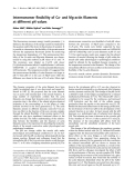
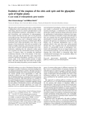
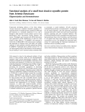
by Fig. 1. Purified of MNSFb SDS ⁄ PAGE (12% polyacrylamide gel) and immunostained for pro- tein. Lanes 1 and 4 contain an aliquot (100 lg of total protein) from liver lysates prepared from Balb ⁄ c mice; lanes 2 and 5 contain an aliquot from the DEAE chromatography purification step; lanes 3 and 6 contain an aliquot from the anti-MNSFb affinity chromatogra- phy purification step; lanes 1 and 2, CBB (Coomassie brilliant blue)- stained; immunostained with antibodies against MNSFb. Mobilities of the 95 and 62 kDa MNSFb adducts and the molecular mass standards (kDa) are indicated to the right and left of the figure, respectively.
The endophilins are a family of proteins identified in a search for SH3 domain-containing proteins. The most extensively studied isoform of the mammalian is endophilins, brain-specific necessary for [11–13]. Endophilin II, which is ubiquitously expressed [14], is a member of the endocytic endophilin family, but its function in regulating endocytosis remains unclear. In this study, we observed that the authentic 45 kDa endophilin II is expressed in various mouse tissues, including brain, testis, and spleen. Interestingly, heter- ogeneous species with molecular masses of about 62, 45 and 25 kDa were observed in liver lysates. We dem- onstrated that MNSFb covalently conjugates to endo- philin II in liver and cells of the macrophage cell line Raw264.7, and that MNSFb–endophilin II complex formation might be implicated in phagocytosis in macrophages.
Results
shown). No clear
Purification of MNSFb adducts from murine liver
trypsin. Selected peptides from the 62 kDa band were subjected to sequence analysis, and the results showed the amino acid sequences of MNSFb and an irrelevant protein (data not results were obtained from the 95 kDa band.
MALDI-TOF MS analysis of MNSFb adducts
that
To identify the target molecule of MNSFb, MALDI- TOF MS was performed by separating the 62 kDa MNSFb adduct by SDS ⁄ PAGE under reducing condi- tions. Bands corresponding to the 62 kDa MNSFb adduct were excised and subjected to in-gel digestion with trypsin as described. Then, the resulting mixtures of peptides were analyzed by MALDI-TOF MS. Table 1 shows the peptide masses observed by MALDI-TOF MS of the 62 kDa MNSFb adduct puri- fied from murine liver. The resulting sets of peptide masses were then used to search the NCBI database for potential matches. Seven of the peptides in the MALDI spectrum matched endophilin II protein, pro- result viding a sequence coverage of 32%. This indicates is an the 62 kDa MNSFb adduct MNSFb–endophilin II protein complex. Endophilin II is a member of the endophilin A family, and is ubiqui- tissues. Endophilin II tously expressed in various possesses the SH3 domain, which is critical for associa- tion with synaptojanin-1 and dynamin. A signal was
In previous studies, we have shown that MNSFb cova- lently conjugates to various target proteins [2,4]. Dur- ing the studies, we observed that several MNSFb adducts were found in liver extracts of mice. To fur- ther study the mechanism of action of MNSFb, we tried to isolate the MNSFb conjugates. When an extract from fresh murine liver was chromatographed through a DEAE–Sepharose column, and various frac- tions were subjected to western blotting by using antibody against MNSFb, several MNSFb antibody- reactive conjugates were eluted from the column by 100 mm NaCl (Fig. 1, lane 5). We further purified these conjugates by MNSFb antibody affinity chroma- tography. The final preparation gave two silver-stained bands on SDS ⁄ PAGE with mobilities corresponding to 62 and 95 kDa under reducing conditions (Fig. 1, lane 3). We observed that antibody against MNSFb recognized these two bands (Fig. 1, lane 6). These MNSFb anti- body-reactive proteins were first subjected to N-termi- nal sequence analysis after electroblotting. Despite repeated attempts, ambiguous signals were obtained from each protein (about 100 pmol). For internal sequencing, the protein bands were digested in-gel with
FEBS Journal 276 (2009) 6355–6363 ª 2009 The Authors Journal compilation ª 2009 FEBS
6356
M. Nakamura and S. Shimosaki MNSFb conjugates to endophilin II
Table 1. Assignment for peptide fragments from a trypsin digest of the 62 kDa MNSFb adduct. The 62 kDa MNSFb adduct was digested by trypsin and subjected to MALDI-TOF MS analysis. The data in the second column are the mass values obtained experimentally, whereas the results in the third column are those calculated from the trypsin fragmentation of the gene products of endophilin II and MNSFb. The fourth column indicates the numbers of the first and last amino acid of the indicated endophilin II and MNSFb peptides, whereas the fifth shows the corresponding amino acid sequences.
Mass (MH+)
Observed Calculated Residues Sequence Protein
Endo-II
919.1 1007.0 1260.5 1389.4 1605.9 2039.9 2385.6 919.0 1007.2 1260.5 1389.6 1605.8 2041.2 2385.7 129–136 68–76 302–312 54–65 137–149 103–123 261–282 DSLDIEVK LTMLNTVSK SMPPLDQPSCK TIEYLQPNPASR QNFIDPLQNLCDK ELGGESNFGDALLDAGESMK EPFELGELEQPNGGFPCAPAPK MNSFb 2096.3 2096.4 7–25 AQELHTLEVTGQETVAQIK
the
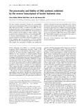
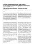
tography. The results of the blotting revealed that anti- body against endophilin II specifically recognized the 95 kDa, MNSFb adduct 62 kDa, but not (Fig. 3A), indicating that the 62 kDa band is a com- plex of MNSFb with endophilin II.
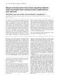
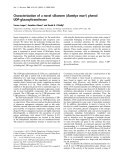
detected at 2096.3 Da that corresponds to amino acids 7–25 of MNSFb. Importantly, a pair of signals was detected at 1161.4 and 1605.0 Da (Table 2). These sig- nals correspond to digestion fragments in which amino acids 291–297 of endophilin II are covalently linked by an isopeptide bond to amino acids 71–74 of MNSFb, and amino acids 291–301 are linked by an isopeptide bond to amino acids 71–74. Collectively, MNSFb may conjugate to endophilin II with a linkage between the C-terminal Gly74 and Lys294. Lys294 is specific for endophilin II among all members of the endophilin family (Fig. 2).
Immunoblot and immunoprecipitation analyses
To confirm that MNSFb covalently conjugates to endophilin II, we performed immunoblotting of samples purified by MNSFb antibody affinity chroma-
Table 2. Isopeptide bonds between the C-terminal glycine of MNSFb and the lysine of endophilin II. The 62 kDa MNSFb adduct was digested by trypsin and subjected to MALDI-TOF MS analysis. The data in the first column are the mass values obtained experi- mentally, whereas the results in the second column are those calculated from the trypsin-fragmented peptide complexes. The third column shows the corresponding amino acid sequences of MNSFb and endophilin II (in bold).
Mass (MH+)
Observed Calculated Sequence
1161.3 1161.4
To further study these interactions, we performed antibodies immunoprecipitation experiments using against endophilin. Cell lysates were prepared from mouse liver and immunoprecipitated with antibodies directed against MNSFb, and associated proteins were analyzed by western blot analysis by using antibodies against endophilin. As shown in Fig. 3B, MNSFb associated with endophilin II. Immunoprecipitation of cell lysates with normal IgG followed by western blot analysis revealed no detectable association of endophi- lin II, indicating the specificity of MNSFb for endo- philin II. In addition, converse immunoprecipitation with antibody against endophilin II and immunoblot analysis with antibody against MNSFb confirmed the association between endophilin II and the MNSFb adduct. Brain extracts were also examined by immuno- precipitation ⁄ western blot. As depicted in Fig. 3C, neither antibody against endophilin I nor antibody against endophilin II recognized the MNSFb adduct. Thus, we concluded that the 62 kDa MNSFb adduct is an MNSFb–endophilin II complex and is specifically expressed in liver. Collectively, the results of immuno- blotting, together with the internal peptide sequences in Table 1 and MALDI-TOF MS analysis, show that MNSFb covalently binds to endophilin II via an isopeptide bond.
1605.2 1605.0 SSDKPIR(291–297) MLGG(71–74) SSDKPIRMPSK(291–301) MLGG(71–74)
We next performed immunoblotting analysis to mea- sure endophilin II levels in various organs of mice. As shown in Fig. 4, the 45 kDa endophilin II is expressed
FEBS Journal 276 (2009) 6355–6363 ª 2009 The Authors Journal compilation ª 2009 FEBS
6357
M. Nakamura and S. Shimosaki MNSFb conjugates to endophilin II
Fig. 2. Primary structure of mouse endophi- lins. End-I, endophilin I; End-II, endophilin II; End-III, endophilin III. The SH3 domain is boxed. In End-II, the Lys294 residue respon- sible for isopeptide formation is in bold. The lengths and numbers of the endophilins are indicated.
cells
zymosan
phagocytized
(1.4-fold)
including
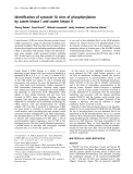
in different mouse tissues, including brain (cerebrum and cerebellum) and testis. It should be pointed out that the 30 kDa truncated form of endophilin II (tEnd-II) was consistently observed in liver (Fig. 4A, lane 4). The N-terminal region of endophilin II might be cleaved, because we employed antibodies against the middle (in this study) and C-terminal (not shown) regions of endophilin II. We examined the expression of endophilin II in the murine Raw264.7 macrophage- like cell line and peritoneal macrophages, because endophilin II is a member of the endocytic endophilin family. We observed two bands (62 kDa MNSFb– endophilin II and 45 kDa endophilin II) in the western lanes 1 and 2). blot of the macrophages (Fig. 4B, the Interestingly, heterogeneous bands, 62 kDa band (MNSFb–endophilin II), were reproduc- ibly observed in liver lysates but not in brain lysates (Fig. 4B, lanes 3 and 4: dialyzed against NaCl ⁄ Pi in the presence of protease inhibitors). This observation is consistent with the results for the formation of the 62 kDa complex of MNSFb and endophilin II in liver (Fig. 3).
Regulation of phagocytosis by endophilin II
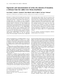
Endophilin II is one of three members of the subgroup endophilin A, but its function in regulating endocytosis remains unclear. It has been reported that endophilin I and endophilin III are implicated in receptor-mediated endocytosis [15]. Thus, we assessed the possible roles of endophilin II in the phagocytosis of zymosan in macrophages. We employed Raw264.7 cells, which have been studied in our laboratory [3]. The 62 kDa MNSFb adduct and endophilin II are expressed in untreated Raw264.7 cells (Figs 4B and 5A). Endophi- lin II small interfering RNA (siRNA), but not control scramble siRNA, specifically reduced the expression of endophilin II but not of a-tubulin (Fig. 5A). It should
be noted that the expression of 62 kDa MNSFb–endo- philin II was also decreased. As can be seen in Fig. 5B, Raw264.7 particles (30.3% ± 3.6%). Cells capturing more than two ops- onized zymosan particles were judged as positive. We examined the effect of endophilin II siRNA on the phagocytosis of opsonized zymosan in Raw264.7 cells. The treatment of Raw264.7 cells with endophilin II siRNA significantly enhanced phagocytosis (1.9-fold) (Fig. 5C). Interestingly, MNSFb siRNA, which has been used in the previous studies [3], showed similar effects on phagocytosis, although the effect was less potent (Fig. 5C). To determine whether MNSFb–endophilin II formation in Raw264.7 cells is involved in the phagocytosis of zymosan, immunoblot- ting was performed with the use of antibodies against endophilin II. We did not observe 62 kDa MNSFb– endophilin II in the cells treated with MNSFb siRNA (Fig. 5A), suggesting that MNSFb siRNA was effec- tive in reducing MNSFb protein levels. Importantly, Raw264.7 cells transfected with endophilin II and MNSFb siRNA (double knockdown) enhanced phago- cytosis to a similar extent as observed for cells treated with endophilin II siRNA alone. Thus, it is unlikely that free MNSFb plays a central role in the phagocy- tosis. The facilitatory effect of MNSFb siRNA was lower than that of endophilin II siRNA. In addition, the expression of 62 kDa MNSFb–endophilin II was much lower than that of endophilin II. Together, these results strongly indicate that MNSFb–endophilin II has a much higher inhibitory activity than endophilin II alone. Transient DNA transfection experiments were performed to confirm this idea. As can be seen in Fig. 5D, transfection with pcDNA3.1–endophilin II resulted in significant inhibition of the phagocytosis in Raw264.7 cells, consistent of zymosan (71.1%) with the data from siRNA knockdown experiments (Fig. 5C). No such inhibition was observed in cells
FEBS Journal 276 (2009) 6355–6363 ª 2009 The Authors Journal compilation ª 2009 FEBS
6358
8
1
2
3
4
5
6
7
M. Nakamura and S. Shimosaki MNSFb conjugates to endophilin II
A 65 kDa-
45 kDa-
-End-II
inhibition of
-tEnd-II
31 kDa-
expressing a mutant endophilin II that has an arginine in place of the lysine at position 294 that is responsible for MNSFb conjugation. Cotransfection of the expres- sion vectors encoding endophilin II and MNSFb the phagocytosis of caused stronger zymosan (75.1%). We observed weak but significant inhibition of zymosan phagocytosis in cells transfected with pcDNA3.1–MNSFb. Immunoblotting analysis showed that the expression of MNSFb–endophilin II
4
1
2
3
B
A
65 kDa-
-MNSF/End-II
-End-II
45 kDa-
- tEnd-II
31 kDa-
B
Fig. 4. Tissue distribution of endophilin II. (A) Tissue homogenates (50 lg of protein each) obtained from the indicated organs were subjected to immunoblotting analysis using antibody against endo- philin II. Lane 1: cerebrum. Lane 2: cerebellum. Lane 3: brain stem. Lane 4: liver. Lane 5: testis. Lane 6: epididymis. Lane 7: prostate gland. Lane 8: seminal vesicle. (B) Lane 1: peritoneal macrophage lysates. Lane 2: Raw264.7 cells. Lane 3: liver lysates dialyzed against NaCl ⁄ Pi in the presence of protease inhibitors. Lane 4: brain lysates dialyzed as liver in lane 3.
was increased in cotransfected cells (Fig. 5E). Collec- tively, these observations demonstrate that Lys294 of endophilin II is the only site of MNSFb conjugation
C
FEBS Journal 276 (2009) 6355–6363 ª 2009 The Authors Journal compilation ª 2009 FEBS
6359
Fig. 3. Western blot (WB) and immunoprecipitation (IP) analysis of MNSFb adducts. (A) Samples purified by anti-MNSFb affinity chro- matography from mouse liver cell lysates were analyzed by wes- tern blot. Lane 1: control rabbit IgG. Lane 2: antibodies against MNSFb. Lane 3: antibodies against endophilin II. (B) Liver cell extracts (300 lg) were immunoprecipitated with antibodies against MNSFb (lane 2) or normal IgG (lane 1). Immunoprecipitates were analyzed by western blot with antibodies against endophilin II (lanes 1 and 2). Conversely, the extracts (300 lg) were immunopre- cipitated with antibodies against endophilin II (lane 4) or normal IgG (lane 3). Immunoprecipitates were analyzed by western blot with antibodies against MNSFb (lanes 3 and 4). (C) Brain cell extracts (300 lg) were immunoprecipitated with antibodies against MNSFb (lanes 1 and 2). Immunoprecipitates were analyzed by western blot with antibodies against endophilin II (lane 1) or endophilin I (lane 2). The cell extracts (5 lg) were analyzed by western blot with anti- bodies against endophilin I (lane 3). In (A) and (B), mobilities of the 62 kDa MNSFb adduct (arrowhead) and the molecular mass stan- dards (kDa) are indicated to the right and left of the figure, respec- tively. In (C), the mobility of 40 kDa endophilin I (End-I) is indicated to the right of the figure.
1
2
3
4
5
M. Nakamura and S. Shimosaki MNSFb conjugates to endophilin II
B
A
Control
End-II siRNA End-II cDNA
MNSF/End-II-
Untreated cells
End-II-
Opsonized zymosan
-tubulin-
D
E
3
C
MNSF/End-II-
**
**
2
1
*
*
End-II-
1
**
**
x e d n i s i s o t y c o g a h P
-tubulin-
x e d n i s i s o t y c o g a h P
0
End-II
–
+
–
+
–
–
0 End-II
–
–
+
+
–
–
–
+
–
+
End-II siRNA
–
–
–
–
+
+
–
–
–
–
+
+
mEnd-II
mEnd-II
–
–
+
+
MNSF siRNA
–
–
+
+
–
+
–
+
+
–
–
+
MNSF
MNSF
Fig. 5. Effect of endophilin II on formation of phagosomes in Raw264.7 cells. (A) Expression of endophilin II and MNSFb–endophilin II were analyzed by western blot after treatment with siRNAs for 72 h. Lanes 1, 3 and 4: transfected with scramble (control) siRNA. Lane 2: endo- philin II siRNA. Lane 5: MNSFb siRNA. Lanes 1, 2, 4 and 5: immunostained with antibody against endophilin II. Lane 3: control rabbit IgG. Mobilities of 62 kDa MNSFb ⁄ endophilin II (open arrowhead) and 45 kDa endophilin II (closed arrowhead) are indicated to the left and right of the figure. (B) Raw264.7 cells were untreated or incubated with IgG-opsonized zymosan particles (Alexa488-labeled) for 30 min. Fluo- rescence was viewed with confocal microscopy. Cells were transfected with siRNA or cDNA for endophilin II as described in Experimental procedures. (C) Raw264.7 cells were treated with siRNAs for 72 h before addition of zymosan particles. The phagocytosis assay was performed as described in Experimental procedures. Cells capturing more than two zymosan particles were regarded as positive cells. Values are shown as the mean ± standard deviation (n = 5). *P < 0.05 versus untreated; **P < 0.01, one-way ANOVA test. (D) Raw264.7 cells were transiently cotransfected with expression vectors encoding either the endophilin II or endophilin II mutant, along with expression vector for MNSFb. The phagocytosis assay was performed after 72 h of transfection as described in Experimental procedures. Values are shown as the mean ± standard deviation (n = 3). *P < 0.05 versus empty vector; **P < 0.01, one-way ANOVA test. (E) Western blot analysis of extracts of cells transfected with the wild-type or mutant endophilin II cDNA. The results of immunoblotting using antibodies against endophilin II and a-tubulin are shown.
and that this post-translational modification may nega- tively regulate phagocytosis in macrophages.
Discussion
endophilin II conjugated by a single molecule of MNSFb (calculated molecular mass, 49.242 Da). One may specu- late that MNSFb might conjugate to at least one lysine in addition to Lys294 in endophilin II. However, we did not observe any candidate fragments with theoretical masses by MALDI-TOF MS. Additionally, experiments with mutants indicate that Lys294 of endophilin II is the only site of MNSFb conjugation (Fig. 5E).
We have previously demonstrated that MNSFb forms an isopeptide bond with Bcl-G, a proapoptotic protein. MNSFb becomes isopeptide-linked via its C-terminal diglycine motif, by analogy with ubiquitin and other ubiquitin-like modifiers, such as SUMO. MNSFb–Bcl-G directly binds to ERKs, and inhibits ERK activation by MAP kinase kinase 1 [3]. Similarly, in this study, we showed that MNSFb covalently binds to endophilin II in liver and Raw264.7 cells. We isolated a 62 kDa MNSFb adduct from mouse liver lysates (Fig. 1). MALDI-TOF MS analysis of the 62 kDa MNSFb adduct showed that MNSFb conjugates to endophilin II with a linkage between the C-terminal Gly74 and Lys294. The molecu- lar mass of 62 kDa was somewhat larger than that of
Endophilins are SH3 domain-containing proteins. Three isoforms of endophilin have been identified: endophilin I, which functions during synaptic vesicle recycling; endophilin II, which is ubiquitously distrib- uted throughout many tissue types; and endophilin III, which is expressed in brain and testis [16]. Although many studies have been focused on endophilin I, the underlying mechanism of action of endophilin II remains unclear. Lua and Low have reported [17] that overexpression of endophilin II enhances epidermal growth factor (EGF)-stimulated receptor endocytosis
FEBS Journal 276 (2009) 6355–6363 ª 2009 The Authors Journal compilation ª 2009 FEBS
6360
M. Nakamura and S. Shimosaki MNSFb conjugates to endophilin II
that monoubiquitination is involved in endocytosis of EGF receptor [23]. In this study, we presented data showing that endophilin II may act in concert with MNSFb to inhibit the phagocytosis of zymosan (Fig. 5). Thus, ubiquitin and ubiquitin-like protein(s) may regu- late endocytosis by modification of target proteins.
Taken as a whole, the present study demonstrates that endophilin II may negatively regulate phagocyto- sis, and that MNSFb–endophilin II formation might be important for potent regulation of phagocytosis in macrophages.
and ERK1 ⁄ 2 phosphorylation. Angers et al. [18] have reported that the E3 ubiquitin ligase Itch ubiquitinates endophilin I and localizes to the endosomal system fol- lowing EGF stimulation. They also mentioned that to germinal center kinase-like endophilin I binds kinase, suggesting a role for endophilin I in the c-Jun N-terminal kinase signaling pathway [19]. Together with our previous studies on the regulation of ERK activity by MNSFb, this indicates that endophilins, ubiquitin and ubiquitin-like protein(s) may be closely involved in MAP kinase pathways. Experiments are underway to isolate E3 ubiquitin ligase-like enzyme(s) involved in MNSFb conjugations.
Experimental procedures
Materials
Rabbit polyclonal antibodies against MNSFb were prepared as previously described [4]. Rabbit polyclonal antibodies against endophilin I and endophilin II were purchased from Santa Cruz Biotechnology (Santa Cruz, CA, USA). Alexa488–zymosan A was purchased from Molecular Probes. The zymosan particles were opsonized by using Opsonizing Reagents (Molecular Probes, Eugene, OR, USA) derived from purified rabbit polyclonal IgG antibodies that are specific for zymosan. Rabbit TrueBlot was obtained from eBioscience (San Diego, CA, USA). DEAE cellulose was purchased from Sigma-Aldrich (Tokyo, Japan).
Purification of the MNSFb adduct
Sugiura et al. [15] mentioned that the variable region of endophilin III is important in regulating transferrin endocytosis. In this study, we showed that MNSFb covalently modifies the Lys294 in the variable region of endophilin II and that this modification is involved in the phagocytosis of zymosan in Raw264.7 cells. Thus, as in the case of endophilin III, the variable region of endophilin II may be important in regulating endocytosis in macrophages. Interestingly, both endo- philin II and endophilin III negatively regulate recep- tor-mediated internalization [15]. It has been reported that endophilin family members bind to synaptojanin and dynamin via a Grb2-like Src homology 3 domain (constant region) [9,20]. In this context, MNSFb may not affect the binding of endophilin II to these part- ners. It should be noted that the residue responsible for MNSFb conjugation (Lys294) is only found in endophilin II (Fig. 2). Indeed, we did not observe MNSFb–endophilin I in brain extracts (Fig. 3C).
in macrophages
In preliminary experiments, we observed that dectin- 1 (b-glucan receptor) involves the mechanism of action of endophilin II on phagocytosis in macrophages. Dec- tin-1-mediated intracellular signaling pathways regulat- ing phagocytosis remain largely unknown. Although Syk involves most of the functions of dectin-1, this kinase is not required for particle uptake in macrophages [21]. Investigations are under- way to clarify whether MNSFb–endophilin II is a mediator in dectin-1-mediated signaling.
It
Mouse liver was obtained from BALB ⁄ c mice. All purifica- tion steps were performed at 4 (cid:2)C. A portion of a mouse liver (8 g) was homogenized in buffer A (20 mm Tris ⁄ HCl, pH 7.5, 50 mm NaCl, 1 mm dithiothreitol) containing a protease inhibitor mixture [1 mm 4-(2-aminoethyl)-benzenesulfonyl fluoride, 10 mgÆL)1 leupeptin, 5 mgÆL)1 aprotinin, and 1 mgÆL)1 pepstatin A], using a tissue blender (3 · 30 s). The homogenate was centrifuged at 13 000 g for 20 min, filtered through gauze, and centrifuged at 13 000 g for another 20 min. The resulting supernatant was further centrifuged at 120 000 g for 60 min to obtain a cytosolic extract, which was incubated overnight with DEAE (Sigma, Tokyo, Japan) pre- equilibrated with buffer A. Following extensive washing with buffer A, bound proteins were eluted in 20 mm Tris ⁄ HCl (pH 7.5) and 100 mm NaCl, and the protease inhibitor mix- ture. Additional purification was achieved by anti-MNSFb affinity chromatography, as previously described [1].
Immunoblotting
Cell extracts in SDS sample buffer were subjected to 12% SDS ⁄ PAGE, and blotted onto poly(vinylidene difluoride) membranes. The membrane was incubated overnight at 4 (cid:2)C in a Tris-buffered saline solution with 5% milk to
is interesting that MNSFb–endophilin II was detected in liver cells in terms of endocytosis. The truncated 30 kDa form may be a consequence of the MNSFb adduct. It is possible that the N-terminal region may be deleted from endophilin II in these tis- sues, because specific antibodies against C-terminal (Fig. 4) and central (not shown) regions detected the truncated form. Investigations are underway to clarify the mechanism of action of endophilin II in liver cells. Monoubiquitination is thought to regulate receptor internalization and endosomal sorting [22]. It is evident
FEBS Journal 276 (2009) 6355–6363 ª 2009 The Authors Journal compilation ª 2009 FEBS
6361
sequences were as
transfection reagent
block nonspecific binding sites. Subsequently, the mem- branes were incubated with anti-MNSFb rabbit IgG in the blocking buffer, after which they were incubated with peroxidase-conjugated anti-rabbit IgG. Detection was performed according to the enhanced chemiluminescence detection system (Amersham Biosciences, Chalfont St Giles, UK). We have previously demonstrated that antibody against MNSFb does not cross-react with ubiquitin [4].
M. Nakamura and S. Shimosaki MNSFb conjugates to endophilin II
In-gel digestion and MALDI-TOF MS
purified by Qiagen, Inc. (Chatsworth, CA, USA). The tar- follows: MNSFb siRNA-437, get 5¢-CCACCCTGCCATGCTAATAAA-3¢ [3]; and endophi- lin II siRNA, 5¢-AAGGTGCTCTAGAAACACTAA-3¢. Scramble siRNA directed against 5¢-GGACTCGACGC AATGGCGTCA-3¢ was the negative control. Cells were treated with siRNA according to the instructions provided with the RNAiFect (Qiagen, Inc.). Raw264.7 cells (1.2 · 105) were treated with 3 lg of siRNA in RPMI-1640 medium supplemented with 10% fetal boo- vine serum in the presence of the RNAiFect transfection reagent. After a 48 h incubation at 37 (cid:2)C, the medium con- taining the mixture of RNAiFect and siRNA was replaced by DMEM containing 10% fetal bovine serum, and cells were incubated for a further 24 h. Mutant endophilin II (K294R) was generated by replacing the codon for Lys294 with the codon for arginine by utilizing the LA PCR in vitro Mutagenesis Kit (Takara Bio, Ohtsu, Japan). In some experiments, Raw264.7 cells were transiently cotrans- fected with expression vectors encoding either the endophi- lin II or endophilin II mutant along with expression vector for MNSFb, as previously described [3].
Collection of peritoneal macrophages
All experimental procedures were described in the previous study [2]. Briefly, silver-stained spots were cut out of the gels for in-gel digestion and digested with trypsin (Sigma). The peptides were extracted from the gel matrix by vortexing for 30 min, and then concentrated using Zip Tips (Millipore Corp., Bedford, MA, USA). Peptide mass fingerprinting was performed using a PerkinElmer ⁄ PerSeptive Biosystems (Framingham, MA, USA) Voyager-DE-RP MALDI-TOF mass spectrometer. The peptide samples were cocrystallized with matrix on a gold-coated sample plate using 0.5 lL of matrix (a-cyano-4-hydroxytranscinnamic acid) and 0.5 lL of sample. Cysteines were treated with iodoacetamide to form carboxyamidomethyl cysteine, and methionine was consid- ered to be oxidized.
Immunoprecipitation
Peritoneal macrophages were obtained using mice injected 4 days previously with 2 mL of a sterile 3% brewer thio- glycolate broth (Difco, Detroit, MI, USA). The cells were collected by centrifugation (at 400 g for 5 min), washed, and resuspended in DMEM containing 10% fetal bovine serum. Lysates of the collected macrophages were used for immunoblotting.
pH 7.4,
Phagocytosis assays
Immunoprecipitation was performed with a horseradish peroxidase-conjugated antibody that recognizes native rabbit IgG (TrueBlot), according to the manufacturer’s instructions. RIPA buffer (50 mm Tris, 1% Nonidet P-40, 0.25% deoxycholate, 150 mm NaCl, 1 mm EDTA, 1 mm phenylmethylsulfonyl containing fluoride, 1 mgÆmL)1 each of the protease inhibitors aprotinin, leu- peptin, and pepstatin) extracts of Raw264.7 cells were pre- cleared with 50 mL of anti-rabbit IgG beads for 1 h on ice. Subsequently, 5 mg of primary antibody against MNSFb or endophilin II was added to precleared lysates and incu- bated on ice for an additional 1 h. Samples were then incu- bated overnight at 4 (cid:2)C with 50 lL of anti-rabbit IgG beads. The beads were washed five times with RIPA buffer, and immunoprecipitates were released from the beads by 10 min of boiling in NuPAGE LDS sample buffer (Invitro- gen). Immunoblotting was performed with antibody against MNSFb or antibody against endophilin II. A rabbit IgG TrueBlot was employed as a second antibody.
Raw264.7 cells were treated with siRNAs as described above. Cells were seeded at 5 · 104 cells per well in four-well cham- ber plates (Nunc, Roskilde, Denmark). Cells were washed, and Alexa488–zymosan A (Molecular Probes) was added (10 particles per cell) and allowed to bind for 1 h at 4 (cid:2)C. Following this incubation, unbound zymosan was removed by washing, and the cells were incubated at 37 (cid:2)C for 30 min to allow particle uptake. To determine the internalization of zymosan, cells capturing zymosan were treated with 0.05% Trypan blue in saline for a few minutes before observations by fluorescence microscopy. To determine the phagocytosis index, we identified > 200 Alexa-positive cells in randomly chosen fields of view, and the percentage of cells capturing more than two zymosan particles was determined.
Cell culture, the siRNAs, and transfection of cells
Acknowledgement
Cells of the Raw264.7 macrophage-like cell line (ATCC TIB-71) was cultured routinely in DMEM with 10% fetal bovine serum and penicillin ⁄ streptomycin at 37 (cid:2)C and 5% CO2. Small interfering RNA duplexes were synthesized and
This study was supported in part by Grants-in-Aid for Scientific Research (19570131 to M. Nakamura).
FEBS Journal 276 (2009) 6355–6363 ª 2009 The Authors Journal compilation ª 2009 FEBS
6362
M. Nakamura and S. Shimosaki MNSFb conjugates to endophilin II
References
late stages in clathrin-mediated synaptic vesicle endo- cytosis. Neuron 24, 143–154.
13 Schmidt A, Wolde M, Thiele C, Fest W, Kratzin H,
1 Nakamura M, Xavier RM, Tsunematsu T & Tanigawa Y (1995) Molecular cloning and characterization of a cDNA encoding monoclonal nonspecific suppressor factor. Proc Natl Acad Sci USA 92, 3463–3467.
Podtelejnikov AV, Witke W, Huttner WB & So¨ ling HD (1999) Endophilin I mediates synaptic vesicle formation by transfer of arachidonate to lysophosphatidic acid. Nature 401, 133–141.
14 Hirayama S, Bajari TM, Nimpf J & Schneider WJ
2 Nakamura M & Tanigawa Y (2003) Characterization of ubiquitin-like polypeptide acceptor protein, a novel pro- apoptotic member of the Bcl2 family. Eur J Biochem 270, 4052–4058.
(2003) Receptor-mediated chicken oocyte growth: differ- ential expression of endophilin isoforms in developing follicles. Biol Reprod 68, 1850–1860.
3 Nakamura M & Yamaguchi S (2006) The ubiquitin-like protein MNSFb regulates ERK–MAPK cascade. J Biol Chem 281, 16861–16869.
4 Nakamura M & Tanigawa Y (1998) Ubiquitin-like
15 Sugiura H, Iwata K, Matsuoka M, Hayashi H, Take- miya T, Yasuda S, Ichikawa M, Yamauchi T, Mehlen P, Haga T et al. (2004) Inhibitory role of endophilin 3 in receptor-mediated endocytosis. J Biol Chem 279, 23343–23348.
16 Araki Y, Ogawa K, Toh Y, Akimutsu N, Hmamoto H,
polypeptide conjugates to acceptor proteins in conca- navalin A- and interferon gamma-stimulated T-cells. Biochem J 330, 683–688.
5 Haas AL, Ahrens P, Bright PM & Ankel H (1987)
Sekimizu K, Matsusue K, Kono A, Iguchi H & Takiguchi S (2005) Direct interaction between metastasis-associated protein 1 and endophilin 3. FEBS Lett 579, 3731–3736.
Interferon induces a 15-kilodalton protein exhibiting marked homology to ubiquitin. J Biol Chem 262, 11315–11323.
17 Lua BL & Low BC (2005) Activation of EGF receptor
6 Raasi S, Schmidtke G, Giuli RD & Groettrup M (1999) A ubiquitin-like protein which is synergistically induc- ible by interferon-c and tumor necrosis factor-a. Eur J Immunol 29, 4030–4036.
7 Kito K, Yeh ETH & Kamitani T (2001) NUB1, a
endocytosis and ERK1 ⁄ 2 signaling by BPGAP1 requires direct interaction with EEN ⁄ endophilin II and a func- tional RhoGAP domain. J Cell Sci 118, 2707–2721. 18 Angers A, Ramjaun AR & McPherson PS (2004) The HECT domain ligase itch ubiquitinates endophilin and localizes to the trans-Golgi network and endosomal system. J Biol Chem 279, 11471–11479.
NEDD8-interacting protein, is induced by interferon and down-regulates the NEDD8 expression. J Biol Chem 276, 20603–20609.
8 Raasi S, Schmidtke G & Groettrup M (2001) The
ubiquitin-like protein FAT10 forms covalent conjugates and induces apoptosis. J Biol Chem 276, 35334–35343.
19 Ramjaun AR, Angers A, Legendre-Guillemin V, Tong XK & McPherson PS (2001) Endophilin regulates JNK activation through its interaction with the germinal cen- ter kinase-like kinase. J Biol Chem 276, 28913–28919.
9 Ringstad N, Nemoto Y & De Camilli P (1997) The
SH3p4 ⁄ Sh3p8 ⁄ SH3p13 protein family: binding partners for synaptojanin and dynamin via a Grb2-like Src homology 3 domain. Proc Natl Acad Sci USA 94, 8569–8574.
20 Gout I, Dhand R, Hiles ID, Fry MJ, Panayotou G, Das P, Truong O, Totty NF, Hsuan J, Booker GW et al. (1993) The GTPase dynamin binds to and is acti- vated by a subset of SH3 domains. Cell 75, 25–36. 21 Underhill DM, Rossnagle E, Lowell CA & Simmons
RM (2005) Dectin-1 activates Syk tyrosine kinase in a dynamic subset of macrophages for reactive oxygen production. Blood 106, 2543–2550.
10 Huttner WB & Schmidt A (2000) Lipids, lipid modifica- tion and lipid–protein interaction in membrane budding and fission – insights from the roles of endophilin A1 and synaptophysin in synaptic vesicle endocytosis. Curr Opin Neurobiol 10, 543–551.
11 Anggono V, Smillie KJ, Graham ME, Valova VA,
22 Haglund K, Shimokawa N, Szymkiewicz I & Dikic I (2002) Cbl-directed monoubiquitination of CIN85 is involved in regulation of ligand-induced degradation of EGF receptors. Proc Natl Acad Sci USA 99, 12191– 12196.
Cousin MA & Robinson PJ (2006) Syndapin I is the phosphorylation-regulated dynamin I partner in synaptic vesicle endocytosis. Nat Neurosci 9, 752–760.
23 Galan JM & Haguenauer-Tsapis R (1997) Ubiquitin lys63 is involved in ubiquitination of a yeast plasma membrane protein. EMBO J 19, 5847–5854.
12 Ringstad N, Gad H, Lo¨ w P, Di Paolo G, Brodin L, Shupliakov O & De Camilli P (1999) Endophi- lin ⁄ SH3p4 is required for the transition from early to
FEBS Journal 276 (2009) 6355–6363 ª 2009 The Authors Journal compilation ª 2009 FEBS
6363