Role of active-site residues of dispersin B, a biofilm-releasing b-hexosaminidase from a periodontal pathogen, in substrate hydrolysis Suba G. A. Manuel, Chandran Ragunath, Hameetha B. R. Sait, Era A. Izano, Jeffrey B. Kaplan and Narayanan Ramasubbu
Department of Oral Biology, University of Medicine and Dentistry of New Jersey, Newark, NJ, USA
Keywords biofilm detachment; dispersin B; enzyme mechanism; hydrolysis; site-directed mutagenesis
Correspondence N. Ramasubbu, Department of Oral Biology, C-634, MSB, UMDNJ, 185 South Orange Ave, Newark NJ 07103, USA Fax: +1 973 972 0045 Tel: +1 973 972 0704 E-mail: ramasun1@umdnj.edu
(Received 28 August 2007, revised 30 September 2007, accepted 1 October 2007)
Dispersin B (DspB), a family 20 b-hexosaminidase from the oral pathogen Aggregatibacter actinomycetemcomitans, cleaves b(1,6)-linked N-acetylglu- cosamine polymer. In order to understand the substrate specificity of DspB, we have undertaken to characterize several conserved and noncon- served residues in the vicinity of the active site. The active sites of DspB and other family 20 hexosaminidases possess three highly conserved acidic residues, several aromatic residues and an arginine at subsite )1. These res- idues were mutated using site-directed mutagenesis and characterized for their enzyme activity. Our results show that a highly conserved acid pair in b-hexosaminidases D183 and E184, and E332 play a critical role in the hydrolysis of the substrates. pH activity profile analysis showed a shift to a higher pH (6.8) in the optimal activity for the E184Q mutant, suggesting that this residue might act as the acid ⁄ base catalyst. The reduction in kcat observed for Y187A and Y278A mutants suggests that the Y187 residue (unique to DspB) located on a loop might play a role in substrate specific- ity and be a part of subsite +1, whereas the hydrogen-bond interaction between Y278A and the N-acetyl group might help to stabilize the transi- tion state. Mutation of W237 and W330 residues abolished hydrolytic activity completely suggesting that alteration at these positions might col- lapse the binding pocket for the N-acetyl group. Mutation of the conserved R27 residue (to R27A or R27K) also caused significant reduction in kcat suggesting that R27 might be involved in stabilization of the transition state. From these results, we conclude that in DspB, and possibly in other structurally similar family 20 hydrolases, some residues at the active site assist in orienting the N-acetyl group to participate in the substrate-assisted mechanism, whereas other residues such as R27 and E332 assist in holding the terminal N-acetylglucosamine during the hydrolysis.
Interestingly, DspB exhibits biofilm-detachment activ- ity not only for A. actinomycetemcomitans, but also species for biofilms produced by several bacterial including Gram-positive species (Staphylococcus epide- rmidis) [2] and Gram-negative species (Actinobacillus
The biofilm attachment and detachment properties of the oral pathogen Aggregatibacter actinomycetemcomi- tans (formerly Actinobacillus actinomycetemcomitans) are mediated by a soluble b-N-acetylglucosaminidase [1]. (dispersin B abbreviated to DspB; EC 3.2.1.52)
doi:10.1111/j.1742-4658.2007.06121.x
FEBS Journal 274 (2007) 5987–5999 ª 2007 The Authors Journal compilation ª 2007 FEBS
5987
Abbreviations DspB, dispersin B; Hex A, human b-hexosaminidase; MuGlcNAc, 4-methylumbelliferyl-b-D-N-Acetylglucosamine; PGA, homopolymer of b(1,6)-linked GlcNAc; pNPGlcNAc, p-nitrophenyl-b-D-N-acetylglucosamine; SpHex, S. plicatus hexosaminidase.
residues
pleuropneumoniae; Escherichia coli, Yersinia pestis and Pseudomonas fluorescens) [3,4]. DspB belongs to fam- ily 20 of the glycoside hydrolase classification scheme, members of which are exo-acting hexosaminidases that cleave terminal monosaccharide residues from the non- reducing end. The substrate of DspB is a hexosamine- containing matrix polysaccharide consisting of a linear homoglycan of N-acetyl-d-glucosamine in [5–9]. DspB catalytic activity b(1,6)-linkages (PGA) has been investigated using PGA from E. coli, which showed that the reaction products included GlcNAc and other unassigned GlcNAc oligomers [4]. In an ear- lier study, we reported the crystal structure of DspB in complex with glycerol and an acetate at the active site, which suggested that the active-site architecture of DspB is similar to that of the family 20 b-hexosami- nidases [10]. However, very little information is avail- able on the mechanistic aspects of DspB.
binding. In b-hexosaminidases, highly conserved tryp- tophan residues at subsite )1 create a binding pocket the terminal GlcNAc, especially the N-acetyl for group. Because there is a clear difference in the sub- strate specificity between DspB [acts on b(1,6)-linked poly(N-acetylglucosamine)] and the other members of the family 20 b-hexosaminidases [act on b(1,4)-linked N-acetylglucosamine], we tested the roles of conserved and nonconserved residues at the active site using site- directed mutagenesis. Our results demonstrate that: (a) DspB follows a substrate-assisted mechanism proposed for the family 20 b-hexosaminidases with participation of the conserved acidic pair D183 and E184; (b) the aromatic residues W237 and Y278 along with D183, play a role in hydrolysis and assist the N-acetyl group to properly orient itself to take part in catalysis; (c) the aromatic residue Y187, which is part of a loop at subsite +1, might play a role in the specificity exhib- ited by DspB; and (d) three residues, R27, E332 and W330 interacting with the GlcNAc at the C4 side pro- vide transition state stabilization during hydrolysis.
S. G. A. Manuel et al. Active site of dispersin B
Results
such
family 56
enzymes (EC 3.2.1.35)
and
+-affinity
using Ni2
stable and retained its activity for
Mutations were performed using the primers listed in Table 1. Wild-type DspB and the mutants were gen- erated with a His6 tag to facilitate purification using metal-affinity chromatography as described previously [10]. SDS ⁄ PAGE analysis of the cell lysates indicated that all the mutant enzymes were present in a solu- ble form and were expressed at levels comparable with wild-type DspB. All proteins were purified to chromatography homogeneity with yields ranging from 20 to 30 mgÆL)1 of culture. Our earlier studies had shown that the half-life of DspB was (cid:2) 3–4 h at 37 (cid:2)C [19]. However, DspB was several months when stored in buffer comprising 50 mm sodium phosphate, pH 5.8, 50 mm NaCl and 50% glycerol at )20 (cid:2)C. Purified enzymes were stored in this buffer until further use.
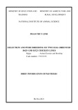
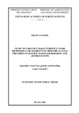
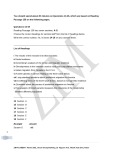
It has been proposed that family 20 and the closely related family 18 b-hexosaminidases follow a retaining mechanism (Fig. 1) with anchimeric assistance pro- vided by the C2-acetamido group of the substrate and an acidic active-site residue acting as the catalytic acid [11,12]. A unique feature of this mechanism is the acet- amido group acting as the nucleophile in catalysis. Interestingly, a substrate-assisted mechanism is opera- tive in other sequentially dissimilar but functionally similar hyaluronid- as family 84 O-GlcNAcase ase (EC 3.2.1.52) [13,14]. Using the crystal structure of DspB enzyme [10], we identified residues at subsite )1 of the active site that are likely to be involved in catal- ysis and ⁄ or substrate binding. We generated a struc- ture-based sequence alignment through superposition of the active-site regions of the reported crystal struc- tures of b-hexosaminidases [15–18] and noted that, in addition to the well-conserved pair of acidic residues D–E at positions 183 and 184 (DspB numbering), acidic residue E332 is also structurally conserved (Fig. 2A,B). Juxtaposition of this residue appears to be suitable for exerting a significant role in substrate
FEBS Journal 274 (2007) 5987–5999 ª 2007 The Authors Journal compilation ª 2007 FEBS
5988
Fig. 1. Hydrolysis mechanism proposed for family 20 b-hexosaminidases. In this substrate-assisted mechanism one acidic residue, Glu184, acts as the acid ⁄ base. The nucleophile is the N-acetyl group of the substrate and is assisted by Asp183.
S. G. A. Manuel et al. Active site of dispersin B
A
Role of active-site acidic residues
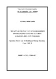
Fig. 2. Sequence and structure alignment of b-hexosaminidases with DspB. (A) Multiple amino acid sequence alignment of DspB and b-hexosaminidases whose crystal structures have been reported. Subsite )1 residues are indicated by an asterisk (*). Note the conserved residues WXE near the C-terminus corresponding to W330 and E332. Although all four aromatic residues, W216, W237, Y278 and W330 are conserved, the sequence homology in the region around Y187 of DspB is weak. This region might correspond to residues in subsite +1 and contribute to the substrate specificity being b(1,6) in DspB. Sequences shown are: DspB, 1yht [10]; chito- biase from Serratia marcescens 1qbb [20]; b-hexosaminidase from Streptomyces plicatus, 1hp5 [17]; human lysosomal b-hexosamini- dase isoform b, 1now [16]; human b-hexosaminidase from placenta, b-chain, 2gjx [30]. The secondary structure as observed in DspB is shown on top. Arrows represent b strands and cylinders represent helices. (B) Superposition of the various crystal structures of b- hexosaminidases using the suite SWISS-PDBVIEWER [31]. The fit of D183, E184 and E332 residues in the proximity of the subsite )1 is shown. Structures shown are: DspB; PDB Code: 1yht (gray) [10]; PDB Code: 2gjx (red) [30]; PDB Code: 1hp5 (green) [17]; PDB Code: 1now (blue) [16]; PDB Code: 1qbb (yellow) [20].
B
Glu184
Glu332
Asp183
Comparison of the amino acid sequence and crystal structure of DspB with other family 20 b-hexosaminid- ases clearly suggests that three acidic residues, D183, E184 and E332, are highly conserved (Fig. 2B). Among these, the residues equivalent to D183 and E184 in other family 20 b-hexosaminidases have been shown to play a critical role in the catalytic reaction the E184 equivalent has been [11,12,17]. Thus, proposed to act as the acid ⁄ base catalyst, whereas the D183 equivalent has been suggested to orient the N-acetyl group [11]. In general, in family 20 b-hexos- aminidases, the E184 equivalent is juxtaposed close to the anomeric center, whereas the D183 equivalent is well positioned to interact with the N- of the acetam- ido group. Many crystal structures also show that a glutamate residue (equivalent to E332) is located in close proximity to the C3- and C4-hydroxyl groups on the opposite side of the bound sugar at subsite )1. To investigate the proposed role of the D183 and E184 acidic residues and the potential role of E332 in DspB, these residues were replaced by either N or Q and their biochemical properties analyzed using the substrate p-nitrophenyl-b-d-N-acetylglucosamine (pNPGlcNAc). All three acidic mutants display significantly reduced specific activities as shown in Table 2. The specific activity of the catalytic residue mutant E184Q was 122-fold less, and that of D183N was 11 000-fold lower than wild-type. The observed results for the mutant E184Q are entirely consistent with loss of the acid–base residue, whereas the significant loss in activ- ity exhibited by D183N is consistent with its proposed
role as a residue that activates ⁄ orients the N-acetyl group to act as a nucleophile [11]. Inhibition studies were carried out with the mechanism-based inhibitor NAG-thiazoline [17]. IC50 for inhibition was found to be 147 ± 5 lm compared with 47 ± 4 lm for the jack bean enzyme. Inhibition of DspB by NAG-thiazoline is consistent with an anchimeric assistance mechanism occurring during catalysis in DspB because NAG- thiazoline is a mechanism-based inhibitor of b-hexos- specific activity aminidases. The (cid:2) 2700-fold lower
FEBS Journal 274 (2007) 5987–5999 ª 2007 The Authors Journal compilation ª 2007 FEBS
5989
S. G. A. Manuel et al. Active site of dispersin B
Table 1. Primers used for mutational analysis of the active-site residues.
Mutation Type Primer sequence (5¢- to 3¢)
R27A
R27K
D183N
E184Q
Y187A
W237A
Y278A
W330Y
E332Q Forward Reverse Forward Reverse Forward Reverse Forward Reverse Forward Reverse Forward Reverse Forward Reverse Forward Reverse Forward Reverse CTGGACATCGCCGCGCATTTTTATTCAC GTGAATAAAAATGCGCGGCGATGTCCAG GCTGGACATCGCCAAACATTTTTATTCACCCG CGGGTGAATAAAAATGTTTGGCGATGTCCAGC GGTGGCAACGAATTTGGTTATTCTGTGG CCACAGAATAACCAAATTCGTTGCCACC GGTGGCGATCAATTTGGTTATTCTGTGG CCACAGAATAACCAAATTGATCGCCACC GATGAATTTGGTGCGTCTGTGGAAAG CTTTCCACAGACGCACCAAATTCATC CCGAATATTGAAATTACTTATGCGAGCTATGATGGCG CGCCATCATAGCTCGCATAAGTAATTTCAATATTCGG CCTATTATCTTGCGATTGTTCCGAAAGC GCTTTCGGAACAATCGCAAGATAATAGG GCAGCATTATCGATCTACGGAGAAGATGC GCATCTTCTCCGTAGATCGATAATGCTGC CATTATCGATCTGGGGACAAGATGCAAAAGC GCTTTTGCATCTTGTCCCCAGATCGATAATG
Table 2. Specific activities and kinetic parameters of DspB and its variants for hydrolysis with pNPGlcNAc. Kinetic assays were performed with pNPGlcNAc as described in Experimental procedures. Reactions were carried out in NaCl ⁄ Pi at 37 (cid:2)C. The molar absorptivity of p-nitro- )1Æcm)1) at 405 nm. Results are the average of three independent experiments. Standard errors for the values of appar- phenol used 9900 (M ent Km and kcat are given in parentheses. ND, not determined because the reaction was too slow to be detected.
)1)
Enzyme Specific activity kcat (min)1) KM (mM) Conc (lM) kcat ⁄ KM (min)1ÆmM
55.6 (2.7) 46.2 (2.7) 0.39 (0.04)
536 (34) – 0.05 (0.01) 4.4 (0.8) 0.2 (0.2) 1.41 (0.2) 0.002 (0.001) 0.073 (0.001) 0.005 (0.001) 19.4 (2.5) 3.7 (0.5) 6.5 (2.3)
0.1 4 25 1 12 16 9 ND ND ND ND ND ND 1.20 3.61 0.00009 0.017 0.0006 ND ND
16.8 (4.4) 1 4 – 3.6 (0.2) 0.43 101.1 (6.7) 0.59 0.03 0.73
35.9 (1.4) 1.13
a No measurable activity for the substrate MuGlcNAc.
pNPGlcNAc as substrate (Table 2). All three mutants exhibited substantially lower Km and significantly reduced kcat values resulting in lower catalytic effi- ciency (kcat ⁄ Km) for the mutant enzymes (71-fold lower for E184Q, 13 333-fold lower for D183N and 2000- fold lower for E332Q) compared with wild-type. All three mutants required higher enzyme concentrations to exhibit measurable enzyme activity compared with
exhibited by E332Q suggests that this residue plays a significant role in catalysis. The location of this resi- due, although away from the anomeric carbon, might be critical in providing necessary stabilization in the transition state while the terminal GlcNAc is undergo- ing conformational changes during catalysis (Fig. 2B). To further substantiate our observations, kinetic parameters were determined for the mutants using
FEBS Journal 274 (2007) 5987–5999 ª 2007 The Authors Journal compilation ª 2007 FEBS
5990
Wild-type pNPGlcNAc MuGlcNAc D183Na E184Qa E332Qa W237Aa W330Ya Y187A pNPGlcNAc MuGlcNAc Y278A pNPGlcNAc MuGlcNAc R27Aa R27Ka 1 4 6.0 5.5 3.1 (1.3) – 0.60 (0.2) 0.31 (0.2) 0.25 (0.007) 0.09 0.023 (0.06) 0.05 (0.01) 14.5 (1.7) 76.2 (11) 0.0013 0.08 0.0005 0.0007
Role of aromatic residues at the active site
wild-type (E184Q, 1 lm; E332Q, 12 lm; and D183N, 25 lm compared with 0.1 lm for wild-type; Table 2). In addition, we also used another substrate, 4-methyl- umbelliferyl-b-d-N-acetylglucosamine (MuGlcNAc), to study the effect of leaving aglycone (Table 2). Interest- ingly, this substrate was also poorly hydrolyzed by the wild-type enzyme and required a higher enzyme con- centration (4 lm), compared with 0.1 lm for pNPGlc- NAc. There was no measurable activity for the mutant enzymes, D183N, E184Q and E332Q.
structures,
the
for
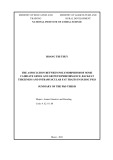
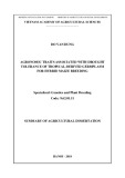
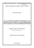
The pH activity profiles of the wild-type and variant enzymes were also determined using pNPGlcNAc and are shown in Fig. 3. The wild-type enzyme exhibited an approximately bell-shaped profile with a pH opti- mum of 5.8, which might be interpreted as resulting from the ionization of two residues with pKa values on either side of the maximum. The apparent pKa value for the basic limb corresponding to the acid ⁄ base of the wild-type profile was estimated to be 6.8 ± 0.1. Interestingly, the pH optimum for the E184Q variant is shifted to 6.8 with an apparent pKa of 8.3 ± 0.1 for the basic limb consistent with the substitution of E184 carboxyl group to an amide. The pH activity profile of E184Q mutant supports its presumed role in catalysis as the acid ⁄ base (Fig. 1). The extremely low activity of the pNPGlcNAc D183N and E332Q variants substrate prevented us from obtaining a pH profile for these mutants.
substrate
the
as
S. G. A. Manuel et al. Active site of dispersin B
15
300
One of the characteristics of family 20 b-hexosaminid- ases is the substrate-binding pocket provided by a number of conserved tryptophan residues. As shown in Fig. 2A, W216, W237 and W330 are well con- served and together create a hydrophobic pocket into which the N-acetyl group of the GlcNAc moiety at subsite )1 binds [15]. Together with the acidic resi- dues, such binding juxtaposes the acetamido group for neighboring-group participation and enables intra- molecular nucleophilic attack at the anomeric center the sugar molecule at subsite )1 [16]. Residue of Y278 occupies a space close to the bound acetyl group and in several crystal including DspB, is known to interact with the O of the acetyl group via a hydrogen bond. Residue Y187 is unique to DspB in that it is located in a mobile loop that is disordered in the crystal structure (PDB code 1YHT) [10] and appears to be present at the entrance to the catalytic site. To investigate the roles of these aro- matic residues in DspB, we designed mutations at these positions (Table 1) and the results of the bio- chemical analysis are given in Table 2. Clearly, muta- tion of tryptophan residues at positions 237 (W237A) and 330 (W330Y), which create the N-ace- tyl-binding pocket, had a drastic effect on enzyme activity. We observed that even at concentrations of 9–16 lm, enzyme activity was undetectable using (Table 2). Both pNPGlcNAc mutants, W237A and W330Y, were also ineffective when MuGlcNAc was used as a substrate.
12
R27K E184Q
9
250
wild type E184Q R27K
6
y t i v i t c A
3
200
0
5
6
8
9
150
7 pH
y t i v i t c A
100
50
0
4
5
6
7
8
9
pH
Mutation of the two tyrosines at the active site (Y187 and Y278) showed lower specific activity than the wild-type (Table 2) by as much as 33-fold (Y187A) or 176-fold (Y278A). Mutant Y187A has a higher Km and a lower kcat in the hydrolysis of pNPGlcNAc (Table 2). The lower kcat ⁄ Km value (40-fold less for pNPGlcNAc and 4.9-fold less MuGlcNAc) for Y187A suggests that the mutation at Y187 affects substrate binding. Involvement of this residue, which is farther away from subsite )1 (6 A˚ ), suggests that DspB might have multiple subsites for substrate binding. Mutant Y278A, by contrast, showed a greater decrease in kcat ⁄ Km value (923-fold for pNPGlcNAc and 45-fold for MuGlcNAc) compared with wild-type suggesting that the hydrogen bond to the acetamido group is sig- nificant for enzyme activity. Because this hydrogen bond involves the hydroxyl group of Y278 and car- bonyl oxygen of the acetamido group, the reduction in the kinetic parameters suggests that Y278 might partic- ipate in orienting the acetyl group in conjunction with the D183 residue.
FEBS Journal 274 (2007) 5987–5999 ª 2007 The Authors Journal compilation ª 2007 FEBS
5991
Fig. 3. pH dependence of wild-type DspB and mutants. The pH profile was measured using pNPGlcNAc as the substrate at pH val- ues of 3.5–10.0. The solid lines refer to the fitted curve through the data points using a Gaussian form using GraphPad PRISM3. (Inset) Bell-shaped curve for E184Q showing the shift in optimum pH for the mutant. Activity values were arbitrarily scaled by a factor of 10 to show the Gaussian fit of the profile. The pH profile for the mutants of D183N and E332Q could not be measured due to their very low activities. Note that no pH shift was observed for the R27K mutant.
Role of R27 at the active site
shown in Fig. 4A,
b-Hexosaminidases have one arginine at the active site, equivalent to R27 of DspB, that is structurally con- served and involved in substrate binding [11,12]. Crystal structure analyses of hexosaminidases with substrate ⁄ inhibitor complexes have shown that R27- equivalent arginine enters into a hydrogen-bonding interaction with C3 and C4 oxygen atoms. In the crys- tal structure of DspB with glycerol at the active site, it was observed that R27 hydrogen bonds with the hydroxyl groups of glycerol [10]. As shown in Table 2, a conservative mutation at position 27 of DspB (R27K) affects the activity less than a nonconservative mutation (R27A). Nevertheless, both substitutions ren- dered the enzyme significantly less active with a 2400- fold reduction in kcat ⁄ Km for the R27K mutant (5.5 lm) and a 1714-fold reduction for R27A (6.0 lm). Both mutants were ineffective against MuGlcNAc.
Biofilm detachment and hydrolytic activities of DspB and its mutants
We previously used the biofilm detachment assay in which the substrate is the PGA polymer to study the
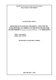
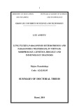
activity of DspB on biofilms of various bacteria [1,2,19]. The PGA polymer from S. epidermidis is a natural substrate with a chain length of (cid:2) 130 GlcNAc residues in b(1,6)-linkages [5]. We used this biofilm- detachment assay to validate the hydrolytic activities of the active-site mutants using biofilm from S. epide- rmidis. As the E184Q mutant showed very low activity that was not measurable even at an enzyme concentration of 1.4 lm. Although both D183N and E332Q variants are less effective than E184Q in the hydrolysis of pNPGlcNAc, these variants behaved differently towards the detachment of S. epi- dermidis biofilm. Both D183N and E332Q mutants exhibited biofilm-detachment activity [time required for 50% detachment, T50, is 7.2 min for E332Q at 0.055 lm (163-fold lower) and 8.0 min for D183N at 0.275 lm (28-fold lower than wild-type; Table 3)]. Mutants W237A and W330Y exhibited biofilm-detach- ment activity with a T50 of (cid:2) 1 min for W330Y and (cid:2) 9 min for W237A, each at an enzyme concentration of 0.275 lm (Fig. 4B). In contrast to pNPGlcNAc hydrolysis, the biofilm-detachment activity of the tyro- sine mutants is not severely affected (Fig. 4C). Thus, the T50 for the mutant Y187A is 5.5 min at 0.022 lm and for the Y278A mutant is (cid:2) 2.8 min at 0.055 lm.
S. G. A. Manuel et al. Active site of dispersin B
E
A
B
30
100
5
5
Efficiency GlcNAc
wild type (0.011 µM) E332Q (0.055 µM) E184Q (1.375 µM) D183N (0.275 µM)
4
4
75
Wild type (0.011 µM) W237A (0.275 µM) W330Y (0.275 µM)
20
3
3
2
2
50
% R e l a t i v e E
1
1
10
m n 0 9 5 t a e c n a b r o s b A
m n 0 9 5 t a e c n a b r o s b A
25
0
f f i c i e n c y
0
) l o m n ( d e s a e l e r c A N c l G
0
1
2
4
6
8
9
10
0
1
2
8
9
10
4
6
3 5 7 Time (minutes)
3 5 7 Time (minutes)
0
0
C
D
5
5
R 27 K
R 27 A
D s p B
Y 187 A
Y 278 A
D 183 N
E 332 Q
E 184 Q
W 330 Y
W 237 A
Wild type (0.011 µM) R27A (0.056 µM) R27K (0.055 µM)
Enzyme
wild type (0.011 µM) Y278A (0.055 µM) Y187A (0.022 µM)
4
4
3
3
2
2
1
1
m n 0 9 5 t a e c n a b r o s b A
m n 0 9 5 t a e c n a b r o s b A
0
0
0
1
2
3
8
9
10 11
0
1
2
3
8
9
10 11
5
6
7
5
6
7
4 Time (minutes)
4 Time (minutes)
FEBS Journal 274 (2007) 5987–5999 ª 2007 The Authors Journal compilation ª 2007 FEBS
5992
Fig. 4. S. epidermidis biofilm detachment activity of DspB and its mutants. (A) Biofilm detachment activity of DspB and its acidic variants D183N, E184Q and E332Q. Biofilms were treated with NaCl ⁄ Pi or the enzyme variant for the indicated time intervals and assayed in 96-well microtiter plates [1,2,19]. Biofilms were then rinsed and stained with crystal violet. The amount of bound crystal violet dye which is propor- tional to the biofilm mass was quantitated by measuring its absorbance at 590 nm. (B) Biofilm detachment activity of the aromatic residue mutants W237A and W330Y. (C) Biofilm detachment activity of the aromatic residue mutants Y187A and Y278A. (D) Biofilm detachment activity of the mutants R27A and R27K. Values plotted correspond to the mean absorbance of triplicate wells. Error bars indicate standard deviations calculated using the program PRISM3 (GraphPad). Legends include the final concentrations of the enzymes used in the detachment assay. (E) Plot of relative efficiency of biofilm detachment (shown on the right y-axis) and the amount of GlcNAc generated (left y-axis) by the various mutants in comparison with wild-type DspB.
S. G. A. Manuel et al. Active site of dispersin B
Table 3. Relative biofilm detachment efficiency of DspB and its mutants with respect to DspB. Values are given in time required for removal of 50% of biofilm. Relative efficiency was calculated as: 100 · (T50 for mutant) · (concentration of for DspB ⁄ T50 DspB ⁄ concentration of mutant).
Enzyme % Relative efficiency T50 (min) Concentration used (lM)
in DspB, at
100
catalyzed attack of the water at the anomeric center. The strongest candidate for the acidic residue that sta- bilizes the transition state and the acid ⁄ base residue has been suggested to be a highly conserved DE or DD pair [21], equivalent to D183 and E184 in DspB. Because of the involvement of this pair, family 20, 56 and 84 enzymes have been suggested to follow a sub- strate-assisted mechanism [11,13–16,20]. Based on superposition of the available crystal structures of vari- ous b-hexosaminidases with that of DspB structure (PDB code 1YHT), subsite )1 in DspB has been deduced to be formed by residues R27, D183, E184, W216, W237, Y278, W330 and E332 (Fig. 5A). Thus, the active-site architecture least at subsite )1, has several salient features that are charac- teristic of b-hexosaminidases exhibiting the substrate- assisted mechanism.
a Data could not be fitted because of very low activity.
Role of acidic residues
the
Interestingly, two mutants R27A and R27K showed biofilm-detachment activity at concentrations 0.055 lm with T50 (cid:2) 5–7 min, which is 20–36-fold less efficient than the wild-type (Fig. 4D). The relative bio- film-detachment activities of the mutants were com- pared with the amount of GlcNAc generated due to hydrolysis of PGA in the biofilm (Fig. 4E). One nota- ble outcome from the GlcNAc-releasing activities of the R27 mutants is that R27K exhibited much higher activity than R27A. Nevertheless, mutation of residues interact with the acetyl group (W237, W330, that D183, Y278) or the catalytic residue E184 showed low activity in both biofilm detachment and pNPGlNAc hydrolysis.
Wild-type D183N E184Q E332Q W237A W330Y Y187A Y278A R27K R27A 0.011 0.275 1.375 0.055 0.275 0.275 0.022 0.055 0.055 0.056 1.3 (0.7) 8.0 (1.4) –a 7.2 (0.3) 9.0 (1.4) 1.0 (0.8) 5.5 (0.2) 2.8 (0.2) 5.2 (0.2) 6.9 (0.3) 0.65 –a 3.6 0.6 5.2 12.0 9.3 5.0 3.8
Discussion
the orientation of
functionally similar enzymes
Among the three acidic residues studied here, D183 is the putative residue that assists the N-acetyl group in neighboring-group participation. Replacement of Asp with Asn at position 183 in DspB rendered this mutant inactive. Mutations of D183-equivalent residues pro- duced very low enzyme activity in human b-hexosamin- idase (Hex A), Streptomyces plicatus b-hexosaminidase and human O-GlcNAcase enzymes. Thus, the mutant D354N (human enzyme) exhibited a kcat value that was only 0.04% that of the wild-type [12], whereas D313N (SpHex enzyme) produced a 560 000-fold reduction in the kcat ⁄ Km value [11] and that of the O-GlcNAcase exhibited a 6727-fold reduction in kcat ⁄ Km [21]. The very low activity of the D313N mutant in SpHex was rationalized by the increased bulk (COO– to CONH2) due to the mutation and ⁄ or by the adaptation of alternate conformations of the N-acetyl group of the terminal GlcNAc residue [11]. In addition to the assistance provided to activate the N-acetyl group, it has been suggested that this Asp might control the C2-acetamido group. The inactivity of the D183N mutant suggests like its counterparts in that residue D183 in DspB, other b-hexosaminidases, participates in cleavage of the glycosidic bond and formation of the intermediate by assisting the acetamido group during catalysis. The conclusion that DspB utilizes a substrate-assisted catal- ysis not only relies on comparison with the work of others, but is also supported by the ability of NAG-thiazoline to inhibit DspB.
It is generally accepted that family 20 hydrolases such as chitobiase from Serratia marcescens and human b-hexosaminidases operate via a retaining mechanism [17]. In such a mechanism, a general acid ⁄ base catalyst plays a dual role by first protonating the departing aglycone and then deprotonating the incoming water molecule (Fig. 1). Because of a lack of nucleophile derived from protein, family 20 b-hexosaminidases and other such family 56 hyaluronidase and family 84 O-GlcNAcase utilize the C2-acetamido group to help stabilize the incipient oxazolinium ion at the anomeric carbon [13,14,17,20]. Formation of the cyclic oxazolinium ion intermediate is assisted by another acidic residue, which interacts with the nitrogen atom of the N-acetyl group and helps stabilize the transition state. The cyclic interme- diate formed is then hydrolyzed by the general base-
Our results support the role of E184 in DspB as an acid ⁄ base catalyst because the E184Q mutant shows significant reduction in kcat ⁄ Km (67-fold lower than the
FEBS Journal 274 (2007) 5987–5999 ª 2007 The Authors Journal compilation ª 2007 FEBS
5993
S. G. A. Manuel et al. Active site of dispersin B
A
R27
E184
E332
GlcNAc
D183
W330
W237
W216
Y278
B
wild-type) and a shift in the apparent pKa for the acid ⁄ base. Mutational studies on E314 from S. plicatus also showed a reduction in the kinetic parameters and a shift in the pH optimum. The acid ⁄ base role for this residue is identical to that of the equivalent gluta- mate ⁄ aspartate residues in family 20 b-hexosaminidas- es and family 56 and family 84 enzymes.
three catalytic residues in human salivary a-amylase and other a-amylases that belong to the family 13 hy- drolases [22,23]. Overall, the acidic residue mutations in DspB tend to destabilize the transition state and the oxazolinium intermediate (Fig. 1) that might be accu- mulated (low Km values), and undergo a slower hydro- lysis (reduced kcat) by a suitably juxtaposed water molecule.
Role of aromatic residues
Although subsite )1 is highly conserved in family 20 b-hexosaminidases, significant differences in the active site of DspB should be noted. For example, the bind- ing pockets created by the aromatic residues for sub- site )1 in DspB and S. plicatus enzyme differ in size and shape (Fig. 5B). It is possible that the difference in pocket shape might lead to subtle differences in the binding of the terminal GlcNAc residue and enable the active site of DspB to be flexible. Differences
Our findings reveal an important role for E332 in DspB and possibly other hexosaminidases as well. As shown in Fig. 2B, E332 of DspB and its equivalent res- idues (residue Glu739 in the human Hex enzyme and residue E444 in the S. plicatus enzyme) in fam- ily 20 b-hexosaminidases occupy the same space on the opposite side of the bound ligand at subsite )1. These residues are positioned in space to interact with the hydroxyl group at C4. Kinetic parameters obtained for the E332Q mutant, reflecting the loss of activity upon mutation, unequivocally show that a loss of transition- state stabilization has occurred. In this regard, E332 residue might be acting similar to D300, one of the
FEBS Journal 274 (2007) 5987–5999 ª 2007 The Authors Journal compilation ª 2007 FEBS
5994
Fig. 5. Active site of DspB. (A) Active-site residues of DspB and their potential interactions with a docked GlcNAc at subsite )1. GlcNAc was docked onto the DspB active site manually using SpHex structure (PDB code: 1hp5) [17] as a reference. Superposition of the crystal structures was carried out using SWISS-MODEL [31]. (B) Superposition of the active-site cavity formed by subsite )1 in DspB (blue) and S. plicatus enzyme (yellow). Two views 90(cid:2) to each other are shown in the left and right panels. Note the difference in the cavity size and shape (207 A˚ 3 for DspB and 222 A˚ 3 for S. plicatus enzyme) suggesting that the binding of the terminal GlcNAc residue might be different in the two enzymes. Cavity was calculated using the server at http://www.bioinformatics. leeds.ac.uk/cgi-bin/pocketfinder/.
[24]. The importance of the aglycone in DspB is in contrast to the existing body of work on other fam- ily 20 enzymes that cleave b(1,4)-linked substrates, wherein the aglycone does not appear to have a signifi- cant impact. Analysis of the reaction mixtures by TLC with chitin oligosaccharides using DspB and the mutants did not generate any hydrolytic products (data not shown) further substantiating the substrate specificity of DspB. The active site of DspB is clearly designed to bind 1,6-linked substrates whereas 1,4- linked substrates and other substrate analogs mimick- ing 1,4-linked substrates either do not bind at all, or bind only poorly.
S. G. A. Manuel et al. Active site of dispersin B
Note that the acetyl group and W237 are involved in a stacking interaction as has been observed in many b-hexosaminidase crystal structures bound to a ligand. Even in DspB, a similar stacking is present between W237 and the bound acetate mimicking the N-acetyl group [10]. In the W237A mutant, binding of the ace- tyl group might have been affected thus reducing the enzyme activity against pNPGlcNAc substrate. By con- trast, the observed activity of the tryptophan mutants W237A and W330Y in biofilm detachment could be explained because the active site is designed to bind the 1,6-linked substrate as present in the biofilm matrix. These results have been further confirmed by total glucosamine assay measuring the hydrolysis rather than the biofilm detachment (Fig. 4E). The rela- tive hydrolytic activity, as measured by the release of GlcNAc (Fig. 4E), might be considered to reflect true activities of these mutations than the use of pNPGlc- NAc as a substrate. It is likely that DspB possesses multiple subsites for GlcNAc residues to bind and pro- vides potential interactions between GlcNAc residues at subsites +1 and ⁄ or +2 and protein atoms.
The role of arginine at the active site
and MuGlcNAc,
comparable
are
and
in the substrate specificity in DspB versus other b-hexosaminidases might arise because of differences in the residues that create subsite +1. In particular, tryptophan residues W685 (human Hex A) and W408 (S. plicatus), respectively, interact with the subsite +1 moiety via hydrophobic-stacking interactions [16,17]. In DspB, an equivalent tryptophan residue is absent. In this regard, the juxtaposition of two loops segment in DspB (residues 185–191 containing Y187) and a sec- ond loop (residues 238–247 containing Q244) might be important. As a result, there are two pockets in the active site, formed by the intrusion of Y187, and unnatural substrates such as pNPGlcNAc may be ori- ented to bind in these pockets, albeit poorly (Fig. 6). Another commonly used substrate, MuGlcNAc, is also poorly cleaved by DspB (Table 2) suggesting that agly- cone might be critical in DspB. The first-order rate constant for this substrate is 39-fold lower than for pNPGlcNAc. No measurable activity was observed for the mutants D183N, E184Q, E332Q, W330Y, W237A, R27A and R27K, whereas values for the other two mutants, Y187A and Y278A are consistent with their proposed roles. In the case of SpHex enzyme, however, the kinetic parameters for the two substrates, pNP- (kcat: GlcNAc 193 ± 3 s)1 versus 180 ± 7 s)1; Km: 0.049 ± 0.004 0.054 ± 0.03 mm; mm kcat ⁄ Km: versus 3300 ± 300 s)1Æmm)1) 3900 ± 400 s)1Æmm)1 versus
The available crystal structure data show that R27 is directly involved in substrate binding and helps to dock a GlcNAc at subsite )1. This is facilitated by bridging interactions with the C3- and C4-hydroxyl groups, as observed in the crystal structures of several b-hexosaminidases reported in the literature. The lower kcat value for mutant R27K suggests that R27 might be involved in stabilization of the transition state [18,25]. Interestingly, distinct differences in GlcNAc- releasing activity from the biofilm have been noted for the two R27 mutants (Fig. 4E). The low activities of the two mutants with respect to pNPGlcNAc hydroly- sis, compared with the wild-type, may be because this substrate does not truly mimic the natural substrate. The difference between substitution with a neutral side
FEBS Journal 274 (2007) 5987–5999 ª 2007 The Authors Journal compilation ª 2007 FEBS
5995
Fig. 6. Representation of the surface cavity in DspB. The large substrate-binding pocket is partitioned by the residues Y187 and Q244 positioned in the middle. The residue W237 provides a wall for a stacking interaction with the bound acetate ⁄ N-acetyl group. The pocket extends on the right and might be utilized to bind b(1,6)-linked GlcNAc polymer to generate multiple subsites.
occurs via a substrate-assisted mechanism. Further- more, the notable differences in the activity of DspB mutant Y187A towards pNPGlcNAc and PGA biofilm from S. epidermidis, suggest that DspB might utilize the loop containing Y187 for its substrate specificity. In conclusion, our mutational analysis has shown that every residue at the subsite )1 in DspB is essential for optimal activity.
S. G. A. Manuel et al. Active site of dispersin B
Experimental procedures
Expression and purification of DspB and mutants
chain (R27A) and a charged side chain (R27K) becomes apparent when one compares biofilm-detach- ment activities. The small increase in efficiency for mutant R27K (Fig. 4D and Table 3) is likely to arise because of the residual potential stabilization of the transition state due to retention of the positive charge (R27 versus K27). However, the biofilm is a complex environment and detachment activity requires action on the surface-attached PGA or PGA very close to the abiotic surface. By contrast, when one compares the GlcNAc-releasing activity of the mutant and wild-type enzymes, charge retention at the transition state in R27K clearly reflects the role of R27 in DspB. Thus, R27K exhibits a significantly higher activity (Fig. 4E) than R27A. Unlike biofilm detachment, GlcNAc release can occur from the exposed chains of PGA in the biofilm each of which consists up to 130 b(1,6)- linked GlcNAc moieties [5] further supporting the idea that GlcNAc-releasing activity might truly reflect the roles of these residues in DspB.
Active-site residues of DspB and their role in the substrate-assisted mechanism
[26]
Isopropyl
DspB and its site-specific mutants were expressed and puri- fied using plasmid pRC3 carrying the dspB gene (encoding amino acids 21–381 fused directly to a hexahistidine metal- binding C-terminal tail located downstream from an iso- thio-b-d-galactoside-inducible tac promoter) [10]. propyl Mutants of DspB were generated using the primers listed in Table 1. Native and the mutant enzymes were expressed as previously described [10]. Briefly, a 2 L Erlenmeyer flask containing 500 mL Luria–Bertani broth supplemented with 30 lg kanamycin per mL was inoculated with 5 mL of an overnight culture of E. coli strain Rosetta(cid:3) (DE3) (Nov- agen, Madison, WI) transformed with the plasmid pRC3 containing the appropriate mutation. The flask was incubated at 37 (cid:2)C with agitation (200 r.p.m.) until thio-b-d-galactoside was A600 ¼ 0.6 (c. 3 h). added to a final concentration of 0.1 mm, and the flask was incubated for 5 h with agitation. Cells were harvested by centrifugation for 15 min at 6000 g.
The cell pellet was resuspended in 20 mL lysis buffer (20 mm Tris ⁄ HCl, pH 8.0, 500 mm NaCl, 1 mm phenyl- methylsulfonyl fluoride and 0.1% Nonidet P-40). The cell suspension was then sonicated on ice for 30 s (· 5 with 2 min intervals) at 30% capacity with a 30% duty cycle using a Branson model 450 sonicator equipped with a microprobe. The cell debris was pelleted by centrifugation (15 000 g for 20 min; 4 (cid:2)C), and the supernatant was loaded onto a 3 mL (bed volume) activated Ni–NTA aga- rose affinity column (Qiagen, Valencia, CA) according to the manufacturer’s instructions. The column was washed with 50 mL wash buffer (20 mm Tris, pH 8.0, 500 mm NaCl) containing 5 mm imidazole. Bound proteins were eluted with 25 mL wash buffer containing 50 mm imidaz- ole. Fractions of the eluate (5 mL) were collected and assayed for the presence of DspB by SDS–PAGE and Coomassie Brilliant Blue R250 staining [27]. Fractions containing pure protein were pooled and dialyzed overnight against water (2 · 4 L changes at 4 (cid:2)C) by using a 12–14 kDa cut-off dialysis membrane (Spectra ⁄ Por2, Spectrum Laboratories, Inc., Rancho Dominguez, CA), and lyophil- lized. The purity and molecular mass of DspB proteins were determined by MS analysis.
The hydrolysis of pNPGlcNAc and the biofilm- detachment activity exhibited by the mutants studied clearly suggest that several residues at subsite )1 play a significant role in the hydrolytic reaction. It is well established that the position of the N-acetyl group is for b-hexosaminidase enzymes to exhibit a critical substrate-assisted mechanism. Whereas earlier reports focused on the individual role played by D183 in this regard, our results show that, in addition to D183, residues such as W237 and Y278 assist in orienting the acetyl group by participating in a critical-stacking or hydrogen-bond interaction with it. Mutation of R27 or E332 has a drastic effect on enzyme activity. Because these residues are on the same side of the bound ligand and interact with C4 oxygen, the stabil- ization provided by the residues in the transition state appears to be critical as the saccharide is undergoing a conformational change. A detailed study on the probable conformational changes during the sub- strate-assisted mechanism has suggested that the ter- minal saccharide changes from a skew conformation in the Michaelis complex, to an envelope (or perhaps half chair) conformation in the transition state, and then to a chair conformation in the intermediate [14]. In this regard, residue W330, also on the side of C4, provides a wall for the GlcNAc-binding pocket and thus might work in concert with R27 and E332 to provide the necessary stabilization in the transition that hydrolysis in DspB state. Our results suggest
FEBS Journal 274 (2007) 5987–5999 ª 2007 The Authors Journal compilation ª 2007 FEBS
5996
Enzyme assays
pH, Vmax is the maximum rate at optimum pH, [H] is the hydrogen ion concentration and Ka and Kb represent the acid dissociation constants. pH dependence was measured on three separate occasions with similar results. Because of the very low activity of D183N and E332Q, no pH optima for these mutants were determined.
substrate
Bacterial strains, media, and growth conditions
S. epidermidis strain NJ9709 (isolated from the surface of infected intravenous catheters removed from patients at University Hospital, Newark, NJ) [2] was streaked onto blood agar plates and incubated for 24 h at 37 (cid:2)C. Plates were stored at 4 (cid:2)C, and bacteria were passaged weekly. Biofilms were grown in Trypticase soy broth (Becton-Dick- inson, Franklin Lakes, NJ) supplemented with 6 g yeast extract and 8 g glucose per L. Isolation and identification of the bacterial strains were part of routine diagnostic pro- cedures at the hospital and were carried out with the under- standing and consent of each subject.
Biofilm growth and detachment assay
pNPGlcNAc (Sigma-Aldrich, St Louis, MO) or MuGlcNAc was used to determine the enzyme activity. Typically, enzyme reactions were carried out in 100-lL mixtures con- taining 50 mm NaCl ⁄ Pi (pH 5.8), 50 mm NaCl, and varying (0.312–10 mm). The final concentrations of enzyme concentrations used are provided in Table 2. Because of the solubility constraints of the substrate, the maximum substrate concentration was restricted to 10 mm. All assays were carried out in 96-well microtiter plates placed in a 37 (cid:2)C incubator. Reactions were terminated at various times by adding 1 lL of 5 m NaOH. The increase in absorption resulting from the release of p-nitrophenol in each well was measured as nitrophenolate with a Bench- mark microplate reader (Bio-Rad, Hercules, CA) set at 405 nm. The specific activities of DspB and its mutants were determined using pNPGlcNAc where 1 unit of enzyme activity was defined as the amount of enzyme needed to hydrolyze 1 lmol of pNPGlcNAc to p-nitrophenol and N-acetylglucosamine per min at pH 5.8 at 37 (cid:2)C in 50 mm NaCl ⁄ Pi containing 50 mm NaCl [11]. Michaelis–Menten kinetic parameters were obtained from double-reciprocal plots (1 ⁄ v versus 1 ⁄ S) using first-order kinetics because of the low solubility of pNPGlcNAc. Inhibition studies were carried out using the mechanism-based inhibitor NAG- thiazoline [16] whose concentration was varied from 0 to 1000 lm. For comparison of the inhibitory efficiency against DspB, jack bean b-hexosaminidase (Sigma-Aldrich) was used. All assays were performed in triplicate on at least three separate occasions, which exhibited similar results with minimal variation among them.
Determination of pH optima
(pH 3.5–6.5), Hepes
The bacterial inoculum was prepared as follows. Two loops of colonies scraped from the surface of an agar plate were transferred to a microfuge tube containing 200 lL fresh medium. Cells were homogenized with a disposable pellet Kontes pestle (Fischer, Itasca, IL) and vortex agitated at high speed for 30 s. Twenty-two microliters was transferred to a 50 mL polystyrene tube with 22 mL fresh medium. The resulting inoculum contained 109–1010 cfuÆmL)1. Biofilms were grown in 96-well tissue-culture-treated polystyrene mi- crotiter plates (Corning model 3595, Sigma-Aldrich). Wells filled with 200 lL inoculum were incubated for 16 h at 37 (cid:2)C. For detachment studies, wells were rinsed by sub- merging the entire plate in a tub of cold, running tap water. Varying concentrations of DspB or mutant enzymes in NaCl ⁄ Pi were added at specific time intervals ranging from 1 to 10 min. Biofilms were washed in tub of cold, running tap water and were stained with crystal violet as previously described [1]. The absorbance values of the well solutions were determined by using a Bio-Rad Benchmark microplate reader set at 590 nm. All assays were performed in triplicate wells on at least three separate occasions, which exhibited similar results with minimal variation among them.
Total hexosamine assay
Confluent biofilms were grown in 100-mm diameter tissue- culture-treated polystyrene Petri dishes. The biofilm that formed on the surface of the plate was rinsed with NaCl ⁄ Pi (3·) and then scraped from the surface into 30 mL of NaCl ⁄ Pi by using a cell scraper. Cells were pelleted by cen- trifugation for 10 min at 2684 g in a Sorvall model RT 7
DspB activity was measured using pNPGlcNAc at various pH values ranging from 3.5 to 10.0. The buffers used were Mes (pH 7.0–8.0) and Tris ⁄ HCl (pH 8.5–10.0). The pH dependence on activity was mea- sured using a final substrate concentration of 5 mm. The molar absorption coefficient of the liberated p-nitrophenol varies with pH from 7280 to 19 000 depending upon the degree of ionization [28]. The appropriate molar absorption coefficient for the tested pH was used in the calculations. p-Nitrophenol release was measured at 400 nm over a time interval after the addition of enzyme to initiate the reaction. The enzyme concentrations used were 100 nm DspB, 5.5 lm R27K, and 1 lm E184Q. The change in absorbance was fitted to a first-order rate equation to yield the pseudo- first order rate constant at each pH value tested. The exper- imental points were fitted to a sigmoid curve using the program prism 3.0 (GraphPad Software, San Diego, CA). The apparent pKa values for the acidic and basic limbs were estimated by fitting the data to the equation v ¼ Vmax ⁄ (1 + [H] ⁄ Ka + Kb ⁄ [H]), where v is rate at a given
FEBS Journal 274 (2007) 5987–5999 ª 2007 The Authors Journal compilation ª 2007 FEBS
5997
S. G. A. Manuel et al. Active site of dispersin B
Cope L et al. (2003) Isolation, structural characteriza- tion, and immunological evaluation of a high-molecu- lar-weight exopolysaccharide from Staphylococcus aureus. Carbohydr Res 338, 903–922.
9 Wang X, Preston JF 3rd & Romeo T (2004) The
(50 mg) was resuspended in centrifuge. The cell pellet NaCl ⁄ Pi containing 5.75 lm of DspB or mutant enzymes and the reaction mixture (200 lL) was incubated at 37 (cid:2)C for 30 min with occasional stirring. After centrifugation, total hexosamine was measured in the supernatant by using the Morgan–Elson colorimetric assay [29]. All assays were performed at least three times with similar results.
pgaABCD locus of Escherichia coli promotes the synthe- sis of a polysaccharide adhesin required for biofilm for- mation. J Bacteriol 186, 2724–2734.
S. G. A. Manuel et al. Active site of dispersin B
Acknowledgements
10 Ramasubbu N, Thomas LM, Ragunath C & Kaplan JB (2005) Structural analysis of dispersin B, a biofilm- releasing glycoside hydrolase from the periodontopatho- gen Actinobacillus actinomycetemcomitans. J Mol Biol 349, 475–486.
11 Williams SJ, Mark BL, Vocadlo DJ, James MN &
This project was supported by the USPHS Grant DE16291 (NR) and DE15124 (JBK). We would like to thank Ms. Samin Nawaz, Ms. Maryam Sheikh and Mr James Rankin for technical assistance. We thank Dr Spencer A. Knapp for generous gift of the NAG-thiaz- oline inhibitor used in this study.
Withers SG (2002) Aspartate 313 in the Streptomyces plicatus hexosaminidase plays a critical role in substrate- assisted catalysis by orienting the 2-acetamido group and stabilizing the transition state. J Biol Chem 277, 40055–40065.
References
1 Kaplan JB, Meyenhofer MF & Fine DH (2003) Biofilm growth and detachment of Actinobacillus actinomyce- temcomitans. J Bacteriol 185, 1399–1404.
12 Hou Y, Vocadlo DJ, Leung A, Withers SG & Mahuran D (2001) Characterization of the Glu and Asp residues in the active site of human beta-hexosaminidase B. Biochemistry 40, 2201–2209.
2 Kaplan JB, Ragunath C, Velliyagounder K, Fine DH & Ramasubbu N (2004) Enzymatic detachment of Staphy- lococcus epidermidis biofilms. Antimicrob Agents Chemo- ther 48, 2633–2636.
3 Izano EA, Sadovskaya I, Vinogradov E, Mulks MH,
13 Sheldon WL, Macauley MS, Taylor EJ, Robinson CE, Charnock SJ, Davies GJ, Vocadlo DJ & Black GW (2006) Functional analysis of a group A streptococcal glycoside hydrolase Spy1600 from family 84 reveals it is a beta-N-acetylglucosaminidase and not a hyaluroni- dase. Biochem J 399, 241–247.
14 Whitworth GE, Macauley MS, Stubbs KA, Dennis RJ,
Velliyagounder K, Ragunath C, Kher WB, Ramasubbu N, Jabbouri S, Perry MB et al. (2007) Poly-N-acetylglu- cosamine mediates biofilm formation and antibiotic resistance in Actinobacillus pleuropneumoniae. Microb Pathol 43, 1–9.
4 Itoh Y, Wang X, Hinnebusch BJ, Preston JF 3rd &
Romeo T (2005) Depolymerization of beta-1,6-N-acetyl- d-glucosamine disrupts the integrity of diverse bacterial biofilms. J Bacteriol 187, 382–387.
Taylor EJ, Davies GJ, Greig IR & Vocadlo DJ (2007) Analysis of PUGNAc and NAG-thiazoline as transition state analogues for human O-GlcNAcase: mechanistic and structural insights into inhibitor selectivity and transition state poise. J Am Chem Soc 129, 635–644. 15 Maier T, Strater N, Schuette CG, Klingenstein R, Sand- hoff K & Saenger W (2003) The X-ray crystal structure of human beta-hexosaminidase B provides new insights into Sandhoff disease. J Mol Biol 328, 669–681.
5 Mack D, Fischer W, Krokotsch A, Leopold K, Hart- mann R, Egge H & Laufs R (1996) The intercellular adhesin involved in biofilm accumulation of Staphylo- coccus epidermidis is a linear beta-1,6-linked glucosami- noglycan: purification and structural analysis. J Bacteriol 178, 175–183.
16 Mark BL, Mahuran DJ, Cherney MM, Zhao D, Knapp S & James MN (2003) Crystal structure of human beta- hexosaminidase B: understanding the molecular basis of Sandhoff and Tay–Sachs disease. J Mol Biol 327, 1093– 1109.
17 Mark BL, Vocadlo DJ, Knapp S, Triggs-Raine BL,
6 Gerke C, Kraft A, Sussmuth R, Schweitzer O & Gotz F (1998) Characterization of the N-acetylglucosaminyl- transferase activity involved in the biosynthesis of the Staphylococcus epidermidis polysaccharide intercellular adhesin. J Biol Chem 273, 18586–18593.
7 Maira-Litran T, Kropec A, Abeygunawardana C, Joyce
J, Mark G, Goldmann DA 3rd & Pier GB (2002) Immunochemical properties of the staphylococcal poly-N-acetylglucosamine surface polysaccharide. Infect Immun 70, 4433–4440.
8 Joyce JG, Abeygunawardana C, Xu Q, Cook JC,
Withers SG & James MN (2001) Crystallographic evi- dence for substrate-assisted catalysis in a bacterial beta- hexosaminidase. J Biol Chem 276, 10330–10337. 18 Mark BL, Wasney GA, Salo TJ, Khan AR, Cao Z, Robbins PW, James MN & Triggs-Raine BL (1998) Structural and functional characterization of Streptomy- ces plicatus beta-N-acetylhexosaminidase by comparative molecular modeling and site-directed mutagenesis. J Biol Chem 273, 19618–19624.
Hepler R, Przysiecki CT, Grimm KM, Roper K, Ip CC,
FEBS Journal 274 (2007) 5987–5999 ª 2007 The Authors Journal compilation ª 2007 FEBS
5998
19 Kaplan JB, Ragunath C, Ramasubbu N & Fine DH
beta-hexosaminidase B. Biochemistry 39, 6219– 6227.
(2003) Detachment of Actinobacillus actinomycetemcomi- tans biofilm cells by an endogenous beta-hexosaminidase activity. J Bacteriol 185, 4693–4698.
20 Tews I, Perrakis A, Oppenheim A, Dauter Z, Wilson
26 Dubendorff JW & Studier FW (1991) Creation of a T7 autogene. Cloning and expression of the gene for bacte- riophage T7 RNA polymerase under control of its cognate promoter. J Mol Biol 219, 61–68.
KS & Vorgias CE (1996) Bacterial chitobiase structure provides insight into catalytic mechanism and the basis of Tay–Sachs disease. Nat Struct Biol 3, 638–648. 21 Cetinbas N, Macauley MS, Stubbs KA, Drapala R &
Vocadlo DJ (2006) Identification of Asp174 and Asp175 as the key catalytic residues of human O-GlcNAcase by functional analysis of site-directed mutants. Biochemis- try 45, 3835–3844.
22 Ramasubbu N, Ragunath C & Mishra PJ (2003) Prob-
27 Sambrook J, Fritsch E & Maniatis T (1989) Molecular Cloning. A Laborartory Manual, 2nd edn. Cold Spring. Harbor Laboratory Press, Cold Spring Harbor, NY. 28 Rauscher E, Neumann U, Schaich E, von Bulow S & Wahlefeld AW (1985) Optimized conditions for deter- mining activity concentration of alpha-amylase in serum, with 1,4-alpha-d-4-nitrophenylmaltoheptaoside as substrate. Clin Chem 31, 14–19.
ing the role of a mobile loop in substrate binding and enzyme activity of human salivary amylase. J Mol Biol 325, 1061–1076.
23 Ramasubbu N, Ragunath C, Mishra PJ, Thomas LM,
29 Strominger JL, Park JT & Thompson RE (1959) Com- position of the cell wall of Staphylococcus aureus: its relation to the mechanism of action of penicillin. J Biol Chem 234, 3263–3268.
Gyemant G & Kandra L (2004) Human salivary alpha-amylase Trp58 situated at subsite-2 is critical for enzyme activity. Eur J Biochem 271, 2517–2529.
30 Lemieux MJ, Mark BL, Cherney MM, Withers SG, Mahuran DJ & James MN (2006) Crystallographic structure of human beta-hexosaminidase A: interpreta- tion of Tay–Sachs mutations and loss of GM2 ganglio- side hydrolysis. J Mol Biol 359, 913–929.
24 Vocadlo DJ & Withers SG (2005) Detailed comparative analysis of the catalytic mechanisms of beta-N-acetyl- glucosaminidases from families 3 and 20 of glycoside hydrolases. Biochemistry 44, 12809–12818. 25 Hou Y, Vocadlo D, Withers S & Mahuran D
31 Schwede T, Kopp J, Guex N & Peitsch MC (2003) SWISS-MODEL: an automated protein homology- modeling server. Nucleic Acids Res 31, 3381–3385.
(2000) Role of beta Arg211 in the active site of human
FEBS Journal 274 (2007) 5987–5999 ª 2007 The Authors Journal compilation ª 2007 FEBS
5999
S. G. A. Manuel et al. Active site of dispersin B