
T
ẠP CHÍ KHOA HỌC
TRƯ
ỜNG ĐẠI HỌC SƯ PHẠM TP HỒ CHÍ MINH
Tập 22, Số 3 (2025): 437-442
HO CHI MINH CITY UNIVERSITY OF EDUCATION
JOURNAL OF SCIENCE
Vol. 22, No. 3 (2025): 437-442
ISSN:
2734-9918
Websit
e: https://journal.hcmue.edu.vn https://doi.org/10.54607/hcmue.js.22.3.4192(2025)
437
Research Article1
FOUR COMPOUNDS FROM DICRANOPTERIS LINEARIS SPORES
Doan Ngoc Anh, Le Thi Phuong Thao, Nguyen Hong Ngoc,
Nguyen Thi Ngoc Duyen, Vuong Boi Phong, Duong Thuc Huy*
Ho Chi Minh University of Education, Vietnam
*Corresponding author: Duong Thuc Huy – Email: huydt@hcmue.edu.vn
Received: April 01, 2024; Revised: October 16, 2024; Accepted: December 11, 2024
ABSTRACT
Dicranopteris linearis, a widely utilized plant species in traditional Vietnamese medicine, has
been chemically investigated. Four compounds (1–4), including syringaresinol (1), shikimic acid (2),
angelicin (3), and 3,4-dihydroxycinnamic acid (4) were isolated and structurally elucidated.
Extensive spectroscopic methods were employed for structural elucidation. These isolates were
subsequently evaluated for their alpha-glucosidase inhibitory activity and inhibitory effect on nitric
oxide production in LPS-stimulated RAW 264.7 cells. Isolated compounds exhibited no inhibitory
activity in either alpha-glucosidase or nitric oxide inhibition assays.
Keywords: alpha-glucosidase; angelicin; Dicranopteris linearis; nitric oxide inhibition;
shikimic acid; syringaresinol
1. Introduction
Comprehensive reviews have underscored ferns as widely recognized sources with
numerous traditional applications. These include hepatoprotective effects,
antihyperglycemic properties, leishmanicidal activity, and trypanocidal activity (Cao et al.,
2017; Kumar et al., 2010). Dicranopteris linearis (Burm. F.) Underw. is a globally
distributed fern species that has been traditionally utilized in East Asian countries for the
treatment of diverse diseases. In Malaysia, it is employed to alleviate fevers, while in
Indochina, it is utilized to combat intestinal worms (Kamisan et al., 2014). In India, it is used
to treat infertility in women, and in Papua New Guinea, it is utilized for wound healing
(Sarker & Hossain, 1970). Various pharmacological properties of D. linearis have been
reported, including anticancer, antibacterial, antioxidant, analgesic, and anti-HIV activities
(Chen et al., 2014; Li et al., 2008; Ponnusamy et al., 2015; Zakaria et al., 2021). A
comprehensive chemical analysis of D. linearis was conducted, revealing the presence of
over 50 compounds, predominantly found in the leaves of the plant (Chen et al., 2014; Duong
et al., 2023, 2024; Li et al., 2008; Ponnusamy et al., 2015; Raja et al., 1995). Numerous
pharmaceutical properties of extracts of D. linearis leaves have been the subject of scientific
Cite this article as: Doan, N. A., Le, T. P. T., Nguyen, H. N., Nguyen, T. N. D., Vuong, B. P., & Duong, T. H.
(2025). Four compounds from Dicranopteris linearis spores. Ho Chi Minh City University of Education Journal
of Science, 22(3), 437-442. https://doi.org/10.54607/hcmue.js.22.3.4192(2025)
HCMUE Journal of Science
Doan Ngoc Anh et al.
438
investigation. Our previous report indicated that the organs of D. linearis may possess potent
alpha-glucosidase inhibitors (Duong et al., 2023). Recently, D. linearis spores have been
chemically investigated using a bioactive-guide procedure based on antioxidant activity,
including DPPH and ABTS (Duong et al., 2024). This study aims to elucidate the chemical
composition and bioactive properties of the spores of D. linearis, with a particular emphasis
on their alpha-glucosidase and nitric oxide inhibitory activities.
O O
O
1
2
3
4
5
6
7
89
10
2' 3'
AB
C
O
O
O
HO
O
O
O
OH
1
OOH
HO OH OH
1
35
1
3
5
1' 3'
5'
7
9
8'
23
O
OH
HO OH 4
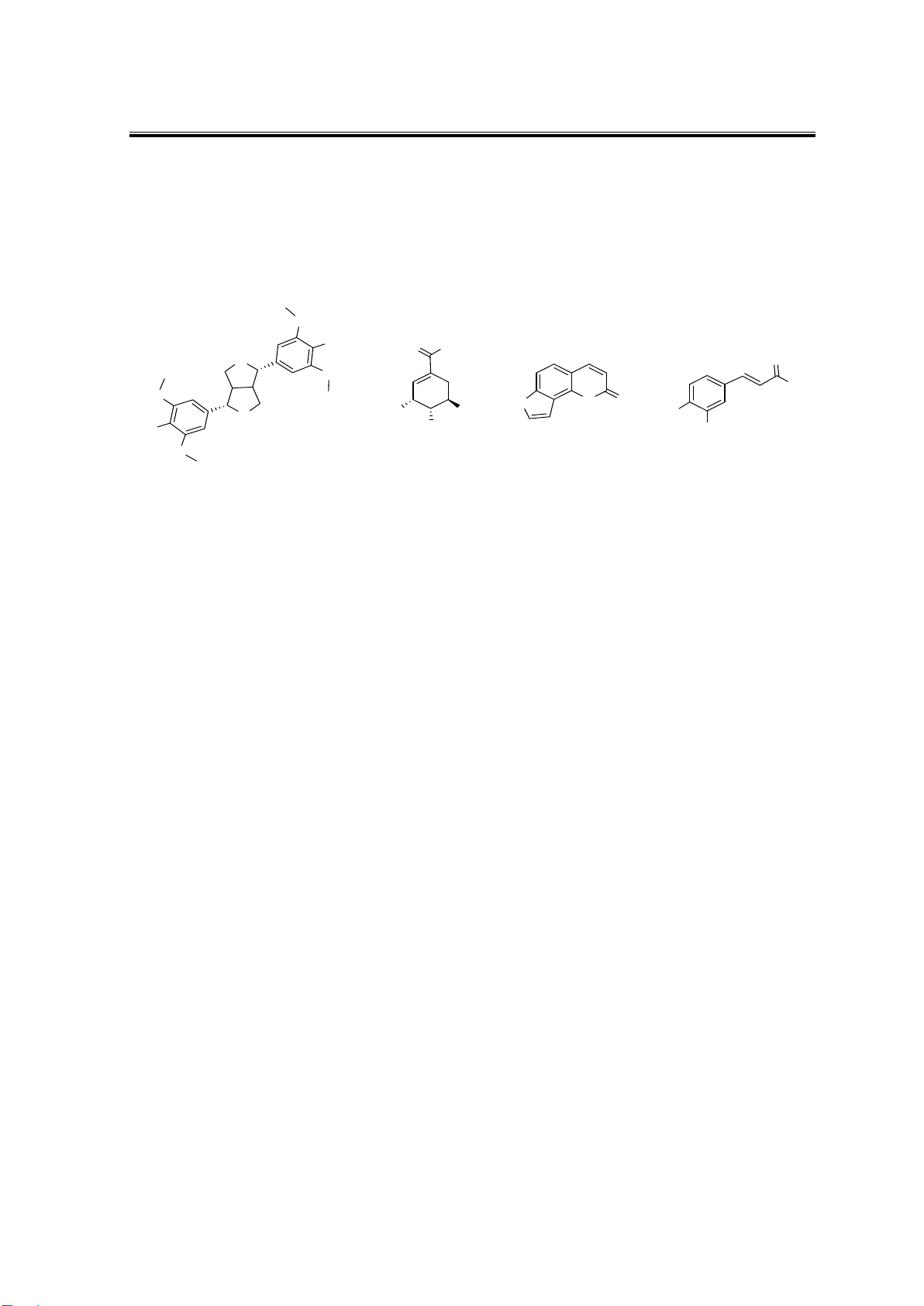
Figure 1. Chemical structures of 1-4
2. Experiments
2.1. General experimental procedures
The NMR spectra were recorded on a Bruker Avance spectrometer (500 MHz for 1H–
NMR and 125 MHz for 13C–NMR) in acetone-d6 and CDCl3. Thin-layer chromatography
was carried out on silica gel 60 (Merck, 40-63 μm), and spots were visualized by spraying
with 10% H2SO4 solution, followed by heating.
2.2. Plant materials
In November 2022, spores of Dicranopteris linearis were collected in Binh Thuan
Province, Vietnam and were authenticated by Assoc. Prof. Van-Son Dang, Institute of
Tropical Biology, Vietnam Academy of Science and Technology (VAST). A voucher
specimen (No. UE-P017A) was deposited in the VNM Herbarium, Institute of Tropical
Biology, VAST.
2.3. Extraction and isolation
The dried powder of D. linearis spores (200 g) was extracted with methanol (10 x 1 L,
each 8 hours) at room temperature using the maceration method. After removing the solvents
using an evaporator, a crude methanol extract (12 g) was obtained. This extract was separated
by silica gel column chromatography (CC) using the gradient system of n-hexane-ethyl
acetate (1:3-0:1, v/v) followed by methanol to afford five fractions EA1-EA5. Fraction EA4
(3.6 g) underwent further purification using silica gel CC, resulting in four subfractions
(EA4.1-EA4.4). Fraction EA4.1 (850 mg) was applied to silica gel CC, isocratically eluted
with EtOAc-MeOH-H2O (100:0:0 then 98:2:0.1, v/v/v) to obtain three fractions T1-T3.
Fraction T1 (67 mg) was further purified using silica gel CC, eluted with EtOAc-MeOH-
H2O (100:0:0 then 95:5:0.1, v/v/v) to yield three compounds 1 (4.0 mg), 3 (3.5 mg), and 4
(21.0 mg). Fraction T3 (255 mg) underwent further purification using silica gel CC, resulting
in compound 2 (11.5 mg).
Syringaresinol (1). Colorless oil; 1H NMR (acetone-d6, 500 MHz): δH 6.70 (2H, s, H-
2/2′ and H-6/6′), 4.68 (2H, d, J = 4.5 Hz, H-7/7′), 3.11 (2H, m, H-8/8′), 4.30 (2H, m, H-9a/9′a),

HCMUE Journal of Science
Vol. 22, No. 3 (2025): 437-442
439
3.90 (2H, dd, J = 3.5, 1.0 Hz, H-9b/9′b), 3.84 (12H, s, 4 x OCH3). 13C NMR (acetone-d6, 125
MHz): 133.1 (C-1/1′), 103.4 (C-2/2′ and C-6/6′), 147.6 (C-3/3′ and C-5/5′), 136.9 (C-4/4′), 85.8
(C-7/7′), 54.0 (C-8/8′), 71.7 (C-9/9′), 56.5 (4 x OCH3).
Shikimic acid (2). White amorphous powder; 1H NMR (methanol-d4, 500 MHz): δH 6.77
(1H, brs, H-2), 4.02 (1H, dd, J = 8.5, 5.5 Hz, H-3), 3.70 (1H, dd, J = 7.3, 4.3 Hz, H-4), 4.40 (1H,
m, H-5), 2.69 (1H, dd, J = 18.0, 5.5 Hz, H-6a), 2.17 (1H, dd, J = 18.0, 4.5 Hz, H-6b). 13C NMR
(methanol-d4, 125 MHz): 171.1 (C=O), 136.8 (C-1), 130.8 (C-2), 66.1 (C-3), 71.5 (C-4), 66.9
(C-5), 30.9 (C-6).
Angelicin (3). White amorphous powder; 1H NMR (acetone-d6, 500 MHz): δH 6.39 (1H,
d, J = 9.5 Hz, H-3), 8.10 (1H, d, J = 9.5 Hz, H-4), 7.55 (1H, d, J = 8.5 Hz, H-5), 7.63 (1H, d, J
= 8.5 Hz, H-6), 7.55 (1H, d, J = 8.5 Hz, H-5), 8.01 (1H, d, J = 2.0 Hz, H-2′), 7.21 (1H, d, J = 2.0
Hz, H-3′). 13C NMR (acetone-d6, 125 MHz): 171.1 (C=O), 136.8 (C-1), 130.8 (C-2), 66.1 (C-
3), 71.5 (C-4), 66.9 (C-5), 30.9 (C-6). 160.8 (C-2), 114.9 (C-3), 145.6 (C-4), 125.4 (C-5), 109.3
(C-6), 158.4 (C-7), 117.4 (C-8), 150.0 (C-9), 114.9 (C-10), 147.6 (C-2′), 104.5 (C-3′).
3,4-Dihydroxycinnamic acid (4). White amorphous powder. 1H NMR (acetone-d6,
500 MHz) and 13C NMR (acetone-d6, 125 MHz) data were consistent with those reported
previously (Oboh et al., 2015).
2.4. Alpha-Glucosidase Inhibition and Nitric oxide inhibition Assays
The alpha-glucosidase inhibitory activity of compounds 1-4 was determined using a
method adapted from previously published protocol (Duong et al., 2024). The samples were
analyzed in triplicate at ten distinct concentrations ranging from the IC50 values, and the
mean values were recorded. NO inhibition of compounds 1-4 were determined using the
same procedure previously reported.(Ngoc Mai et al., 2024; Sukandar et al., 2023) L-
NMMA was used as a positive control. Each sample was analyzed in triplicate at five
different concentrations around the IC50 values, and the mean values were recorded.
3. Results and discussion
Compound 1 was obtained as colorless oil. The 1H-NMR spectrum of 1 showed the
presence of a symmetric benzene ring characterized by four aromatic proton signals at δH 6.70
(4H, s, H-2/H-6 and H-2′/H-6′). In addition, the 1H-NMR spectrum showed the signals of two
oxymethine protons at δH 4.68 (2H, d, J = 4.5 Hz, H-7/H-7′), two methine protons at δH 3.11
(2H, m, H-8/H-8′), four oxymethylene protons at δH 4.30 (2H, m, H-9), 3.90 (2H, dd, J = 3.5,
1.0 Hz, H-9′) and four methoxy groups at δH 3.84 (12H, s). These findings indicated that 1 is a
symmetric lignan. The 13C-NMR spectral data of 1 showed the presence of 12 aromatic methine
carbons at δC 133.1 (C-1/C-1′), 103.4 (C-2/C-2′), 147.6 (C-3/C-3′ and C-5/C-5′), 136.9 (C-4/C-
4′), 103.5 (C-6/C-6′); 2 oxymethine carbon at δC 85.8 (C-7/C-7′); 2 carbon methine at δC 54.0
(C-8/C-8′); 2 methylene groups at δC 71.8 (C-9/C-9′) and 4 methoxy groups at δC 56.5.
Comparison of the NMR data of 1 and those of syringaresinol showed the high similarity, thus
suggesting that the structure of 1 is syringaresinol (Ban et al., 2020).
Compound 2 was obtained as a white amorphous powder. The 1H-NMR (500 MHz,
methanol-d4) spectrum of 2 showed the presence of an olefinic methine at δH 6.77 (1H, brs, H-

HCMUE Journal of Science
Doan Ngoc Anh et al.
440
2), three oxymethines at δH 4.02 (1H, dd, J = 12.0, 5.5 Hz, H-3), 3.70 (1H, dd, J = 7.3, 4.3, H-
4), and 4.40 (1H, m, H-5), a methylene group at δH 2.17 (1H, dd, J = 18.0, 5.5 Hz, H-6) and 2.69
(1H, dd, J = 18.0, 4.5 Hz, H-6). The 13C-NMR (125 MHz, methanol-d4) spectrum of 2 showed
the presence of seven carbon signals including: a carboxyl carbon at δC 168.7, a substituted
olefinic carbon at δC 138.6 (C-1), an olefinic methine carbon at δC 130.1 (C-2), three oxymethine
carbons at δC 130.1 (C-3), 66.7 (C-4), 72.3 (C-5), and a methylene carbon at δC 31.4 (C-6).
Comparison of the NMR data of 2 and those of shikimic acid showed the high similarity, thus
suggesting that the structure of 2 is shikimic acid (Bochkov et al., 2011).
Compound 3 was obtained as a white amorphous powder. The 1H NMR spectrum showed
the presence of two aromatic protons [δH 7.55 (1H, d, J = 8.5 Hz, H-5) and δH 7.63 (1H, d, J =
8.5 Hz, H-6)], four olefinic protons [δH 6.39 (1H, d, J = 9.5 Hz, H-3), 8.10 (1H, d, J = 9.5 Hz,
H-4), 8.01 (1H, d, J = 2.0 Hz, H-2′), 7.21 (1H, d, J = 2.0 Hz, H-3′)]. The 13C NMR data in
accordance with HSQC spectrum exhibited 11 carbon signals: a carbonyl carbon at δC 160.8 (C-
2); four substituted olefinic carbons at δC 158.4 (C-7), 117.4 (C-8), 150.0 (C-9), and 114.9 (C-
10); two aromatic methine carbons at δC 125.4 (C-5), and 109.3 (C-6), four olefinic methine
carbons at δC 114.9 (C-3), 145.6 (C-4), 147.6 (C-2′), and 104.5 (C-3′). HMBC correlations of
proton H-3 to C-4 (δC 145.6), of H-4 to C-2 (δC 160.8), of proton H-5 to C-10 (δC 114.9)
supported the structure of the A-ring. In addition, HMBC correlations of protons H-6 and H-2′
to carbons C-9 (δC 150.0) and C-7 (δC 158.4) and of proton H-3′ to C-7 (δC 158.4) indicated the
connection between the B-ring and C-ring. A comparative analysis of the NMR data of 3 and
those of angelicin revealed a striking degree of similarity, strongly suggesting that the structure
of 3 is identical to that of angelicin (Mar et al., 2001).
To the best of our knowledge, compounds 1-3 were initially identified in the plant. Compounds
1-4 were evaluated for their alpha-glucosidase inhibitory activity and inhibitory effect on
nitric oxide production in LPS-stimulated RAW 264.7 cells. Isolated compounds exhibited
no inhibitory activity in either alpha-glucosidase or nitric oxide inhibition assays. None of
the compounds exhibited any activity.
4. Conclusions
From Dicranopteris linearis spores, four compounds, syringaresinol (1), shikimic acid
(2), angelicin (3), and 3,4-dihydroxycinnamic acid (4), were isolated from the MeOH extract
of Dicranopteris linearis. To the best of our knowledge, compounds 1-3 were first reported in
the plant Dicranopteris linearis. Further studies on this species are in progress.
Conflict of Interest: Authors have no conflict of interest to declare.
Acknowledgments: This research is funded by Vietnamese Ministry of Education and
Training under grant number B2023-SPS-06.

HCMUE Journal of Science
Vol. 22, No. 3 (2025): 437-442
441
REFERENCES
Ban, N. K., Truong, L. H., Linh, T. M., Mai, N. C., Yen, D. T. H., Van Doan, V., Nhiem, N. X.,
Tai, B. H., & Van Kiem, P. (2020). Phenolic compounds from Trigonostemon honbaensis and
their cytotoxic activity. Vietnam Journal of Chemistry, 58(6), 759-764.
https://doi.org/10.1002/vjch.202000068
Bochkov, D. V., Sysolyatin, S. V., Kalashnikov, A. I., & Surmacheva, I. A. (2011). Shikimic acid: Review
of its analytical, isolation, and purification techniques from plant and microbial sources. Journal
of Chemical Biology, 5(1), 5-17. https://doi.org/10.1007/s12154-011-0064-8
Cao, H., Chai, T.-T., Wang, X., Morais-Braga, M. F. B., Yang, J.-H., Wong, F.-C., Wang, R., Yao,
H., Cao, J., Cornara, L., Burlando, B., Wang, Y., Xiao, J., & Coutinho, H. D. M. (2017).
Phytochemicals from fern species: Potential for medicine applications. Phytochemistry
Reviews, 16(3), 379-440. https://doi.org/10.1007/s11101-016-9488-7
Chen, J., Chen, J.-J., & Gao, K. (2014). Chemical constituents and biological activities of
Dicranopteris linearis. Chemistry of Natural Compounds, 49(6), 1129-1131.
https://doi.org/10.1007/s10600-014-0839-6
Duong, T.-H., Tran, T.-M.-D., To, P.-M., Phan, N.-H.-N., Nguyen, T.-P., Le, H. T., & Sichaem, J.
(2024). Potential antioxidant compounds from the spores of Dicranopteris linearis and the
branches of Averrhoa bilimbi. Antioxidants, 13, 1319-1333.
Duong, T.-H., Vu, Y. T., Long, N. P., Phan, N.-H.-N., Pham, N.-K.-T., Sichaem, J., Kieu, N.-K.-D.,
Duong, C.-B., Nguyen, T.-T., Dang, V.-S., & Nguyen, H. T. (2023). Bioactive-guided
phytochemical investigations, in vitro and in silico alpha-glucosidase inhibition of two
vietnamese medicinal plants Dicranopteris linearis and Psychotria adenophylla.
Pharmaceuticals, 16(9), Article 1253. https://doi.org/10.3390/ph16091253
Kamisan, F. H., Yahya, F., Mamat, S. S., Kamarolzaman, M. F. F., Mohtarrudin, N., Kek, T. L.,
Salleh, M. Z., Hussain, M. K., & Zakaria, Z. A. (2014). Effect of methanol extract of
Dicranopteris linearis against carbon tetrachloride- induced acute liver injury in rats. BMC
Complementary and Alternative Medicine, 14(1), Article 123. https://doi.org/10.1186/1472-
6882-14-123
Kumar, A., Fernández, H., & Revilla, M. A. (Eds.). (2010). Working with Ferns: Issues and
Applications. Springer New York. https://doi.org/10.1007/978-1-4419-7162-3
Li, X.-L., Tu, L., Zhao, Y., Peng, L.-Y., Xu, G., Cheng, X., & Zhao, Q.-S. (2008). Terpenoids from
two Dicranopteris species. Helvetica Chimica Acta, 91(5), 856-861.
https://doi.org/10.1002/hlca.200890089
Mar, W., Seo, E.-K., & Je, K.-H. (2001). Cytotoxic constituents of Psoralea corylifolia. Arch.
Pharm. Res., 24(3), 211-213.
Ngoc Mai, T. T., Minh, P. N., Phat, N. T., Chi, M. T., Duong, T. H., Nhi Phan, N. H., Minh An, T.
N., Dang, V.-S., Van Hue, N., Hong Anh, N. T., & Tri, M. D. (2024). In vitro and in silico
docking and molecular dynamic of antimicrobial activities, alpha-glucosidase, and anti-
inflammatory activity of compounds from the aerial parts of Mussaenda saigonensis. RSC
Advances, 14(17), 12081-12095. https://doi.org/10.1039/D4RA01865F
Oboh, G., Agunloye, O. M., Adefegha, S. A., Akinyemi, A. J., & Ademiluyi, A. O. (2015). Caffeic
and chlorogenic acids inhibit key enzymes linked to type 2 diabetes (in vitro): A comparative
study. Journal of Basic and Clinical Physiology and Pharmacology, 26(2), 165-170.
https://doi.org/10.1515/jbcpp-2013-0141


![Câu hỏi ôn thi Hóa lý dược [năm] chuẩn nhất](https://cdn.tailieu.vn/images/document/thumbnail/2025/20250714/kimphuong1001/135x160/4391752479246.jpg)


![Tài liệu giảng dạy Sinh học và di truyền [mới nhất]](https://cdn.tailieu.vn/images/document/thumbnail/2026/20260323/hoatudang2026/135x160/42181774414220.jpg)

![Giáo trình Công nghệ vi sinh (Nghề Công nghệ sinh học TC/CĐ) - Trường Cao đẳng Đà Lạt [Mới nhất]](https://cdn.tailieu.vn/images/document/thumbnail/2026/20260224/hoacattuong2026/135x160/87621772161812.jpg)


![Giáo trình Vi sinh vật học môi trường Phần 1: [Thêm thông tin chi tiết nếu có để tối ưu SEO]](https://cdn.tailieu.vn/images/document/thumbnail/2025/20251015/khanhchi0906/135x160/45461768548101.jpg)


