
BioMed Central
Page 1 of 7
(page number not for citation purposes)
World Journal of Surgical Oncology
Open Access
Research
Alveolar soft part sarcoma: clinicopathological findings in a series of
11 cases
Adrien Daigeler*1, Cornelius Kuhnen2, Joerg Hauser1, Ole Goertz1,
Daniel Tilkorn1, Lars Steinstraesser1, Hans-Ulrich Steinau1 and
Marcus Lehnhardt1
Address: 1Department of Plastic Surgery, Burn Center, Hand surgery, Sarcoma Reference Center, BG-University Hospital Bergmannsheil, Ruhr
University Bochum, Buerkle-de-la-Camp-Platz 1, 44789 Bochum, Germany and 2Institute for Pathology, BG-University Hospital Bergmannsheil,
Ruhr-University Bochum, Bürkle-de-la-Camp-Platz 1, 44789 Bochum, Germany
Email: Adrien Daigeler* - adrien.daigeler@rub.de; Cornelius Kuhnen - cornelius.kuhnen@rub.de; Joerg Hauser - joerg.hauser@rub.de;
Ole Goertz - ole.goertz@rub.de; Daniel Tilkorn - daniel.tilkorn@web.de; Lars Steinstraesser - lars.steinstraesser@rub.de; Hans-
Ulrich Steinau - hans-ulrich.steinau@bergmannsheil.de; Marcus Lehnhardt - marcus.lehnhardt@rub.de
* Corresponding author
Abstract
Background: Alveolar sarcoma of the soft parts (ASPS) represents a very rare entity of soft tissue
sarcoma with special features such as young peak age incidence and frequent metastasis to the
brain. The aim of this study was a clinicopathological analysis with special reference to treatment
and outcome.
Methods: From the database of the BG-University Hospital Bergmannsheil, 1597 soft tissue
sarcoma (STS) cases were reviewed and 11 consecutive patients with ASPS were isolated. Data was
acquired from patients' charts and contact to patients, their relatives or general practitioners, with
special reference to treatment and clinical course. The average follow up time from the time of the
definite operation for the primary tumor was 6.5 years. Kaplan-Meier method was used to calculate
survival.
Results: Patients with localized disease who received complete resection and adjuvant radiation
and who did not develop recurrence or metastatic disease within 2 years after surgery had a
positive outcome. The size of the tumor, its localization, and the time of untreated growth before
treatment did not influence the long-term results. All patients who developed recurrent disease
also suffered from distant metastasis, reflecting the aggressive biology of the tumor. All patients
with distant metastasis had the lungs and the brain affected.
Conclusion: Due to the limited number of patients with ASPS, prospective studies would have to
span decades to gather a significant collective of patients; therefore, it is not possible to comment
meaningfully on a possible benefit of neoadjuvant or adjuvant therapy.
We recommend wide surgical excision and, in the absence of data telling otherwise, adjuvant
radiation. In cases with recurrent disease or metastasis, the prognosis is bad and further treatment
will be restricted to palliation in most cases.
Published: 1 July 2008
World Journal of Surgical Oncology 2008, 6:71 doi:10.1186/1477-7819-6-71
Received: 25 March 2008
Accepted: 1 July 2008
This article is available from: http://www.wjso.com/content/6/1/71
© 2008 Daigeler et al; licensee BioMed Central Ltd.
This is an Open Access article distributed under the terms of the Creative Commons Attribution License (http://creativecommons.org/licenses/by/2.0),
which permits unrestricted use, distribution, and reproduction in any medium, provided the original work is properly cited.

World Journal of Surgical Oncology 2008, 6:71 http://www.wjso.com/content/6/1/71
Page 2 of 7
(page number not for citation purposes)
Background
Alveolar soft part sarcoma (ASPS) is a very rare type soft
tissue sarcoma (STS), with several unusual features, such
as a very young peak age incidence and frequent meta-
static spread to the brain [1]. Accounting for less than 1%
of STS, it presents at almost every part of the body with a
predominance of the trunk and the proximal extremities
[2-5] and usually affects patients younger than 40 years
[5]. The name "alveolar" was derived from its pseudo-
alveolar appearance with clustered polygonal cells lacking
central cohesion [4]. Recent cytogenetic studies revealed
chromosome rearrangements at t(X;17)(p11;q25) result-
ing in the ASPL-TFE3 fusion gene, but the origin of ASPS
still remains unclear, in fact, it seems that a normal cellu-
lar counterpart for this sarcoma does not exist [6-9]. Due
to its rarity, its unusual clinical course and an indolent
progression of disease diagnosis and treatment has been
proven to be a challenge for the pathologist and the sur-
geon as well. We reviewed our single center experience
with ASPS over a period of 16 years with special reference
to the clinical course and outcome and assessed our find-
ings against the background of the existing literature.
Methods
From 1991 to 2007, 11 out of 1597 patients that were
treated at our institution for STS were diagnosed with
alveolar sarcoma of the soft parts (ASPS). Data for this
case series were acquired retrospectively from the sarcoma
database of BG-University Hospital Bergmannsheil. Addi-
tional information regarding the clinical course and out-
come was collected from the patients' charts, and phone
calls to the patients, their relatives and their general prac-
titioners. Follow-up data were available for all patients
and consisted of clinical examination, chest X-ray or com-
puted tomography, abdominal ultrasound and CT or MRI
of the tumor site and the brain in three cases. Local recur-
rence was defined as tumor occurrence after treatment at
a site of previous operation. Metastasis was diagnosed
when the tumor occurred at any other site. Summary sta-
tistics were obtained using the Kaplan-Meier method for
calculating survival. Because of the low number of
patients we refrained from further statistical analysis.
Four patients were female, seven were male, and the aver-
age age at time of diagnosis was 32 years (range: 19–49).
The follow-up time from the time of the definite opera-
tion for the primary tumor was 78 months/6.5 years (5–
156 months).
Histopathological examination
In all cases the diagnosis of ASPS was confirmed by a
review of the pathology slides by experienced soft tissue
pathologists of our institution. In two cases (patient 5 and
9), tissue specimens were sent in for second opinion to
another experienced soft tissue pathologist. In both cases,
the primary diagnosis (ASPS) of our institution was con-
firmed.
Results
With 11 cases of alveolar sarcoma of the soft parts out of
1597 patients with soft tissue sarcoma, ASPS accounted
for 0.7% of STS in our data base. Ten patients could
remember the period of time the tumor was growing
before definite diagnoses was made. This time ranged
from one month (patient 11) to 20 years (patient 5). A
correlation between the duration of untreated tumor
growth and outcome could not be detected.
The site most often affected by the tumor was the thigh (n
= 4) followed by the lower leg (n = 2) and the thoracic
wall (n = 2), the upper arm, the forearm, and the foot in
one case each. All tumors were located intramuscular or
subfascial with an average of 6.8 cm in largest diameter
(range: 2.9–13.5 cm). All patients with an unfavorable
outcome had tumors below the average size (table 1).
At time of primary diagnosis no metastatic disease was
detected in any patient. A definite tumor grading accord-
ing to accepted grading classifications (Coindre classifica-
tion) was not applied due to the difficulty of using grading
as prognostic factor in ASPS. In one case, however, the
tumor was designated a poorly differentiated variant of
ASPS (patient 5).
Two patients (patient 1 and 5) had received neoadjuvant
therapy (chemotherapy with etoposide, vincristine, adri-
amycin, ifosfamide and isolated limb perfusion with mel-
phalan and TNF-alpha) prior to surgery because of a large
tumor mass adjacent to crucial structures, leaving 70%
and 30% of the tumor mass viable in the resection speci-
men. These two patients were alive with no evidence of
disease at follow-up. Incisional biopsy was performed in
six cases; fine needle biopsy in one case. Four patients
were primarily resected with microscopically positive
margins at other institutions and referred to BG-Univer-
sity Hospital Bergmannsheil for curative surgery after his-
tological diagnosis. In all but one case (patient 8), who
died of disseminated disease subsequently, free surgical
margins were achieved by definite surgery.
All but three patients received adjuvant radiation therapy
of the primary tumor site with a dose between 60 and
70Gy. One of these (patient 8) who additionally was not
completely resected in the definite operation died of his
disease (table 2).
Three patients (patients 2, 3, 8) developed metastases in
the lung and the brain (n = 3), the liver (n = 2) and the soft
tissue (n = 1). Two of those (3 and 8) developed subse-
quent recurrent disease 29 and 11 months after surgery,

World Journal of Surgical Oncology 2008, 6:71 http://www.wjso.com/content/6/1/71
Page 3 of 7
(page number not for citation purposes)
which was treated with chemotherapy (patient 3) in one
case and lower leg amputation (patient 8) in the other
case. Two patients were operated on for their metastases.
One patient had the soft tissue metastases (R0) and 21
pulmonary metastases (R2) resected (patient 2); the other
one (patient 3) was operated on for his brain metastasis
(R1). All intracranial metastases were also treated with
adjuvant radiation (30Gy), as well as the one soft tissue
metastasis (60Gy). In addition, all patients with meta-
static disease received several chemotherapeutics, but
unfortunately they all died from disseminated disease
after 48, 79, and 97 months. The progression free interval
in these patients was 7, 9, and 12 months, respectively
(table 3). All other patients were alive with no evidence of
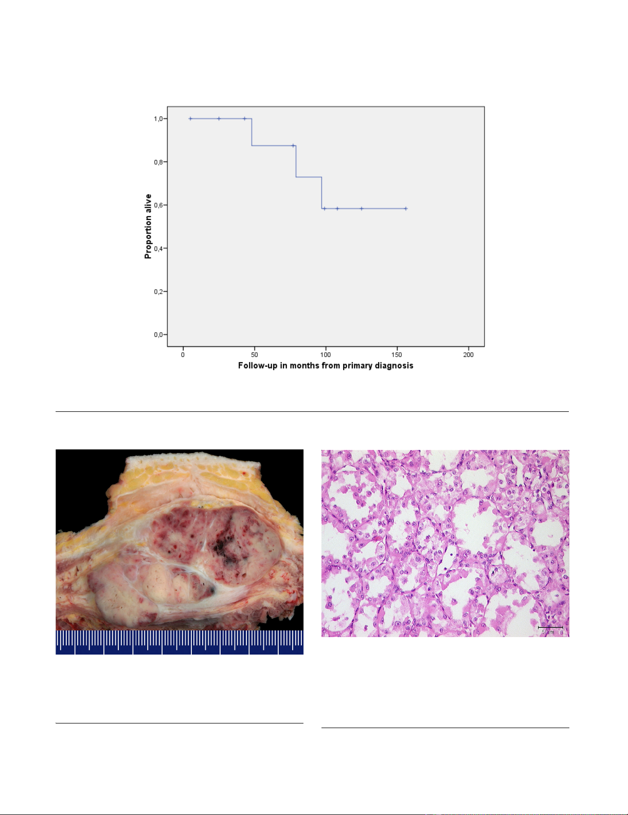
disease. The 2 year survival was calculated at 88%, the 5
year survival was calculated 58% (figure 1).
Histopathology
The firm, well vascularized tumors (figure 2) depicted a
characteristic alveolar (or pseudo-alveolar) growth pat-
tern (figure 3); the tumor cells being epitheliod and polyg-
onal with eosinophilic cytoplasm, vesicular nuclei and
prominent nucleoli. Rhomboid crystalline inclusions
could be detected cytoplasmatically. A vascular invasion
as a typical finding in alveolar soft part sarcoma was evi-
dent in 5 of 11 tumors (figure 4).
In general, most of the tumors showed no or only faint
coagulation (tumor) necrosis. Mitotic activity was low (up
to 3 mitoses in 10 high power fields), except one case
(patient 7) which was characterized by 16 mitoses in 10
high power fields, including atypical ones.
Two patients (patient 1, 5) had been treated systemically
before tumor resection; these tumors showed regression
ranging from 30–40% vital tumor tissue (patient 5: regres-
sion grade IV according to Salzer-Kuntschik) to more than
70% vital tumor (patient 1: regression grade V according
to Salzer-Kuntschik).
Crystalline inclusions could be detected in 5 of 11 alveo-
lar soft part sarcomas. Using immunohistochemistry, var-
iable immunohistochemical reactions could be observed
with reactivity for S-100 in 2 of 5 examined tumors, focal
reactivity for desmin in 4 of 6 tumors, reactivity for actin
in 1 of 7 tumors, and weak reactivity for NSE in 1 exam-
ined ASPS specimen. No reactivity could be obtained in
any tumor for cytokeratins, HMB 45, myogenin, CD 31,
CD 34, factor VIII, and synaptophysin.
Discussion
Histopathology
The tumor harbors a specific chromosomal translocation
at der(17)t(X;17)(p11;q25), often with a loss of the chro-
mosomal region 17q25 (2,5). This translocation results in
a fusion of TFE-3-gene (coding for a transcription factor)
on chromosome Xp11 and the ASPL (RCC17)-gene of
chromosome 17q25. The resulting ASPL-TFE3-oncopro-
tein causes activation of aberrant transcription [3]. A
strong positive immunoreaction against TFE3 (nuclear
staining) is characteristic for alveolar soft part sarcoma
[1]. Other chromosomal abnormalities like trisomy 7,
monosomy 8 and monosomy 18 have also been
described [6].
As differential diagnosis especially, metastasis of renal cell
carcinoma has to be considered: this possibility can be
Table 1: Summarized tumor data.
Patient Localisation Size in cm TNM classification Comments Status
1 left thigh, intramuscular 10 × 8 × 4 ypT2 N0 M0 70% vital tumor in resection specimen: none
responder
alive, NED
2 right thigh, intramuscular 5.5 × 4 pT2 N0 M0 2 tumor free lymph nodes in primary
specimen, lymphangiosarcomatis
DOD
3 right upper arm, intramuscular 5.6 × 4.7 × 3.3 pT2 N0 M0 - DOD
4 left thigh, intramuscular 8 × 6,5 × 5 pT2 N0 M0 3 tumor free lymph nodes in primary
specimen, second expert opinion by Dr.
Mentzel (Friedrichshafen, Germany) and
Prof. Fletcher (Boston, USA)
alive, NED
5 right lower leg, intramuscular 13,5 × 9,5 × 8,3 ypT2b N0 M0 - alive, NED
6 right dorsum, intramuscular 3,5 × 2,9 × 1.9 pT1 N0 M0 - alive, NED
7 left thigh, intramuscular 10.5 × 7.3 × 5.3 pT2 N0 M0 - alive, NED
8 left heel, subfascial 4 × 3 × 2.5 pT1 N0 M0 - DOD
9 right lower leg, intramuscular 3.5 × 2.7 × 1.7 pT1b N0 M0 Inguinal dissection because of suspicious
lymph nodes, ruling out lymph node
metastasis, second expert opinion by Prof.
Katenkamp, Jena (Germany)
alive, NED
10 left thorax, subscapular, intramuscular 7.8 × 2.5 × 1.3 pT2b N0 M0 - alive, NED
11 right forearm, intramuscular 2.9 × 2.4 × 2 pT1b N0 M0 - alive, NED

World Journal of Surgical Oncology 2008, 6:71 http://www.wjso.com/content/6/1/71
Page 4 of 7
(page number not for citation purposes)
excluded by history and further clinical and radiological
examination, whereby metastasic renal cell carcinoma
usually is positive for cytokeratin and vimentine in immu-
nohistochemistry. Other differential diagnoses for the his-
topathologist include paraganglioma, adrenal cortical
carcinoma, hepatocellular carcinoma, alveolar rhab-
domyosarcoma, malignant melanoma and granular cell
tumor. Paraganglioma may show alveolar structures as
well, but, in contrast, is positive by immunohistochemis-
try for chromogranin and synaptophysin (neuroendo-
Table 2: Summarized patient's and treatment data
Patient Age/Sex Untreated
tumor
growth
Initial
procedure
Neodjuvant
treatment for
primary
Definite
procedure
Ajuvant
radiation to
primary
Local and
recurrence
treatment
Metastasis and
treatment
Status
1 30/F 3 months incisional
biopsy
etoposide,
vincristine,
adriamycin,
ifosfamide
R0
resection
68 Gy - - alive, NED
2 40/M 2 years fine needle
biopsy
R0
resection
60 Gy - right lower leg: R0
resection +
radiation 60 Gy lung:
R2 resection of 21
metastases brain:
radiation 2 × 30Gy
DOD
3 21/M 3 years incisional
biopsy
R0
resection
60 Gy +/
adriamycin,
ifosfamide
lung: adriamycin,
ifosfamide brain:
radiation 30 Gy
liver: -
DOD
4 26/F 70 years incisional
biopsy
R0
resection
no - - alive, NED
5 30/M 20 years incisional
biopsy
ILP: Melphalan
+ TNF-alpha
R0
resection
60 Gy - - alive, NED
619/F3 years R1
resection
R0
resection
no - - alive, NED
7 30/M 2 years incisional
biopsy
R0
resection
65 Gy - - alive, NED
8 48/M n/a R1
resection
R1
resection
no +/R0
resection
lung: epirubicin,
ifosfamide brain: R1
resection liver: 5-
FU, cisplatin
DOD
9 24/M 6 months R1
resection
R0
resection
66 Gy - no alive, NED
10 49/M 6 months R1
resection
R0
resection
66 Gy - no alive, NED
11 34/F 1 month incisional
biopsy
R0
resection
70,4 Gy - no alive, NED
(F: female, M: male, R0-resection: complete resection with free margins, R1-resection: resection with microscopically positive margins, R2-
resection: tumor masses remaining in situ, ILP: isolated limb perfusion, NED: no evidence of disease, DOD: died of disease)
Table 3: Time elapsed: Time is calculated from primary diagnosis.
Patient Time to metastasis
(months)
Time to local recurrence
(months)
Progression free survival
(months)
Follow-up (months) Time to death (months)
1 - - 156 156 n/a
2 9 - 9 48 48
312 29 12 79 79
4- - 43 43 n/a
5- - 99 99 n/a
6- - 77 77 n/a
7 - - 125 125 n/a
87 11 7 97 97
9 - - 108 108 n/a
10 - - 25 25 n/a
11 - - 5 5 n/a
World Journal of Surgical Oncology 2008, 6:71 http://www.wjso.com/content/6/1/71
Page 5 of 7
(page number not for citation purposes)
Overall survival after primary diagnosis of ASPSFigure 1
Overall survival after primary diagnosis of ASPS. The tick marks indicate the last follow-up.
Macroscopic appearance of an alveolar soft part sarcoma, showing a quite solid tumor mass located within the soft tis-suesFigure 2
Macroscopic appearance of an alveolar soft part sarcoma,
showing a quite solid tumor mass located within the soft tis-
sues. Necrosis is not a striking macroscopic appearance of
this sarcoma.
Histologic appearance of an alveolar soft part sarcoma: tumor growth characterized by a central loss of cohesion in cell lobules, depicting a pseudoalveolar archictectureFigure 3
Histologic appearance of an alveolar soft part sarcoma:
tumor growth characterized by a central loss of cohesion in
cell lobules, depicting a pseudoalveolar archictecture. Round
tumor cell lobules, delineated by fibrovascular septa, contain-
ing round to polygonally shaped tumor cells with eosinophilic
cytoplasm, vesicular nuclei and prominent nucleoli.


![Báo cáo seminar chuyên ngành Công nghệ hóa học và thực phẩm [Mới nhất]](https://cdn.tailieu.vn/images/document/thumbnail/2025/20250711/hienkelvinzoi@gmail.com/135x160/47051752458701.jpg)


