Barley polyamine oxidase isoforms 1 and 2, a peculiar case of gene duplication Manuela Cervelli1, Marzia Bianchi1, Alessandra Cona1, Cristina Crosatti2, Michele Stanca2, Riccardo Angelini1, Rodolfo Federico1 and Paolo Mariottini1
1 Dipartimento di Biologia, Universita` ‘Roma Tre’, Rome, Italy 2 Istituto Sperimentale per la Cerealicoltura, Sezione di Fiorenzuola d’Arda (PC), Italy
Keywords biochemical characterization; enzyme isoform; gene duplication; polyamine oxidase; tissue specificity
Correspondence P. Mariottini, Dipartimento di Biologia, Universita` degli Studi ‘Roma Tre’, Viale Guglielmo Marconi 446, 00146 Roma, Italy Fax: +39 06 55176321 Tel: +39 06 55176359 E-mail: mariotpa@bio.uniroma3.it
(Received 09 May 2006, revised 23 June 2006, accepted 30 June 2006)
doi:10.1111/j.1742-4658.2006.05402.x
Polyamine oxidases (PAOs, EC 1.5.3.11) are key enzymes responsible for the terminal catabolism of polyamines in plants, bacteria and protozoa. In barley, two PAO isoforms (HvPAO1 and HvPAO2) have been previously analyzed as regards their tissue expression and subcellular localization. Only the major isoform HvPAO2 has been biochemically characterized up to now. In order to study the ear-specific expression of the HvPAO1 iso- form in detail, RT-PCR analysis was performed in barley on the whole ear and on various ear tissues. Moreover, HvPAO1promoter::GUS transient expression was examined in barley developing caryopses at 30-day postfer- tilization. Results from these analyses have demonstrated that the HvPAO1 gene is specifically expressed in all the ear organs analyzed (i.e. basal lemma, rachis, awn, embryo-deprived caryopsis, embryo and sterile spike- lets), at variance with the HvPAO2 gene, which is expressed at high levels in sterile spikelets and at very low levels in embryos. We purified HvPAO1 from barley immature caryopses and characterized its catalytic properties. Furthermore, we carried out in vitro synthesis of HvPAO1 protein in a cell-free translation system. The HvPAO1 enzymes purified from immature caryopses and in vitro synthesized showed the same catalytic properties, in particular, an optimum at pH 7.0 for Spd and Spm oxidation and compar- able Km values for both substrates, i.e. 0.89 · 10)5 m and 0.5 · 10)5 m for Spd and Spm, respectively. It has been found that HvPAO1 enzyme activ- ity significantly differs in substrate specificity and pH optimum when com- pared with the major isoform HvPAO2. As a whole, these data strongly suggest that, in barley, the two PAO genes evolved separately, after a duplication event, to code for two distinct tissue-specific enzymes, and they are likely to play different physiological roles.
and HvPAO2,
formerly BPAO1
1,3-diaminopropane
and H2O2
Plant polyamine oxidase (PAO), a flavin adenine dinu- cleotide-containing enzyme, catalyzes the oxidation of spermidine (Spd) and spermine (Spm) to 4-aminobut- anal and N-(3-aminopropyl)-4-aminobutanal, respect- [1–3]. ively, plus Because these compounds cannot be converted directly to other polyamines, plant PAO is considered to be
involved in the terminal catabolism of polyamines. Two barley (Hordeum vulgare) paralogous PAO genes (HvPAO1 and BPAO2) code for two protein isoforms which share 73% amino acid identity [4]. In particular, HvPAO2 isoform has been purified, characterized and compared with the maize (Zea mays) counterpart PAO, ZmPAO,
Abbreviations HvPAO1, barley polyamine oxidase 1; HvPAO2, barley polyamine oxidase 2; PAO, polyamine oxidase; Spd, spermidine; Spm, spermine; Ubi, ubiquitin; ZmPAO1, maize polyamine oxidase 1; ZmPAO2, maize polyamine oxidase 2.
FEBS Journal 273 (2006) 3990–4002 ª 2006 The Authors Journal compilation ª 2006 FEBS
3990
M. Cervelli et al.
Barley polyamine oxidase isoform 1 gene expression
and
amino
almost
identical
have been underestimated. A recent immunogold ultra- structural study has in fact shown that ZmPAO could be detected in the cytoplasm of differentiating xylem and rhizodermal cells of young root tissues [17]. This localization has been correlated with two possible additional functions, reactive oxygen species-induced programmed cell death of xylem elements [17] and hydrogen peroxide-dependent cross-linking of polysac- charides within the secretory pathway [13,17]. In par- ticular, the early cross-linking of hemicellulose and pectin which occurs in young cells or tissues could result in the formation of large coagula that would have a loosening effect within the cell wall due to their scarce interactions with the cellulose microfibrils, this being diverse from apoplastic polymer cross-linking, which is thought to strengthen the cell wall inasmuch as it occurs after the hemicelluloses and pectins have already bonded to cellulose microfibrils [18]. Under this view, it is reasonable to hypothesize that, in bar- ley, different PAO isoforms, specifically expressed dur- ing development in different tissues and organs, could play different physiological roles.
tissues and internodes. Constructs
that HvPAO1
indicating
This article describes a detailed analysis of HvPAO1 and HvPAO2 gene expression in barley ear and the characterization of the main biochemical features of purified and in vitro synthesized HvPAO1 enzyme. RT-PCR analysis was also carried out on different ear containing HvPAO1, HvPAO2, ZmPAO1 and ZmPAO2 promoter fused to the b-glucuronidase gene sequences [4,9], (GUS), were transiently expressed in roots, leaves and ears of barley, and in roots and leaves of maize with the aid of a biolistic delivery system. In this study, we present evidence that barley HvPAO1 and HvPAO2 genes represent an interesting evolutionary case of gene duplication, since the orthologous HvPAO1 coding sequence corresponds to ZmPAO1-3 genes, while the paralogous HvPAO2 coding sequence could be consid- ered as a more recently evolved gene with a different physiological role.
Results
the best characterized plant member of this class of enzymes [2,5–8]. The maize PAO gene family is repre- sented by a small number of copies; three genes enco- ding polyamine oxidase (ZmPAO1, ZmPAO2 and ZmPAO3, formerly MPAO1, MPAO2 and MPAO3) and their upstream regions have been previously char- acterized [9]. They show a highly conserved gene organization acid sequences, indicating that they originated from dupli- cation events. Molecular modeling of HvPAO2 shows the same global fold of ZmPAO, but the two proteins have different catalytic properties [4]. Both precursor enzymes include a cleavable N-terminal leader; more- over, HvPAO2 has an eight-residue-long carboxyl extension (DELKAEAK) that directs this protein to the vacuole [10]. Thus this C-terminus is responsible for the different subcellular localization observed in leaf tissues between the two enzymes, as HvPAO2 is symplastic in mesophyll cells, at variance with ZmPAO, which is apoplastic in the same tissue [4,10,11]. While HvPAO2 transcript is the major form detectable in all barley plant tissues analyzed so far, HvPAO1 gene transcription is tissue specific, being observed by RT-PCR analysis only in the ear [4]. The presence of the N-terminal signal peptide in HvPAO1 indicates the transit of this protein in the secretory pathway, possibly targeting this protein to extracellular compartment, as in the case of ZmPAO protein. The amino acid identity shared by these two enzymes is 84% higher than the one shared by HvPAO2 and ZmPAO (73%), and ZmPAO1-3 genes are orthologous [4]. The physiologi- cal roles ascribed to ZmPAO relate mainly to poly- amine homeostasis, as well as hydrogen peroxide biosynthesis in the apoplast [3]. The latter functional implication arises from the analysis of several experi- mental results concerning: (i) the high specific activity in extracellular fluids [12]; (ii) the correlation of PAO expression with photomorphogenic growth regulation and the hypersensitive response [13–15]; (iii) the inhibi- tion of hydrogen peroxide release by PAO activity inhibitors [16]; and (iv) the histochemical and ultra- structural studies that demonstrated the association of this enzyme with the cell wall [16,17].
Accumulation of HvPAO1 and HvPAO2 mRNAs in different barley tissues
specific
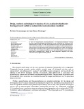
The transcription level of HvPAO1 and HvPAO2 mRNAs has been examined in different stem inter- nodes and whole ear at 30-days postfertilization and in various ear tissues by RT-PCR analysis (Fig. 1). PCR-amplified mRNAs have been probed with for HvPAO1 and HvPAO2 primer-pairs range of PCR isoforms and within the
linear
Many of these studies highlighted the physiological implications of PAO-mediated hydrogen peroxide syn- thesis in the apoplast related to peroxidase-catalyzed reactions or as a compound triggering signal transduc- tion leading to hypersensitive response. However, the vacuolar localization of HvPAO2 and the predicted apoplastic localization of HvPAO1 draw a new scen- ario to the possible role multiplicity of this enzyme family. In fact, even in maize, this multiplicity may
FEBS Journal 273 (2006) 3990–4002 ª 2006 The Authors Journal compilation ª 2006 FEBS
3991
M. Cervelli et al.
Barley polyamine oxidase isoform 1 gene expression
A
B
Fig. 1. HvPAO1 and HvPAO2 transcript detection by RT-PCR in different barley tis- sues. (A) Total RNA isolated from different stem internodes, whole ear and various ear tissues was analyzed by RT-PCR amplifica- tion procedure using specific primers for HvPAO1, HvPAO2 and, as a control, rp-S12 transcripts in the linear range of amplifica- tion (25 cycles). (B) cDNA from embryo was analyzed in saturating PCR condition (35 cycles). The PCR products were fractionated by 1.2% agarose gel electrophoresis. Expec- ted size of PCR fragments are indicated at right.
In details,
(Fig. 1A,
amplification conditions. the HvPAO1 transcript is accumulated only in the ear (Fig. 1A, left panel); even with saturating conditions there were no detectable PCR amplified products in any stem internodes (not shown), thus confirming previ- ous results reported by Cervelli et al. [4]. On the contrary, HvPAO2 mRNA is accumulated in all the tissues examined, including ear. A further RT-PCR analysis has been carried out on different ear tissues (Fig. 1A, right panel) and interestingly the accumula- tion pattern shown by HvPAO1 transcript indicates that this gene is expressed in basal lemma, rachis, awn, embryo-deprived caryopsis, embryo and sterile spikelets, at variance with the HvPAO2 gene, which is expressed at a comparable level only in sterile right panel). The transcription spikelets the ribosomal protein S12 accumulation pattern of
(rp-S12) mRNA has been also analyzed as a control housekeeping gene for RNA stability and the quan- tity processed for each sample. As HvPAO2 promo- ter was capable of driving GUS expression in barley developing caryopsis, exclusively in the embryo (see section below on HvPAO1, HvPAO2, ZmPAO1 and ZmPAO2::GUS expression in different plants and organs), to detect the presence of any low level of HvPAO2 mRNA accumulation in the barley embryo, a further PCR analysis was carried out in the non- linear range of amplification (up to 35 cycles) using the same cDNA sample. The results are shown in Fig. 1(B); a PCR product of faint intensity specific for HvPAO2 transcript was visible only after 35 cycles, demonstrating the presence of the HvPAO2 mRNA in embryonic organs albeit in relatively small amounts.
FEBS Journal 273 (2006) 3990–4002 ª 2006 The Authors Journal compilation ª 2006 FEBS
3992
M. Cervelli et al.
Barley polyamine oxidase isoform 1 gene expression
Cis-elements search in upstream region of HvPAO2 gene
stress response element present in Arabidopsis thaliana but not in rice [22]. To summarize, in spite of the fact that some sequence motifs are shared by HvPAO1, HvPAO2, ZmPAO1 and ZmPAO2 genes, a comparat- ive sequence analysis did not show any evident com- mon cis-acting elements organization in their upstream regions.
HvPAO1, HvPAO2, ZmPAO1 and ZmPAO2::GUS expression in different plants and organs
the
GUS expression was obtained in developing caryopses, roots and leaves of barley and in roots and leaves of maize by means of biolistic inoculations with the GUS gene under control of different promoters. Figure 2(B) shows a schematic representation of the chimerical constructions used in these experiments. Twenty-four hours after bombardment, the different organs were assayed for GUS activity (Figs 3 and 4).
The 5¢-flanking region of HvPAO2 gene was cloned by inverse PCR using specific oligonucleotides designed from HvPAO2 partial gene sequences [4] and analyzed by searching for putative cis-acting elements known in plants by using the PLACE cis-element database [19] (http://www.dna.affrc.go.jp/PLACE/). We found a well-conserved TATA-box, located 25 nucleotides upstream of the putative transcription start point, an I-box localized at )191 ⁄ )186, a G-box at )303 ⁄ )296 and a CCAAT-box at )405 ⁄ )409 (Fig. 2A). These putative light-response elements [20,21] are also present in the promoter regions of HvPAO1, ZmPAO1 and ZmPAO2 genes [4,9]. A sequence motif that is shared only by HvPAO2 and HvPAO1 upstream regions is the MYB1AT-box (consensus 5¢-WAACCA-3¢), located at )348 ⁄ )353 (Fig. 2A). This is a dehydration-
A
B
C
Fig. 2. Gene promoter::GUS constructs util- ized in transient GUS expression experi- ments. A. 5¢-flanking region of HvPAO2 gene. Upstream and exon nucleotide sequences are indicated with lower-case and upper-case letters (start translation codon in bold), respectively. Putative promo- ter sequence motifs are indicated with bold underlined letters and marked. The restric- tion site HindIII used in inverse PCR experi- ments is indicated with italics underlined letters. Exon sequence is numbered from the putative tsp (+ 1), upstream sequence is indicated by negative numbers. B. Schemes of pHTT515 and pHTT-PAOs construct vec- tors. C. Schematic representation of the constructs utilized in particle bombardment and detection (+) or absence (–) of their expression in different organs and plants. Numbering refers at the promoter sequences (open boxes) jointed to the UB intron region (grey boxes).
FEBS Journal 273 (2006) 3990–4002 ª 2006 The Authors Journal compilation ª 2006 FEBS
3993
M. Cervelli et al.
Barley polyamine oxidase isoform 1 gene expression
right. Photographs are representative of
Fig. 3. Histochemical localization of glucoronidase (GUS) activity in different barley tissues after gene bombardment. HvPAO1, HvPAO2, ZmPAO1 and ZmPAO2promoter::GUS (pHTT-PAOs) and UBpromoter::GUS (pHTT515) expression vectors were biolistically delivered to bar- ley roots, leaves and ears. Organs were histochemically reacted with X-Glu and examined for blue staining assessment with a Zeiss stereo- microscope and photographed. Longitudinal section of ears is enlarged at three different experiments each performed in triplicate.
constructs,
ZmPAO2promoter::GUS
Transient expression of HvPAO1, HvPAO2, ZmPAO1 using and pHTT515 plasmid (UBpromoter::GUS) as a control (Fig. 2B), revealed that only barley immature caryopsis for the HvPAO1 promoter driving were competent GUS expression, especially in the embryo, aleurone layer and endosperm. On the contrary, it was inactive in roots and leaves of both barley and maize. Interest-
ingly, HvPAO2 promoter was capable of driving GUS expression in barley roots and leaves, as expected, but also in developing caryopses, albeit exclusively in the embryo, as well as in roots and leaves of maize. Fur- thermore, ZmPAO1, ZmPAO2 and UB promoters transpired to be active in all analyzed organs of barley and maize. Results obtained in the transient GUS expression experiments are summarized in Fig. 2(C).
FEBS Journal 273 (2006) 3990–4002 ª 2006 The Authors Journal compilation ª 2006 FEBS
3994
M. Cervelli et al.
Barley polyamine oxidase isoform 1 gene expression
the crude homogenate through a centrifugation of fractionation in 70% saturated ammonium sulfate and two chromatographic steps (hydroxylapatite and SP-sepharose columns). By this procedure, a 58-fold purification of the enzyme was achieved (Table 1). No detectable HvPAO2 activity was revealed overall dur- ing the entire procedure of HvPAO1 purification, nei- ther in the purification steps reported in Table 1 (1–4 fractions), nor in hydroxylapatite and SP-sepharose flow-through and washing fractions. In western blot analysis, the SP-sepharose eluate (fraction 4; Table 1) showed a band of (cid:2)53 kDa molecular mass, corres- ponding to the HvPAO1 expected mass (Fig. 5) when probed against polyclonal anti-ZmPAO antibodies; it has already been demonstrated that these cross-react with the less conserved HvPAO2 protein [4].
In vitro HvPAO1 protein synthesis
In order to confirm that the PAO activity present in the SP-sepharose eluate (fraction 4; Table 1) could be ascribed to the HvPAO1 isoform, we carried out the in vitro synthesis of HvPAO1 protein utilizing the pET17b-HvPAO1 plasmid as a template in three different cell-free translation systems and precisely RTS-100 Roche (Roche Diagnostics, Monza, Italy), Escherichia coli T7 S30 Extract System for circular DNA (Promega Italia, Milano, Italy) and Pure-System Classic (Post Genome Institute, Tokyo, Japan), as des- cribed in the Methods section. The highest in vitro syn- thesized HvPAO1 protein yield (0.1 U) was obtained with the RTS-100 Roche translation system, as detec- ted by enzymatic assay. Moreover, western blot analy- sis of the in vitro translated product probed against polyclonal anti-ZmPAO antibodies, showed a band of (cid:2)53 kDa molecular mass, thus confirming the presence of HvPAO1 protein (Fig. 5).
ZmPAO, HvPAO2 and HvPAO1 protein catalytic properties
localization of glucoronidase (GUS) activity Fig. 4. Histochemical in different maize tissues after gene bombardment. HvPAO1, HvPAO2, ZmPAO1 and ZmPAO2promoter::GUS (pHTT-PAOs) and UBpromoter::GUS (pHTT515) expression vectors were biolistically delivered to maize roots and leaves. Organs were histochemically reacted with X-Glu and examined for blue staining assessment with a Zeiss stereomicroscope and photographed. Photographs are representative of three different experiments each performed in triplicate.
Purification of the HvPAO1 protein from developing caryopses
HvPAO1 was extracted from immature caryopses with a high ionic strength salt solution. The enzyme was then partially purified from supernatant obtained after
ZmPAO and HvPAO2 showed pH optima and Km val- ues for Spd and Spm oxidation (Table 2) identical to those previously reported by Cervelli et al. [4]. Cata- lytic properties of the HvPAO1 enzyme purified from immature caryopsis resulted identical to those exhib- ited by the in vitro synthesized HvPAO1 recombinant enzyme for both Spd and Spm substrates. In partic- ular, HvPAO1 enzymatic activity showed an optimum at pH 7.0 for Spm and Spd oxidation and Km values of 0.89 · 10)5 m and of 0.50 · 10)5 m for Spd and Spm substrates, respectively (Fig. 6; Table 2). More- over, the Vmax ratio for Spd and Spm at pH 7.0 was
FEBS Journal 273 (2006) 3990–4002 ª 2006 The Authors Journal compilation ª 2006 FEBS
3995
M. Cervelli et al.
Barley polyamine oxidase isoform 1 gene expression
Table 1. Purification of the HvPAO1 protein from barley developing caryopses. HvPAO1 purification was performed from developing caryop- ses at 30 days postfertilization, as described in the Methods section. The enzyme was partially purified from supernatant obtained after cen- trifugation of the crude homogenate (fraction 1), through a fractionation in 70% saturated ammonium sulfate (fraction 2) and two chromatographic steps (fraction 3 and 4).
Total volume (mL)
Purification step
Total protein (mg)
Total activity (U)
Purification fold
Recovery (%)
Protein (mgÆmL)1)
Activity (UÆmL)1)
Specific activity (UÆmg)1Æprotein)
236 104
0.94 1.24
221.84 128.96
0.002 0.004
0.472 0.416
0.002 0.003
1.0 1.5
100.0 88.1
10.56
Crude extract (fraction 1) (NH4)2SO4 70% sat. precipitation (fraction 2) Hydroxylapatite eluate (fraction 3) SP eluate (fraction 4)
44 15
0.24 0.043
0.644
0.005 0.005
0.220 0.075
0.021 0.116
10.5 58.0
46.6 15.9
Fig. 5. Western blot analysis of ZmPAO, HvPAO2 and HvPAO1. ZmPAO and HvPAO2 were purified as previously described [4,5]. HvPAO1 was purified and in vitro synthesized as described in Experimental procedures. Analysis was performed running: 0.1 U of ZmPAO and HvPAO2; 0.002 U of ear purified and RTS-100 Roche TS produced HvPAO1. Proteins were reacted, after deglyco- sylation, with anti-ZmPAO polyclonal antibodies [4]. M, protein molecular weight marker (Fermentas).
found to be 1.3. The HvPAO1 biochemical features exhibited by the in vitro synthesized recombinant pro- tein and the native partially purified enzyme from bar- ley ears are congruent, demonstrating that the PAO activity detected in the SP-sepharose eluate (fraction 4; Table 1) could be reasonably ascribed to the exclusive presence of HvPAO1 enzyme with respect to HvPAO2 in developing caryopses. In fact, the presence of any detectable HvPAO2 activity in the SP-sepharose eluate (Table 1) would result in different PAO catalytic parameters, with Km and pH optimum values for Spd and Spm intermediate between those of purified HvPAO2 and in vitro synthesized HvPAO1. HvPAO1 catalytic properties were very similar to those of ZmPAO (Table 2), moreover HvPAO1 showed com- parable affinity and identical pH optima values for both Spd and Spm substrates (pH 7.0); analogously ZmPAO showed comparable affinity and identical pH optima values for both substrates (pH 6.5). On the contrary, HvPAO1 enzymatic features differ from the ones of HvPAO2 that preferentially oxidizes spermine at pH 5.5 and spermidine at pH 8.0 with a ten-fold lower Vmax [4].
Discussion
Table 2. Km values for ZmPAO, HvPAO2 and HvPAO1. For the determination of the Km values, ZmPAO and HvPAO2 were purified as previously described [4,5]. HvPAO1 was synthesized in vitro as described in Experimental procedures. Data were obtained at 25 (cid:2)C with Spd and Spm as substrates at the specific pH optimum. Km values concerning Spd and Spm oxidation by ZmPAO and HvPAO2 were within the standard error of the values previously reported by Cervelli et al. [4]. Ear purified and RTS-100 Roche TS produced HvPAO1 showed identical Km values. All Km values, calculated from Lineweaver-Burk plots, are means of three different experiments, each performed in duplicate. SD was 8%.
Substrate
pH
Enzyme
Km (M)
lemma,
6.5 6.5 8.0 5.5 7.0 7.0
Spd Spm Spd Spm Spd Spm
ZmPAO ZmPAO HvPAO2 HvPAO2 HvPAO1 HvPAO1
1.0 · 10)5 a,b 2.7 · 10)5a,b 56.0 · 10)5a,b 0.48 · 10)5a,b 0.89 · 10)5a 0.50 · 10)5a
aPresent work; bCervelli et al. [4].
Our results definitely support the identification of the HvPAO1 enzyme as the major product of HvPAO gene expression in barley ear (Fig. 1A, left panel). In fact, HvPAO1 mRNA was detectable by standard RT-PCR analysis only in the ear ([4] and this work), indicating that HvPAO1 gene expression is ear-specific. Further ear-tissue dissection demonstrated that in all the samples examined (basal rachis, awn, embryo-deprived caryopsis and embryo) by RT-PCR, the HvPAO1 gene is expressed at comparable levels (Fig. 1A, right panel). The only tissue where it was possible to detect HvPAO2 gene expression with stand- ard PCR conditions (25–30 cycles) resulted to be the
FEBS Journal 273 (2006) 3990–4002 ª 2006 The Authors Journal compilation ª 2006 FEBS
3996
M. Cervelli et al.
Barley polyamine oxidase isoform 1 gene expression
Fig. 6. HvPAO1 catalytic parameters for Spd and Spm oxidation. HvPAO1 was purified from developing caryopses at 30-day post- fertilization, as described in Experimental procedures. Data reported are the average of three different experiments, each with two replicates. SD was 8%. (A) HvPAO1 cat- alytic activity pH optima were determined at 25 (cid:2)C, in 0.2 M sodium phosphate buffer (pH range 4.5–8.5) with Spd or Spm as sub- strates. PAO activity is expressed as per- cent of the maximum value. (B) HvPAO1 (1 · 10)3 U) Km values were determined at 25 (cid:2)C, with Spd and Spm as substrates at the respective pH optimum and then calcu- lated from Lineweaver-Burk plots.
this
sterile spikelets. Using a higher number of PCR cycles, we were also able to detect a very small amount of the HvPAO2 transcript in the embryo tissue (Fig. 1B). Thus, the HvPAO2 gene is also transcriptionally active in the embryo, albeit at a very low level, probably rep- resenting a basal transcriptional activity. This is in line with the transient GUS expression experiments that confirmed the specific and strong expression of the HvPAO1 gene in barley ear, at variance with the weak expression of the HvPAO2 gene, exclusively localized in the embryo (Figs 2C and 3). As expected, the HvPAO1 gene is silent in roots and leaves of maize (Figs 2C and 3). Interestingly HvPAO2, ZmPAO1 and ZmPAO2 promoters exhibit the same transcription pattern, being able to drive GUS expression in all the organs and plants analyzed in this study (Figs 2C, 3 and 4). Moreover, the ZmPAO1-2 promoters are also active in barley embryo, albeit at a very low level, like the HvPAO2 gene; it seems that the barley embryo is able to allow a basal transcriptional level of these promoter sequences. Sequence analysis of 5¢ flanking regions of HvPAO2 (Fig. 2A), ZmPAO1 and ZmPAO2 genes albeit sharing some potential cis-acting elements, do not show any obvious common promoter architec- ture as expected by their identical transcription pattern [4,9]. Furthermore, there are no evident sequence fea- tures in the HvPAO1 and HvPAO2 promoters that could explain their different gene expression profiles. So, we are facing a puzzling gene duplication event that occurs in barley, since the paralogous HvPAO2 and ZmPAO1-3 genes share a common tissue expres- sion, which is at variance with the orthologous
HvPAO1 and ZmPAO1-3 genes that show a different tissue regulation. The very similar catalytic properties shown by HvPAO1 and ZmPAO could be ascribed to the closer phylogenetic relationship existing between them (84% identity), as compared with that between HvPAO2 and ZmPAO1 (73% identity) (Table 2). It is interesting to recall that, even if the global fold and the flavin adenine dinucleotide-binding pocket are well conserved in HvPAO1, HvPAO2 and ZmPAO, the substitution of Phe403 of ZmPAO by a tyrosine resi- due in HvPAO2 could probably play a relevant role in the different substrate specificity and kinetic parame- isoform [4]. Furthermore, ters observed for HvPAO1 amino acid sequence shows a different C-ter- minus when compared with the HvPAO2 coding sequence, which has an extra eight-residue long tail (DELKAEAK) responsible for the symplastic localiza- tion of this isoform [10]. On the contrary, the higher similarity determined between HvPAO1 and ZmPAO, strongly suggests an apoplastic localization of the HvPAO1 isoform. Indeed, recent results have shown that ZmPAO is also present at high levels in the cyto- plasm, most probably in the secretory pathway of young tissues undergoing or destined to programmed cell death, such as developing xylem vessels and xylem parenchyma of both the root and mesocotyl as well as root cap cells [17]. Later, during cell maturation, ZmPAO is found mainly in the cell wall [17]. On the basis of these results, the authors hypothesized that ZmPAO could play a dual role in these tissues being involved either in programmed cell death or cell wall reaction differentiation through the action of
its
FEBS Journal 273 (2006) 3990–4002 ª 2006 The Authors Journal compilation ª 2006 FEBS
3997
M. Cervelli et al.
Barley polyamine oxidase isoform 1 gene expression
products, hydrogen peroxide and aminoaldehydes and ⁄ or modulation of polyamine levels [3,13,17]. One in barley PAO tissue-specific can hypothesize that functions are associated with the distinct isoforms HvPAO1 and HvPAO2, an event that arose in the course of evolution of C3 cereals. According to the tis- sue distribution of HvPAO1 and HvPAO2, the prefer- ential HvPAO2 substrate Spm has been detected at higher level than Spd in barley leaves [23], whereas in the developing grains a higher level of Spd than Spm has been found [24]. Figure 7 summarizes the evolu- tionary relationship among the cereal PAO genes stud- ied in this work.
Is there any specific role for HvPAO1 in the ear tissues and in particular in embryonic tissues and aleurone, where the expression of HvPAO1 gene is prominent, if not exclusive, compared with HvPAO2? The available data suggest that HvPAO1 could have a specific role in the aleurone cells. This is a tissue that plays a key role during the germination of cereal seeds. Aleurone cells secrete, under the stimulus of embryo-synthesized gibberellin, amylase and other involved in endosperm reserve hydrolytic enzymes
mobilization. Gibberellin also induces programmed cell death in these cells, a process that is mediated by hydrogen peroxide [25,26]. Thus the accumulation of HvPAO1 during the development of barley caryopsis could be functional to the production of hydrogen peroxide needed in the programmed cell death process taking place in the aleurone during germination. However it should be recalled that hydrogen peroxide production during germination could also have a general protective role against microbial pathogens. Alternatively, HvPAO1 could have a role in the regu- lation of polyamine levels in the aleurone cells and in it has been recently the embryo as well. Indeed, reported that DNA synthesis early in development and the advance in cell cycle ⁄ endocycle are tempor- ally and spatially related to polyamine catabolism and vascular development [27]. Moreover, polyamines are active in triggering the synthesis of nitric oxide in specific tissues of Arabidopsis thaliana seedlings [28]. This molecule is known to delay programmed cell death in aleurone cells and also to have pleiotropic effects on many facets of plant development and defense [29].
Experimental procedures
Chemicals
HvPAO2
HvPAO1
LEAVES
ROOTS
EARS ? ?
ZmPAO1,2
Orthologous gene
Italy).
Paralogous gene
Extracellular localization
HvPAO1 promoter
HvPAO2, ZmPAO1,2 promoters
Fig. 7. Schematic representation of the evolutionary relationship among cereal PAO genes. Arrow width represents the promoter expression level according to both RT-PCR analysis and biolistic delivering experiments. The promoter boxes color reflects the tissue specific expression.
FEBS Journal 273 (2006) 3990–4002 ª 2006 The Authors Journal compilation ª 2006 FEBS
3998
Restriction and DNA-modifying enzymes and protein molecular weight marker were purchased from MBI Fer- mentas (MBI Fermentas, St. Leon-Rot, Germany). Sper- midine (Spd) and spermine (Spm), horseradish peroxidase, 4-aminoantipyrine and 3,5-dichloro-2-hydroxybenzenesulf- onic acid were purchased from Sigma-Aldrich-Fluka (Sigma, Milano, Italy). TRIZOL reagent was from Invitro- gen (Invitrogen, Milano, Italy). pGEM-Teasy vector and the E. coli T7 S30 Extract System for circular DNA were from Promega (Promega Italia, Milano, Italy). pET17b vector and E. coli BL21 DE3 competent cells were from Novagen (Novagen Inc., Madison, WI, USA). CHU(N6) medium was from Duchefa (Duchefa Biochemie B. V., Haarlem, the Netherlands). Hydroxylapatite and gold par- ticles (1.0 lm in diameter) were from Bio-Rad (Bio-Rad, Milano, from Amersham SP-sepharose was Biosciences (Amersham Biosciences, Milano, Italy). Carb- oxymethylcellulose was from Whatman (Whatman, Maid- stone, UK). Peroxidase-conjugated goat antirabbit IgG was from Vector Laboratories (Burlingame, CA, USA). The RTS-100 Roche translation system was from Roche (Roche Diagnostics, Monza, Italy). The Pure-System Clas- sic Mini Kit was from the Post Genome Institute (Post Genome Institute, Tokyo, Japan). Other chemicals came from Sigma-Aldrich-Fluka, Bio-Rad and J. T. Baker (Baker Italia, Milano, Italy).
M. Cervelli et al.
Barley polyamine oxidase isoform 1 gene expression
Plant material
DNA methodology and construction of expression plasmids
forward DNA with the oligonucleotides HvPAO2-C, 5¢-AAAAAGCTTACCAAAACTTGTGTAAACTT-3¢, and HvPAO2-D, reverse 5¢-TTTAGATCTGCCCTGCTCTCC GGCCCTGT-3¢, containing the HindIII and the BglII sites, respectively. The PCR-amplified product was cloned in the pGEM-Teasy vector. The promoter gene sequence has been deposited in the EMBL database under EMBL accession number AM231701.
regions
RT-PCR analysis of HvPAO1 and HvPAO2 gene expression in different tissues
Seedlings and adult plants of barley (H. vulgare) cultivar Aura [30] were grown at the ‘Istituto Sperimentale per la Cerealicoltura, Sezione di Fiorenzuola d’Arda, Italy’. Seeds of barley cultivar Aura and of maize (Z. mays) cultivar Corona (Monsanto Agricoltura, Italy) were soaked for 12 h in aerated tap water, germinated at 20 (cid:2)C in the dark and grown aseptically in a growth chamber with a 16 : 8 h light–dark cycle on Magenta vessels (Sigma-Aldrich-Fluka, Milano, Italy) containing 0.8% agar. For ZmPAO and HvPAO2 purification, maize and barley seeds were germi- nated with 1 cm of fertile soil, at 20 (cid:2)C in the dark. Two- day-old barley seedlings were exposed to natural light for 4 days before harvesting; maize seedlings were kept in the dark before protein extraction. Thirty-day postfertilization ears were utilized for RT-PCR experiments, transient expression assays and HvPAO1 purification.
for
reverse forward
HvPAO2 promoter isolation
reverse Total RNA was isolated from different barley tissues by TRIZOL reagent, according to the manufacturer’s instruc- tions. Oligonucleotides utilized as primers specific amplification of HvPAO isoforms were: HvPAO-N, reverse 5¢-GTTATTACTTAGTACCTCTTAAT-3¢, HvPAO-O, for- ward 5¢-GACGGAGATCTCCCACTC-3¢ and HvPAO-P, reverse 5¢-GGTTGTCCGACTGCTGCTC-3¢ for HvPAO1; 5¢-CTCGTCGGCGCGGTCCAT-3¢, HvPAO-Q, 5¢-GAGGGGAGAATTGAAGA HvPAO-R, GAG-3¢ and HvPAO-S, reverse 5¢-GTCGTAGAGGCC ACCGCT-3¢ for HvPAO2 as already described [4]. Oligo- nucleotides for the control barley ribosomal protein S12 were: RPS12-A, reverse 5¢-ATTCTTCACCATAGTCCT-3¢, RPS12-B, forward 5¢-GTGAGCCAATGGACTTGATG-3¢ and RPS12-C, 5¢-ATGCAAGAGCAGCCTAC AAC-3¢ [4].
[4], 5¢-TACTGTGTTAGCACTGCTAGC-3¢, forward
FEBS Journal 273 (2006) 3990–4002 ª 2006 The Authors Journal compilation ª 2006 FEBS
3999
The methods described by Sambrook et al. [31] were used for the manipulation of plasmid DNAs and general DNA in vitro procedures. In order to amplify HvPAO1, HvPAO2, ZmPAO1 and ZmPAO2 promoter ([4,9] and present work), gene-specific oligonucleotides containing HindIII and the BglII sites were designed spanning from the 5¢ end of the promoter sequence down to the transcrip- tion start site [primer sequences used are available on request from the first author (MC)]. HvPAO2, ZmPAO1 and ZmPAO2 promoter PCR products were cloned into the expression vector pHTT515 utilizing the HindIII and the BglII sites and replacing the original ubiquitin (Ubi) promoter sequence. To clone HvPAO1 promoter sequence [4], we used a different procedure because of the presence of a BglII site within the gene sequence. A HvPAO1 pro- moter sequence subclone inserted in pGEM-Teasy vector was digested with EcoRI and filled in at its extremities, then cloned in pHTT515 previously cut with HindIII and BglII ended. The Ubipromoter::GUS expression and blunt pHTT515 vector was used as a control plasmid expres- sing the GUS reporter gene driven by the house-keeping promoter Ubi. The HvPAO1, HvPAO2, ZmPAO1 and ZmPAO2promoter::GUS expression plasmids were se- quenced on both strands using the automated fluorescent dye terminator technique (Perkin Elmer ABI model 373 A). In order to clone the HvPAO1 cDNA (GenBank accession number AJ298131) by PCR amplification, the full-length cDNA was generated possessing modified 5¢- and 3¢-ends. In particular, the two following synthetic oligonucleotides were used to introduce NdeI and XhoI restriction sites at the 5¢- and 3¢-ends of HvPAO1 cDNA: HvPAO1cdna-DIR, and 5¢-CATATGGCCGGCCCCAGGGTCATCATC-3¢ HvPAO1cdna-REV, 5¢-CTGGAACTCGAGCTAGTCAA ACTTGCCCGG-3¢, respectively. The amplified PCR prod- uct was restricted by NdeI and XhoI and ligated with the restricted NdeI ⁄ XhoI pET17b vector, to obtain the genetic construct encoding the mature form of HvPAO1 protein, named pET17b-HvPAO1. The recombinant cDNA con- struct was resequenced to check the accuracy of the nucleo- tide sequence and then utilized to transform E. coli BL21 DE3 (Novagen, Madison, WI, USA) competent cells. Barley DNA was extracted and purified as described in Cervelli et al. [4]. To clone 5¢- and 3¢-flanking regions of HvPAO2 gene, amplified products were obtained by inverse PCR using specific oligonucleotides designed from HvPAO2 in particular, HvPAO2-A, partial gene sequences reverse and 5¢-GAGGGGAGAATTGAAGA HvPAO2-B, GAG-3¢, specific for the 5¢-end region. The gene-specific primer couples were utilized on different samples of purified barley total DNA, previously digested with HindIII and self-ligated. A direct PCR to obtain the corresponding HvPAO2 promoter sequence was performed on total
M. Cervelli et al.
Barley polyamine oxidase isoform 1 gene expression
Transient expression assay
ZmPAO, HvPAO2 and HvPAO1 enzyme purification
from each treated plant three samples Barley and maize transient transformation was performed by particle bombardment with the expression plasmids described above. In this study, the Biolistic PDS-1000 ⁄ He Particle Delivery System from Bio-Rad (Bio-Rad, Milano, Italy) was used. Cartridges containing plasmid-coated gold microcarriers were prepared according to the manufac- turer’s procedure with modification according to the plant organ bombarded. Twenty-five micrograms of HvPAO1, HvPAO2, ZmPAO1 and ZmPAO2promoter::GUS expres- sion plasmids and the pHTT515 (Ubipromoter::GUS) con- trol plasmid were precipitated onto 1.8 mg of gold particles (1.0 lm in diameter, Bio-Rad) with ethanol. DNA-coated gold particles were prepared according to the procedure described by Sessa et al. [32] and finally suspended in 30 lL of ethanol. Ten-microliter aliquots were used for one bom- bardment. The barrel liner provided by the Biolistic PDS- 1000 ⁄ He system determined the distance from the gene gun to the sample. This distance was 6.0 cm. Gold particles coated with the plasmid of interest were used for bombard- ment with a firing pressure of 900 psi for maize and barley for barley roots and leaves and maize roots, 1.100 psi developing caryopsis at 30-days postfertilization. After bombardment, barley organs were placed on callus-induced medium (4.3 gÆL)1 Murashige & Skoog medium (MS), 30.0 gÆL)1 maltose, 1.0 mgÆL)1 thiamine, 0.25 gÆL)1 myo- inositol, 1.0 gÆL)1 hydrolyzed casein, 0.69 gÆL)1 l-proline, 2.5 mgÆL)1 dicamba, 5.0 mgÆL)1 bialaphos, 3.5 gÆL)1 phyta- gel), while maize organs on 4.0 gÆL)1 CHU(N6) medium (Duchefa Biochemie B. V), then bombarded material was incubated for 1-day at 25 (cid:2)C in a growth chamber with a 16 : 8 h light–dark programme. Three separate bombardment experiments were carried out and a minimum of tissue was examined.
Histochemical GUS activity assay
Enzyme assay, protein determination and western blot analysis
All purification procedures were performed at 0–5 (cid:2)C. Low- pressure chromatographic steps were carried out using the GradiFrac System (Amersham Biosciences). As the three ZmPAO genes encode for identical translation products [9], it is not possible to distinguish the contribution of each iso- form within the PAO enzymatic activity detectable in maize and no numbering is used for ZmPAO enzymes. ZmPAO protein was purified from shoots of 10-day-old etiolated maize seedlings, as previously described [5] and HvPAO2 isoform was purified from shoots of 6-day-old de-etiolated barley seedlings as previously described [4]. HvPAO1 pro- tein was purified from 30-day postfertilization developing caryopsis as follows. Plant material (100 g) was homogen- ized in a Waring blender with three volumes of cold 0.5 m NaH2PO4 and the homogenate was filtered through nylon mesh cloth. Filtrate was centrifuged at 20 000 g for 30 min and the supernatant (236 mL, fraction 1) was fractionated with 70% saturated ammonium sulfate and left 2 h at 4 (cid:2)C. The suspension was centrifuged at 20 000 g for 30 min. The pellet was dissolved in 0.2 m NaH2PO4, dialyzed overnight against the same buffer and then centrifuged at 20 000 g for 30 min (104 mL, fraction 2). The dialyzed sample was applied to a hydroxylapatite column equilibrated with the dialysis buffer. The column was washed with 0.2 m NaH2PO4 containing NaCl 0.1 m, until the A280 decreased to zero. The enzyme was eluted with 0.5 m sodium phos- phate buffer, pH 6.5, containing 0.1 m NaCl (44 mL, frac- tion 3) and dialyzed overnight against 50 mm sodium phosphate buffer, pH 5.0. The dialyzed sample was applied to an SP-sepharose column equilibrated with the dialysis buffer. The column was washed with 50 mm sodium phos- phate buffer, pH 5.0, containing 0.1 m NaCl, until the A280 decreased to zero. The enzyme was eluted with 50 mm sodium phosphate buffer, pH 5.0, containing 0.3 m NaCl (15 mL, fraction 4).
FEBS Journal 273 (2006) 3990–4002 ª 2006 The Authors Journal compilation ª 2006 FEBS
4000
The histochemical localization of GUS in transiently trans- formed plant organs was performed essentially as described by Jefferson [33]. Plant samples from barley and maize were immersed in a histochemical reaction mixture containing 3 mgÆmL)1 X-Glu (5-bromo-4-chloro-3-indolyl b-d-glucuro- nide) in 100 mm sodium phosphate buffer, pH 7.0, 0.06% Triton X-100, 5 mm potassium ferrocyanide. The histo- chemical reaction was performed in the dark at 37 (cid:2)C until a blue indigo dye color as precipitate appeared at the site of the enzymatic cleavage after 2–15 h. Roots and barley immature caryopsis were rinsed several times in 50 mm phosphate buffer to stop the reaction, finally kept in the same buffer. After incubation, stained leaves were cleared of chlorophyll by v ⁄ v 70% ethanol fixation. Samples were examined for blue staining assessment with a Zeiss stereo- microscope and photographed. Enzyme activity was measured spectrophotometrically by following the formation of a pink adduct (e515 nm ¼ 2.6 · 104 m)1.cm)1) as a result of the H2O2-dependent oxidation of 4-aminoantipyrine catalyzed by horseradish peroxidase and the subsequent condensation of oxidized 4-aminoantipyrine with 3,5-dichloro-2-hydroxybenzenesulf- onic acid [34]. The measurements were performed in 0.2 m sodium phosphate buffer, pH 6.5, with Spd as a substrate in 0.2 m sodium phosphate buffer, pH 5.5, for ZmPAO; in 0.2 m sodium with Spm as a substrate for HvPAO2; phosphate buffer, pH 7.0, with Spd as a substrate for HvPAO1. Enzyme activity was expressed in IU. Protein content was estimated by the method of Bradford [35]
M. Cervelli et al.
Barley polyamine oxidase isoform 1 gene expression
Acknowledgements
This research was partially supported by the grant PRIN 2005 from ‘Ministero dell’Istruzione, dell’Uni- versita` e della Ricerca’ (MIUR).
with bovine albumin as a standard. SDS ⁄ PAGE was performed according to the method of Laemmli [36]. West- ern blot analysis was performed after protein deglycosyla- tion [37], in order to eliminate cross-reactivity due to glycan moieties, using a rabbit polyclonal anti-ZmPAO antiserum [38].
References
Analysis of catalytic properties
1 McIntire WS & Hartman C (1993) Principle and Appli- cation of Quinoproteins (Davison, VL, eds), pp. 97–171. Marcel Dekker Inc., New York. 2 Seiler N (1995) Polyamine oxidase, properties and func- tions. Prog Brain Res 106, 333–344.
3 Cona A, Rea G, Angelini R, Federico R & Tavladoraki P (2006) Functions of amine oxidases in plant develop- ment and defence. Trends Plant Sci 11, 80–88.
from 3 4 Cervelli M, Cona A, Angelini R, Polticelli F, Federico R & Mariottini P (2001) A barley polyamine oxidase isoform with distinct structural features and sucellular localization. Eur J Biochem 268, 3816–3830. 5 Federico R, Alisi C & Forlani F (1989) Properties of
Reaction conditions for in vitro HvPAO1 protein synthesis
polyamine oxidase from the cell wall of maize seedlings. Phytochemistry 28, 45–46. ZmPAO, HvPAO2 and HvAO1 pH optima for catalytic activity were determined at 25 (cid:2)C, in 0.2 m sodium phos- phate buffer (pH range 4.5–8.5) with Spd or Spm as sub- strates. Km values were determined for the three enzymes (1 · 10)3 U) at 25 (cid:2)C, with Spd and Spm as substrates at the respective pH optimum and calculated from Lineweav- er-Burk plots. Spd or Spm concentrations range was from 4.0 to 20.0 · 10)6 m for ZmPAO enzyme and from 2.0 to 8.0 · 10)6 m for HvPAO1 enzyme. In the case of HvPAO2 enzyme, Spd concentration range was to 20 · 10)4 m, while Spm concentration range was from 5.0 to 60.0 · 10)7 m. Data here reported are the average of three different experiments, each with two replicates. sd was 8%.
6 Tavladoraki P, Schinina` ME, Cecconi F, Di Agostino S, Manera F, Rea G, Mariottini P, Federico R & Angelini R (1998) Maize polyamine oxidase: primary structure from protein and cDNA sequencing. FEBS Lett 426, 62–66.
7 Binda C, Coda A, Angelini R, Federico R, Ascenzi P & long U-shaped catalytic tunnel
Mattevi A (1999) A 30 A˚ in the crystal structure of polyamine oxidase. Structure 7, 265–276.
8 Sˇ ebela M, Radova´ A, Angelini R, Tavladoraki P, Fre´ - bort I & Peˆ c P (2001) FAD-containing polyamine oxi- dases: a timely challenge for researchers in biochemistry and physiology of plants. Plant Sci 160, 197–207.
9 Cervelli M, Tavladoraki P, Di Agostino S, Angelini R, Federico R & Mariottini P (2000) Isolation and charac- terization of three polyamine oxidase genes from Zea mays. Plant Physiol Biochem 38, 667–677.
10 Cervelli M, Di Caro O, Di Penta A, Angelini R, Fede- rico R, Vitale A & Mariottini P (2004) A novel C-term- inal sequence from barley polyamine oxidase is a vacuolar sorting signal. Plant J 40, 410–418.
11 Li ZC & McClure JW (1989) Polyamine oxidase of pri- mary leaves is apoplastic in oat but symplastic in barley. Phytochemistry 28, 2255–2259.
12 Federico R & Angelini R (1986) Occurrence of diamine oxidase in the apoplast of pea epicotyls. Planta 167, 300–302.
FEBS Journal 273 (2006) 3990–4002 ª 2006 The Authors Journal compilation ª 2006 FEBS
4001
13 Cona A, Cenci F, Cervelli M, Federico R, Mariottini P, Moreno S & Angelini R (2003) Polyamine oxidase, a hydrogen peroxide-producing enzyme, is up-regulated by light and down-regulated by auxin in the outer For in vitro protein synthesis, we followed the standard reaction conditions that were basically described in the manufacturer’s instructions. In the RTS-100 (Roche Diag- nostics, Monza, Italy) case, the standard reaction solution (50 lL) contained the following components: 12 lL E. coli lysate, 10 lL reaction mix, 12 lL amino acids, 1 lL methi- onine, 5 lL reconstitution buffer and 2.5 lg of circular template (pET17b-HvPAO1 plasmid) in 10 lL of water. The reaction mixtures were incubated at 30 (cid:2)C for 3 h. The E. coli T7 S30 Extract System for circular DNA (Promega Italia, Milano, Italy) contained the following components: 4 lg of DNA template, 5 lL amino acid mixture, 20 lL S30 premix, 15 lL T7 S30 extract, and nuclease free water, which came to a final volume of 50 lL. The reaction incu- bation was carried out at 37 (cid:2)C for 3 h. In the Pure-System Classic Mini Kit (Post Genome Institute, Tokyo, Japan), the reaction mixtures were prepacked in two tubes, Solu- tions A and B. Before the reaction, we thawed both tubes on ice and, using new tubes we gently mixed 0.5 lg of DNA template, 25 lL of Solution A, 10 lL of Solution B and added RNase free water to a final volume of 50 lL. The tubes were then placed in an incubator for 3 h at 37 (cid:2)C. In order to improve the enzyme activity we add fla- vin adenine dinucleotide (15 mgÆL)1 final concentration) in each tube. In vitro synthesized HvPAO1 protein was ana- lyzed for enzymatic activity and catalytic properties as des- cribed above.
M. Cervelli et al.
Barley polyamine oxidase isoform 1 gene expression
tissues of the maize mesocotyl. Plant Physiol 131, 803– 813.
26 Palma K & Kermode AR (2003) Metabolism of hydro- gen peroxide during reserve mobilization and pro- grammed cell death of barley (Hordeum vulgare L.) aleurone layer cells. Free Radic Biol Med 35, 1261– 1270. 14 Cooley T & Walters D (2002) Polyamine metabolism in barley reacting hypersensitively to the powdery mildew fungus Lumeria graminis f.sp. hordei. Plant Cell Environ 25, 461–468. 15 Yoda H, Yamaguchi Y & Sano H (2003) Induction of
27 Paschalidis KA & Roubelakis-Angelakis KA (2005) Sites and regulation of polyamine catabolism in the tobacco plant. Correlations with cell division ⁄ expansion, cell cycle progression, and vascular development. Plant Physiol 1382174, –2184. hypersensitive cell death by hydrogen peroxide produced through polyamine degradation in tobacco plants. Plant Physiol 132, 1973–1981. 28 Tun NN, Santa-Catarina C, Begum T, Silveira V, 16 Laurenzi M, Rea G, Federico R, Tavladoraki P &
Handro W, Floh EI & Scherer GF (2006) Polyamines induce rapid biosynthesis of nitric oxide (NO) in Arabi- dopsis thaliana seedlings. Plant Cell Physiol 47, 346–354.
Angelini R (1999) De-etiolation causes a phytochrome- mediated increase of polyamine oxidase expression in outer tissues of the maize mesocotyl: a role in the photomodulation of growth and cell wall differentiation. Planta 208, 146–154.
29 Beligni MV, Fath A, Bethke PC, Lamattina L & Jones RL (2002) Nitric oxide acts as an antioxidant and delays programmed cell death in barley aleurone layers. Plant Physiol 129, 1642–1650.
17 Cona A, Moreno S, Cenci F, Federico R & Angelini R (2005) Cellular re-distribution of flavin-containing poly- amine oxidase in differentiating root and mesocotyl of Zea mays L. seedlings. Planta 221, 265–276. 30 Delogu G, Terzi V, Cattivelli L & Stanca AM (1993) Le Varieta` Di Orzo Cultivate in Italia, Editoriale Bortolazzi Stei, San Giovanni Lupatoto, Verona.
31 Sambrook J, Fritsch EF & Maniatis T (1989) Molecular Cloning: a Laboratory Manual, 2nd edn. Cold Spring Harbor Laboratory, Cold Spring Harbor, NY. 32 Sessa G, Borello U, Morelli G & Ruberti I (1998) A 18 Fry SC, Willis SC & Paterson AE (2000) Intraproto- plasmic and wall-localised formation of arabinoxylan- bound diferulates and larger ferulate coupling-products in maize cell-suspension cultures. Planta 211, 679– 692. 19 Higo K, Ugawa Y, Iwamoto M & Higo H (1998)
transient assay for rapid functional analysis of transcrip- tion factors in Arabidopsis. Plant Mol Biol Report 16, 191–197. PLACE: a database of plant cis-acting regulatory DNA elements. Nucleic Acids Res 26, 358–359. 33 Jefferson RA, Kavanaghi TA & Bevan MW (1987) 20 Argu¨ ello-Astrolla GR & Herrera-Estrella LR (1998)
Evolution of light-regulated plant promoter. Annu Rev Plant Physiol Plant Mol Biol 49, 525–555. GUS fusions: beta-glucuronidase as a sensitive and ver- satile gene fusion marker in higher plants. EMBO J 6, 3901–3907. 34 Artiss JD & Entwistle WM (1981) The application of a 21 Terzaghi W & Cashmore AR (1995) Light-regulated transcription. Annu Rev Plant Physiol Plant Mol Biol 46, 445–474.
sensitive uricase-peroxidase couple reaction to a centrifugal fast analyser for the determination of uric acid. Clinica Chimica Acta 116, 301–309.
35 Bradford MM (1976) A rapid and sensitive method for the quantitation of microgram quantities of protein uti- lising the principle of protein-dye binding. Anal Biochem 72, 248–254. 22 Yazaki K, Shimatani Z, Hashimoto A, Nagata Y, Fujii F, Kojima K, Suzuki K, Taya T, Tonouchi M, Nelson C et al. (2004) Transcriptional profiling of genes respon- sive to abscisic acid and gibberellin in rice: phenotyping and comparative analysis between rice and Arabidopsis. Physiol Genomics 17, 87–100.
36 Laemmli UK (1970) Cleavage of structural proteins dur- ing assembly of the head of bacteriophage T4. Nature 277, 680–685. 23 Walters D, Cowley T & Mitchell A (2002) Methyl jas- monate alters polyamine metabolism and induces sys- temic protection against powdery mildew infection in barley seedlings. J Exp Bot 53, 747–756. 24 Asthir B, Duffus CM, Smith RC & Spoor W (2002)
37 Woodward MP, Young WW & Bloodgood RA (1985) detection on monoclonal antibodies specific for carbo- hydrate epitopes using periodate oxidation. J Immunol Methods 78, 143–153. Diamine oxidase is involved in H2O2 production in the chalazal cells during barley filling. J Exp Bot 369, 677– 682. 38 Federico R, Pini C, Raiola A, Tinghino R & Angelini 25 Bethke PC & Jones RL (2001) Cell death of barley
FEBS Journal 273 (2006) 3990–4002 ª 2006 The Authors Journal compilation ª 2006 FEBS
4002
aleurone protoplasts is mediated by reactive oxygen spe- cies. Plant J 25, 19–29. R (1995) Immunological cross-reactivity of plant glyco- proteins: maize polyamine oxidase, a cell wall protein. Physiol Mol Biol Plants 1, 5–12.