doi:10.1111/j.1432-1033.2004.04434.x
Eur. J. Biochem. 271, 4696–4708 (2004) (cid:1) FEBS 2004
1,5-Diamino-2-pentyne is both a substrate and inactivator of plant copper amine oxidases
Zbyneˇ k Lamplot1, Marek Sˇ ebela1, Michal Malonˇ 2, Rene´ Lenobel2, Karel Lemr3, Jan Havlisˇ 4, Pavel Pecˇ 1, Chunhua Qiao5 and Lawrence M. Sayre5 1Department of Biochemistry, 2Laboratory of Growth Regulators, and 3Department of Analytical Chemistry, Faculty of Science, Palacky´ University, Olomouc, Czech Republic; 4Department of Analytical Chemistry, Faculty of Science, Masaryk University, Brno, Czech Republic; 5Department of Chemistry, Case Western Reserve University, Cleveland, OH, USA
mass secondary product of turnover. The compound pro- vided positive reactions with ninhydrin, 2-aminobenzalde- hyde and Kovacs’ reagents, suggesting the presence of an amino group and a nitrogen-containing heterocyclic struc- ture. The secondary product was separated chromato- graphically and was found not to irreversibly inhibit GPAO. MS indicated an exact molecular mass (177.14 Da) and molecular formula (C10H15N3). Electrospray ionization- and MALDI-MS/MS analyses yielded fragment mass patterns consistent with the structure of a dihydropyridine derivative of DAPY. Finally, N-(2,3-dihydropyridinyl)-1,5-diamino-2- pentyne was identified by means of 1H- and 13C-NMR experiments. This structure suggests a lysine modification chemistry that could be responsible for the observed inacti- vation.
Keywords: amine oxidase; diamine; mechanism-based inhi- bition; nuclear magnetic resonance; oxidation.
1,5-Diamino-2-pentyne (DAPY) was found to be a weak substrate of grass pea (Lathyrus sativus, GPAO) and sainfoin (Onobrychis viciifolia, OVAO) amine oxidases. Prolonged incubations, however, resulted in irreversible inhibition of both enzymes. For GPAO and OVAO, rates of inactivation of 0.1–0.3 min)1 were determined, the apparent KI values (half-maximal inactivation) were of the order of 10)5 M. DAPY was found to be a mechanism-based inhibitor of the enzymes because the substrate cadaverine significantly pre- vented irreversible inhibition. The N1-methyl and N5-methyl analogs of DAPY were tested with GPAO and were weaker inactivators (especially the N5-methyl) than DAPY. Pro- longed incubations of GPAO or OVAO with DAPY resul- ted in the appearance of a yellow–brown chromophore (kmax ¼ 310–325 nm depending on the working buffer). Excitation at 310 nm was associated with emitted fluores- cence with a maximum at 445 nm, suggestive of extended conjugation. After dialysis, the color intensity was substan- tially decreased, indicating the formation of a low molecular
Copper-containing amine oxidases (CAOs, EC 1.4.3.6) play a crucial role in the metabolism of primary amines. These enzymes are widely distributed in nature [1]. In micro-
Correspondence to M. Sˇ ebela, Department of Biochemistry, Faculty of Science, Palacky´ University, Sˇ lechtitelu˚ 11, CZ-783 71 Olomouc, Czech Republic. Fax: + 420 5856 34933; Tel.: + 420 5856 34927; E-mail: sebela@prfholnt.upol.cz Abbreviations: ABA, 2-aminobenzaldehyde; ACA, 6-aminocaproic acid; BEA, 2-bromoethylamine; CAO, copper-containing amine oxidase; DABY, 1,4-diamino-2-butyne; DAPY, 1,5-diamino-2-pen- tyne; DDD, 3,5-diacetyl-2,6-dimethyl-1,4-dihydropyridine; DMAB, 4-(dimethylamino)benzaldehyde; DMAC, 4-(dimethylamino)cinna- maldehyde; ESI, electrospray ionization; GPAO, grass pea (Lathyrus sativus) amine oxidase; HABA, 2-(4-hydroxyphenylazo)benzoic acid; IT, ion trap; LSAO, lentil (Lens esculenta) amine oxidase; MALDI, matrix-assisted laser desorption/ionization; OVAO, sainfoin (Onobrychis viciifolia) amine oxidase; PSAO, pea (Pisum sativum) seedling amine oxidase; PSD, post source decay; Q, quadrupole; TNBS, 2,4,6-trinitrobenzenesulfonic acid. Enzyme: copper-containing amine oxidase (EC 1.4.3.6). Note: A website is available at http://prfholnt.upol.cz/biochhp (Received 24 August 2004, revised 23 September 2004, accepted 13 October 2004)
organisms, CAOs have a nutritional role in the utilization of primary amines as the sole nitrogen and carbon source. In mammals and plants, CAOs appear to be tissue specific, and are implicated in wound healing, detoxification, cell growth, signaling and apoptosis [1]. The oxidative deamination of amine substrates catalyzed by CAOs yields the correspond- ing aldehydes with the concomitant production of hydrogen peroxide and ammonia [2].
The reaction proceeds through a transamination mech- anism mediated by an active site cofactor topaquinone. The cofactor is derived from the post-translational self-process- ing of a specific tyrosine residue that requires both active site copper and molecular oxygen [2]. The key step in the oxidative deamination is conversion of the initial substrate Schiff base (quinoimine) to a product Schiff base (quino- aldimine) facilitated by Ca proton abstraction via a conserved aspartate residue acting as a general base at the active site [3]. This step is followed by hydrolytic release of the aldehyde product and the reduced cofactor is finally reoxidized by molecular oxygen with the release of H2O2 +. The reduced topaquinone exists in two forms. and NH4 The first is an aminoresorcinol derivative coexisting with Cu(II), which is in equilibrium with the second form, Cu(I)- semiquinolamine radical [3]. The role of copper in the reoxidation step has not been sufficiently elucidated for
DAPY inactivates plant amine oxidases (Eur. J. Biochem. 271) 4697
(cid:1) FEBS 2004
most of the CAOs. For the amine oxidase from Hansenula polymorpha, it seems likely that cofactor reoxidation involves electron transfer from substrate-reduced topa- quinone to oxygen that is bound at a site separate from copper [2].
lecular peak of m/z 99.1 providing fragment peaks of m/z 82.0 and 70.0. N5-Methyl-1,5-diamino-2-pentyne dihydro- chloride was prepared as for DAPY, substituting metha- nolic methylamine in the penultimate step. N1-Methyl-1,5- diamino-2-pentyne dihydrochloride was prepared by reac- tion of 1,5-dichloro-2-pentyne with aqueous methylamine, and then with methanolic ammonia. Synthetic details and characterization of these analogs are given elsewhere [16]. 3,5-Diacetyl-2,6-dimethyl-1,4-dihydropyridine
and 2,4,6-trinitrobenzenesulfonic
(DDD) was prepared using the Hantzsch synthesis [17]. 2-Amino- benzaldehyde (ABA), bicinchoninic acid solution (Cat. No. B9643), 4-(dimethylamino)benzaldehyde (DMAB), 4-(di- methylamino)cinnamaldehyde (DMAC), 3-hydroxypyri- dine, pyrrole acid (TNBS) solution (5%, w/v) were from Sigma (St. Louis, MO, USA). Deuterium oxide (D2O, 99.96%) and d4- methanol (CD3OD, 99.95%) were from Aldrich (Milwau- kee, WI, USA). 6-Aminocaproic acid (ACA) and NADH were supplied by Fluka (Buchs, Switzerland). 2-(4-Hy- droxyphenylazo)benzoic acid (HABA) was from Bruker Daltonik GmbH (Bremen, Germany). All other chemicals were of analytical purity grade.
Enzymes
Plant CAOs prefer diamine substrates like putrescine and cadaverine, hence they are also called diamine oxidases [4]. Inhibitors of these enzymes have recently been reviewed [5]. Among them, a special place is reserved for mechanism- based inhibitors, which undergo turnover-dependent con- version to electrophilic products capable of covalent binding to an active-site nucleophile resulting in inactivation. Two reported strategies in designing such inhibitors are the incorporation of either halogen or unsaturation at the b-position of amine substrates, examples being 2-bromo- ethylamine (BEA) [6] and 1,4-diamino-2-butyne (DABY) [7], respectively. Namely, the inactivating effect of DABY (as a putrescine analog) on plant CAOs has been studied in detail at the molecular level [8–10]. Various b-unsaturated compounds were tested in the reaction with bovine plasma CAO [10–13]. Propargylic and chloroallylic diamines were highly potent inhibitors of the enzyme, more so than simple allylic diamines [11–13]. A recent study showed that the homopropargyl amine, 1-amino-3-butyne, is also a potent inactivator of certain CAOs [14]. For this reason, and because it is an analog of cadaverine (pentane-1,5-diamine), the best known substrate of plant CAOs [4], it seemed important to determine the potential inactivating properties of the higher DABY homolog, 1,5-diamino-2-pentyne (DAPY). The unsymmetrical DAPY comprises both a propargyl and homopropargyl amine.
Plant diamine oxidases from grass pea (Lathyrus sativus, GPAO) and sainfoin (Onobrychis viciifolia, OVAO) seed- lings were prepared in homogeneous forms following published protocols [18,19]. Specific activities assayed with cadaverine as a substrate were 50 and 120 UÆmg)1, respect- ively. Bovine liver catalase (2000 UÆmg)1) and horseradish peroxidase (100 UÆmg)1) were commercial products from Fluka. Protein content in enzyme samples was estimated using a standard method with bicinchoninic acid [20].
Kinetic measurements
DAPY was synthesized and tested as a substrate of two plant CAOs. DAPY acts as a mechanism-based inhibitor of the enzymes, causing their modification with the concom- itant inactivation. However, in comparison with the effect of DABY previously published [8], the modification extent is considerably decreased. Only a few amino acid side chains seem to be modified as a result of the reaction. A major part of DAPY oxidation product, aminopentynal, after the conjugate addition of an unreacted DAPY molecule, is converted to a free nitrogenous heterocyclic compound, whose dihydropyridine-derived structure was determined using various analytical methods.
Materials and methods
Chemicals
CAO assay was carried out following a previously published protocol [9]. The guaiacol spectrophotometric method was used, which is based on a coupled reaction of horseradish peroxidase [21]. Kinetic parameters of time-dependent inactivation of the enzymes by DAPY were evaluated according to the literature on mechanism-based inhibition [11,22]. Various 0.1 M potassium phosphate buffers in the pH range 5.0–8.0 were used in experiments performed to describe the influence of pH on the inhibition potency of DAPY. To assess the influence of ionic strength on the reaction, 0.1 M Britton–Robinson buffer, pH 7.2, containing variable potassium chloride was used. Rapid scanning of absorption spectra of GPAO or OVAO mixed with DAPY under admission of air was carried out by means of a DU-4500 spectrophotometer (Beckman, Fullerton, CA, USA) essen- tially as described previously [9]. Aerobic scans at longer time intervals (10–120 min) after mixing GPAO or OVAO with DAPY (1 : 100) were carried out using a Lambda 11 spectrophotometer (Perkin–Elmer, Ueberlingen, Germany).
TLC of DAPY oxidation product
The previously unreported DAPY dihydrochloride was synthesized from the known 1,5-dichloro-2-pentyne [15] by displacement of the activated propargylic chloride with methanolic ammonia in a pressure bottle [11], tert-butoxy- carbonyl protection of the introduced primary amine group, displacement of the less reactive homopropargylic chloride with methanolic ammonia in a pressure bottle (accompan- ied by an elimination side reaction), and finally HCl- mediated deprotection. Elemental analysis showed 33.38% C, 7.41% H and 14.01% N (calculated 33.01, 7.08, and 13.18%, respectively). Melting point: 177–179 (cid:2)C; 13C-NMR spectrum (deuterium oxide): d 17.0, 29.4, 38.0, 74.3 and 82.8 p.p.m.; electrospray ionization ion trap mass spectrometry (ESI-IT-MS) and MS/MS: a single quasimo- TLC of the GPAO-DAPY reaction mixture was carried out using commercial TLC plastic sheets (4 · 8 cm) with a layer of Silica gel 60 F254 (Merck, Darmstadt, Germany);
4698 Z. Lamplot et al. (Eur. J. Biochem. 271)
(cid:1) FEBS 2004
n-propanol/MeOH/saturated sodium acetate solution (40 : 3 : 60 v/v/v) was used as a mobile phase. Primary, secondary and tertiary amino groups were detected using ninhydrin, sodium nitroprusside and Dragendorff’s rea- gents, respectively. Aldehyde groups were detected using Schiff’s reagent.
Spectrofluorimetry
Two buffers were used to optimize results: 0.1 M ammo- nium bicarbonate, pH 7.8, and 0.1 M Bistris/HCl, pH 7.0. To evaluate the reactivity of the initial product aminoalde- hyde, the reaction was also carried out in the presence of 5 mM ACA as a trapping nucleophilic reagent. After incubation at 30 (cid:2)C for a sufficiently long time interval (6–24 h), the reaction mixtures were separated by ultrafil- tration using the Microcon centrifugal cartridge as given above. The filtrate was properly diluted using methanol before measurements. Mass spectra were obtained using an ion trap mass spectrometer Finnigan MAT LCQ (Thermo Electron Corp., San Jose, CA, USA) equipped with an ESI interface. All samples were directly introduced to the electrospray interface of the mass spectrometer by a syringe at a flow rate of 5 lLÆmin)1. The ionization mode used produced positively charged quasimolecular ions [M+H]+. Parameters of the electrospray were as follows: source voltage 5.6 kV, sheath gas flow 20 units, cone voltage 33.43 V, capillary temperature 250 (cid:2)C.
Enzymatic microscale production of DAPY oxidation product A solution of DAPY (5 mM) in 20 mM potassium phos- phate buffer, pH 7.0, was oxidized by an excess of GPAO at 23 (cid:2)C for 12 h. After that, the reaction mixture was filtered using a centrifugal cartridge Microcon (Millipore, Bedford, MA, USA), 0.5 mL, equipped with a 10 kDa cut-off filter. The filtrate was used for spectrofluorimetry. Fluorescence emission spectra of the DAPY oxidation product and model compounds (DDD, 3-hydroxypyridine, NADH and pyrrole) were obtained by means of an LS50B spectroflu- orimeter (Perkin–Elmer, Boston, MA, USA). The oxidized DAPY was measured with a fixed excitation at 310 nm. Similarly, for the model compounds, the respective wave- lengths of maximal absorption were taken as excitation wavelengths.
Colorimetric trapping of DAPY oxidation product
The larger quantity of DAPY oxidation product required for further characterization was prepared by a cyclic flux of DAPY solution through a hydroxyapatite column (1 · 10 cm) containing immobilized GPAO and catalase. After GPAO (10 mkat) and catalase (10 mkat) were loaded in 10 mM ammonium bicarbonate, pH 7.8, the column was washed with the same buffer. Then 50 mL of 5 mM DAPY in 10 mM ammonium bicarbonate was left to circulate through the column at 21 (cid:2)C using a peristaltic pump at a flow rate of 1 mLÆmin)1 for 24 h. After stopping the cyclic flux, the column was additionally washed with 20 mL of 10 mM ammonium bicarbonate, and the eluate was added to the solution of oxidized DAPY. The combined solution was then filtered using an ultrafiltration cell (100 mL) equipped with a 10 kDa cut-off filter (Amicon, Danvers, MA, USA). Water was removed on a rotary vacuum evaporator Rotavapor R-200 (Bu¨ chi, Switzerland) at 70 (cid:2)C. The remaining solid was extracted by methanol (2 · 1 mL), the extract transferred to a test tube and the solvent spontaneously evaporated at 21 (cid:2)C. Alternatively, an excess of GPAO was added to 10 mL of 20 mM DAPY solution in 20 mM potassium phosphate buffer, pH 7.0, and the resulting mixture incubated at 37 (cid:2)C for 24 h. During that time, GPAO was added twice more at 4-h intervals. After ultrafiltration, the sample was processed as above.
RP-HPLC separation of DAPY oxidation product
For the various methods listed, absorption spectra were recorded on Lambda 11 spectrophotometer against a blank without DAPY. (a) Reaction with ABA [23]: DAPY (2.5 mM) was oxidized by an excess of GPAO (500 nkat) in 0.1 M potassium phosphate buffer, pH 7.0, in the presence of catalase (200 U) and 2.5 mM ABA. After 1 h of incubation at 30 (cid:2)C, 1 mL of 15% trichloroacetic acid was added to the reaction mixture of the total volume 5 mL; (b) Reaction with ninhydrin [24]: an aliquot (1 mL) of GPAO/ DAPY mixture (5 lM GPAO, 4 mM DAPY; initial con- centrations) in 0.1 M potassium phosphate buffer, pH 7.0, was taken out after 2 h of incubation at 23 (cid:2)C and mixed with the same volume of warm ninhydrin reagent [24]. This was followed by the addition of acetic acid (1.5 mL). The sample was kept in a boiling water bath for 30 min to develop the color. It was then cooled and 2.5 mL of acetic acid was added to make up the volume to 6 mL. (c) Reac- tion with Kovacs’ reagent: an aliquot (1 mL) of GPAO/ DAPY mixture (2 lM GPAO, 0.2 mM DAPY; initial concentrations) in 0.1 M potassium phosphate buffer, pH 7.0, was removed after 90 min of incubation at 23 (cid:2)C and mixed with 2 mL of the original Kovacs’ reagent containing DMAB [7,8,25] (or its alternative contaning DMAC [8]), incubated at 50 (cid:2)C for 30 min and cooled on ice bath. In an alternative experiment, the GPAO/DAPY reaction mixture was first separated by ultrafiltration using the Microcon centrifugal cartridge as described above and only the ultrafiltrate mixed with Kovacs’ reagent. Three model compounds (DDD, pyrrole and NADH) were used to compare spectral properties of their DMAB-adducts with that of the DAPY oxidation product.
MS of DAPY reaction mixture
Samples for ESI-IT-MS were prepared by the oxidation of 5 mM buffered DAPY solution with an excess of GPAO. The isolated DAPY oxidation product was dissolved in 0.3% (v/v) triethylamine acetate, pH 7.0. It was then separated by RP-HPLC on a Supelcosil LC18 column, 25 cm · 4.6 mm i.d., 5 lm particles (Supelco, Bellefonte, PA, USA) connected to a Gold Nouveau 125 NM HPLC system equipped with a diode array detector Model 168 operating at 200–600 nm (Beckman, Fullerton, CA, USA). The buffers used were as follows: A, 0.3% (v/v) triethyl- amine acetate, pH 7.0; B, 0.3% (v/v) triethylamine acetate, pH 7.0, containing 60% (v/v) acetonitrile. Separations at a flow rate of 1 mLÆmin)1 were run isocratically in the
DAPY inactivates plant amine oxidases (Eur. J. Biochem. 271) 4699
(cid:1) FEBS 2004
same amount of GPAO was added again and the incubation proceeded for an additional 12 h. Before 1H- and 13C-NMR measurements, the resulting solution was ultrafiltered as described above and the filtrate was lyophilized. The NMR sample was prepared by extracting the lyophilizate with 0.5 mL of CD3OD.
Determination of free primary amino groups
beginning (10 min), then with an increasing linear gradient from 0 to 100% B for 20 min and isocratically at 100% B for an additional 20 min. This was followed with a decreasing linear gradient from 100 to 0% B in 8 min and a short final isocratic step to give the total time 60 min. Fractions showing highest absorption at 310 nm were pooled, frozen and lyophilized. For MALDI-MS and ESI-MS analyses, the obtained solids were extracted by methanol; for 1H-NMR experiments the extraction was performed using D2O.
MS of HPLC-separated DAPY oxidation product
Primary amino groups in GPAO were determined by modification of the established TNBS method [27,28]. A sample of the enzyme (0.1 mL of a buffered solution containing 10–20 mgÆmL)1) was added to 0.9 mL of 4% (w/v) sodium bicarbonate, pH 8.5, in a test tube and mixed using a vortex. Later, 0.5 mL of 0.01% TNBS was added with mixing and incubation at 40 (cid:2)C in the dark for 1 h. ACA (1 mgÆmL)1) was used as a standard to construct the corresponding calibration curve (10–50 lg). After incuba- tion, all samples were measured at 345 nm against a blank containing water instead of the enzyme. To determine free amino groups in DAPY-reacted GPAO (100 : 1; 2 h of incubation at 30 (cid:2)C), the reacted enzyme was exhaustively dialyzed against 20 mM potassium phosphate, pH 7.0, before an aliquot was processed as given above.
spectrometer Chromatofocusing, quinone staining
MALDI-TOF-MS and MALDI-PSD-TOF-MS (PSD, post source decay) were carried out using an Axima CFR mass spectrometer (Kratos Analytical, Manchester, UK) equipped with a nitrogen laser wavelength of 337 nm. Peak power was 6.0 mW: positive mode with pulsed extraction was used. MALDI probes were prepared by mixing 0.5 lL of a sample diluted by acetonitrile with 0.5 lL of saturated HABA in the same solvent. Acquired spectra were processed by Kratos Axima CFR software KOMPACT v. 2.1.1. Exact mass measurements to determine the elemental composition of the DAPY oxidation product were performed using ESI-Q-TOF-MS on a Q-Tof microTM mass (Micromass, Manchester, UK). The collision-induced dissociation was used to get MS/MS data. All samples were directly introduced to the electrospray interface of the instrument by a syringe at a flow rate of 5 lLÆmin)1. The ionization mode used produced positively charged quasimolecular ions [M+H]+. Parame- ters of the electrospray were as follows: source voltage 2.5 kV, cone voltage 15 V, source temperature 80 (cid:2)C, desolvation temperature 120 (cid:2)C. Acquired spectra were processed by MASSLYNX v. 4 software (Micromass).
NMR spectroscopy of DAPY oxidation product Chromatofocusing was performed on a Mono P HR 5/20 column (Amersham Biosciences) connected to a BioLogic Duo Flow liquid chromatograph (Bio-Rad, Hercules, CA, USA). Loading buffer: 25 mM Tris/HCl, pH 8.2; elution buffer: Polybuffer 96 (Amersham Biosciences, 3 mL) was mixed with Polybuffer 74 (Amersham Biosciences, 7 mL), diluted with water, adjusted to pH 5.0 with acetic acid and then filled to a final volume of 100 mL. All samples were dialyzed against the loading buffer before separation. Redox-cycling quinone staining on nitrocellulose membrane was carried out as described previously [29].
Results
Kinetic measurements
The oxidative conversion of DAPY was studied using two plant CAOs after these enzymes had been isolated from grass pea and sainfoin seedlings. Initial rates measured with 2.5 mM DAPY showed that the compound is a weak substrate. For GPAO, the initial rate reached 5% of the value measured for putrescine at the same concentration. For OVAO, the initial rate with DAPY was 10% towards that of cadaverine as the best substrate for this enzyme. However, because OVAO prefers cadaverine to putrescine by a factor 2.7 (and such a property is unique among plant CAOs) [19], this value may be recalculated as 27% towards that of putrescine.
First, GPAO-catalyzed oxidation of DAPY was carried out in 20 mM D2O-potassium phosphate buffer, pD 7.0, similarly to previous work performed with a CAO and agmatine [26]. DAPY.2HCl (3 mg) was dissolved in 0.5 mL of D2O. Control 1H- and 13C-NMR spectra were recorded at 27 (cid:2)C on a Bruker AVANCE 300 MHz NMR spectro- meter (Bruker Analytik, Rheinstetten, Germany), using tetramethylsilane as internal standard. After this measure- ment, the DAPY solution was pipetted into a test tube containing a GPAO sample lyophilized from 0.5 mL of a homogeneous GPAO (16 mgÆmL)1) in 20 mM potassium phosphate buffer, pH 7.0. The mixture was shaken well on a vortex and incubated at 30 (cid:2)C for 12 h. The enzyme protein was then removed using the Microcon centrifugal cartridge as described above, and the filtrate was used for recording 1H- and 13C-NMR spectra. 1H-NMR spectra in D2O were also measured with the DAPY oxidation product extracted from a lyophilizate obtained after RP-HPLC separation of the GPAO/DAPY reaction mixture.
Longer incubations of both studied enzymes with DAPY led to a significant decrease in their catalytic activity toward (cid:1)normal(cid:2) substrates. The inhibition was time- and concen- tration-dependent and irreversible, as the activity could not be restored by dilution or dialysis. Pseudo-first-order inhibition kinetics were observed at 30 (cid:2)C with DAPY concentrations ranging from 5 to 40 lM; Fig. 1 shows Another NMR experiment was carried out as follows: DAPY (5 mM) in 2 mL of 20 mM potassium phosphate buffer, pH 7.0, was mixed with an excess of GPAO (5 mg, added as a concentrated solution in the same buffer) and the mixture was incubated at 30 (cid:2)C for 12 h. After that, the
4700 Z. Lamplot et al. (Eur. J. Biochem. 271)
(cid:1) FEBS 2004
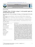
Fig. 2. Partition ratio plot for inactivation of GPAO by DAPY. Residual GPAO activities after 1 h of incubation with DAPY were plotted against the corresponding values of the concentration ratio [DAPY]/[GPAO]. Activity was measured with 70 nM enzyme in 0.1 M potassium phosphate buffer, pH 7.0, at 30 (cid:2)C using the guaiacol spectrophotometric method [21].
Fig. 1. Effect of incubation time on inactivation of GPAO by DAPY. The semilogarithmic plot was constructed for the following DAPY concentrations: 5 (j), 10 (m), 20 (r) and 40 lM (d). Activity was measured with 70 nM enzyme in 0.1 M potassium phosphate buffer, pH 7.0, at 30 (cid:2)C by means of the guaiacol spectrophotometric method [21]. The inset shows the corresponding Kitz–Wilson replot for the determination of kinact and KI values.
inactivation event, was determined to be 120 for DAPY/ GPAO (Fig. 2) and 200 for DAPY/OVAO.
semilogarithmic plots for GPAO, where the slope for each regression line represents the observed rate constant kobs. The kinetic constants describing the inactivation of GPAO and OVAO were determined from the corresponding Kitz– Wilson replots (1/kobs vs. 1/[DAPY]; see inset in Fig. 1 as an example). From these plots kinact, the maximal rate of inactivation, is 1/y ) intercept, and KI, the concentration required for half-maximal inactivation, is )1/x ) intercept. The determined values for GPAO were similar when measured in 0.1 M potassium phosphate or Bistris/HCl buffers, pH 7.0 (Table 1). Comparatively, for OVAO, inactivation by DAPY is slower, but the KI is lower.
GPAO and OVAO (both 70 nM in 0.1 M potassium phosphate buffer, pH 7.0) were each individually incubated with seven different concentrations of DAPY varying from 1 to 50 lM at 30 (cid:2)C for 1 h. Remaining activity was determined by the ratio of the measured activity of the inactivated enzyme to the control enzyme incubated without DAPY. A plot of the remaining activity (%) vs. [DAPY]/ [GPAO] or [DAPY]/[OVAO] was constructed. Extrapola- tion of the linear portion of the data at lower [DAPY] gave the partition ratio (turnover number minus one). This ratio, the number of molecules leading to product per each
Table 1. Inactivation kinetics. Experiments were performed at 30 (cid:2)C in 0.1 M potassium phosphate buffer, pH 7.0, except where noted.
kinact (min)1)
KI (lM)
t1/2 at saturationa (min)
Inhibitor/enzyme
The inhibition strength of DAPY is dependent on pH. GPAO (70 nM) was incubated with 50 lM DAPY in 0.1 potassium phosphate buffers of different pH values over the range 5.0–8.0 at 30 (cid:2)C for 1 h. The obtained remaining activity values were then plotted against pH. DAPY showed a maximal inhibition effect at pH 7.5. The extent of GPAO inhibition by DAPY is also influenced by ionic strength. The reaction was performed in 0.1 M Britton–Robinson buffer, pH 7.2, where ionic strength had been adjusted with KCl to reach values from the range 0.085–0.4. The percentage of remaining activity after 1 h of incubation of the reaction mixture (70 nM GPAO, 50 lM DAPY) at 30 (cid:2)C increases with increasing ionic strength (not shown). Enzymes are protected against mechanism-based inhi- bitors by their substrates and competitive inhibitors. These compounds bind at the active site and compete with binding of the inhibitor. Inactivation of the enzyme is therefore slowed down. GPAO (2 lM) was incubated with 100 lM DAPY in the absence and presence of 1 mM cadaverine as a substrate. At chosen time intervals, aliquots of the reaction mixtures were taken out for activity assay. The protective effect of cadaverine was significant. For example, after 15 min of incubation, the remaining activity was 15% in the reaction mixture with cadaverine and only 8% without.
Due to the potential information that might be provided about the mechanism of enzyme inactivation by DAPY, we also determined the kinetics of inactivation of GPAO by the two possible N-monomethyl analogs of DAPY. As shown in Table 1, N1-methyl-DAPY and especially N5-methyl- DAPY were weaker inactivators relative to DAPY itself.
0.27 0.31 0.13 0.11
45 50 10 45
2.2 1.9 4.5 6.3
0.05
36
13.9
DAPY with GPAOb DAPY with GPAO DAPY with OVAO N1-Methyl-DAPY with GPAO N5-Methyl-DAPY with GPAO
a Time required for half of the enzyme to become inactivated in the presence of saturating concentration of inhibitor. b In 0.1 M Bistris/ HCl buffer, pH 7.0.
Spectrophotometry and spectrofluorimetry, TLC
Substrates of CAOs are known to disturb the characteristic absorption spectrum of the enzymes [4]. Under anaero- biosis, the topaquinone cofactor maximum at 500 nm is bleached after the substrate addition and replaced by a complex spectrum of the Cu(I)-semiquinolamine radical showing maxima at 360, 435 and 465 nm. This is supple- mented with a peak at 315 nm that is thought to reflect the
DAPY inactivates plant amine oxidases (Eur. J. Biochem. 271) 4701
(cid:1) FEBS 2004
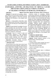
Fig. 3. Spectrophotometric studies on the reactions of GPAO and OVAO with DAPY. (Upper) Difference absorption spectrum of GPAO (20 lM) after the addition of DAPY (final concentration 1 mM) in air-saturated 0.1 M potassium phosphate buffer, pH 7.0. The spectrum was recorded 2 s after mixing the reactants at 30 (cid:2)C, using the rapid-scan- ning technique [9]. (Lower) Time-dependent development of the DAPY oxidation product as observed in difference absorption spectra. The spectra were recorded using a solution of OVAO (20 lM) in air-saturated 0.1 M potas- sium phosphate buffer, pH 7.0, after adding 0.1 M DAPY (1 mM final concentration) at 30 (cid:2)C. Intervals between scans: 15 s, total time: 10 min.
presence of the aminoresorcinol form of topaquinone (the fully reduced cofactor), which is in equilibrium. Using rapid-scanning techniques, the mentioned spectral features are observable also in the presence of air [9].
485 and 520 nm) when excited at 310 nm. For model compounds, the following fluorescence characteristics were obtained: DDD, solvent water, emission maximum at 503 nm (excitation at 410 nm); 3-hydroxypyridine, solvent water, emission maximum at 460 nm (excitation at 310 nm); NADH, solvent water, emission maximum at 465 nm (excitation at 340 nm); pyrrole, solvent water, emission maximum at 360 nm (excitation at 290 nm).
TLC experiments revealed the presence of a free primary amino group in the DAPY oxidation product obtained by the reaction of GPAO (positive ninhydrin spot, Rf ¼ 0.62); DAPY itself showed Rf ¼ 0.33 in the same system. Staining for tertiary amines (Draggendorff’s reagent) was also positive for the GPAO/DAPY reaction mixture (an orange spot, Rf ¼ 0.62). Staining for aldehydes using Schiff’s reagent was negative.
Colorimetric detections of DAPY oxidation product
As shown in Fig. 3 (upper), rapid scanning after the aerobic addition of DAPY to a purified GPAO in 0.1 M potassium phosphate buffer, pH 7.0, revealed the formation of a spectrum identical to that of a substrate-reduced CAO. In addition, the reaction of the studied plant CAOs with DAPY gave rise to an oxidation product providing near UV/visual absorption with a maximum at 310–315 nm, which increased in intensity with temperature. Absorbances measured after 90 min of incubation of GPAO/DAPY reaction mixtures at 50 (cid:2)C were almost three times higher than those measured after the incubation at 37 (cid:2)C. Figure 3 (lower), shows an increasing development of the product within the first 10 min after mixing OVAO with DAPY in 0.1 M potassium phosphate buffer, pH 7.0. The same spectrum was also observable in the GPAO/DAPY reaction mixture. In 0.1 M Bistris/HCl buffer, pH 7.0, the product absorption maximum was shifted to 325 nm (not shown). GPAO and OVAO were fully inactivated by incubation with an excess of DAPY and no activity could be recovered by dialysis. The inactivated enzymes after dialysis were still faint yellow due to a broad absorption below 330 nm, but the color intensity was substantially decreased.
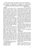
After separation of the enzyme protein by ultrafiltration, the GPAO/DAPY reaction mixture exhibited a fluorescence emission spectrum with a maximum at 445 nm (shoulders at Kovacs’ reagent containing DMAB (detects indoles and pyrroles [25]) was previously used for the visualization of pyrrole derivatives formed by the oxidation of DABY by plant amine oxidases [7,8]. Reaction mixtures of the studied CAOs with DAPY reacted positively with Kovacs’ reagent after a period of incubation and provided a red soluble adduct upon heating. The red color intensity increased with increasing temperature in the range 30–50 (cid:2)C. A typical absorption spectrum of the adduct with a maximum at 520 nm (shoulder at 500 nm) is shown in Fig. 4 (upper).
4702 Z. Lamplot et al. (Eur. J. Biochem. 271)
(cid:1) FEBS 2004
to form the corresponding aminoaldehydes, which sponta- neously cyclize to 1-pyrroline and 1-piperideine, respect- ively. The latter cyclic imines condense with ABA to generate the corresponding substituted dihydroquinazolin- ium compounds. The DAPY oxidation product obtained by the reaction of GPAO provided an adduct with ABA characterized by an absorption maximum at 430 nm (not shown).
The ninhydrin reagent described for activity assay of CAOs by Naik et al. [24] was also tested to trap the DAPY oxidation product in the reaction mixture with GPAO. The same cyclic imines above (1-pyrroline and 1-piperideine) react with ninhydrin in strongly acidic medium to form colored compounds of unknown structure with absorption maxima at 440 and 515 nm, respectively [30]. The DAPY oxidation product displayed a broad absorption between 400 and 550 nm with a maximum at 465 nm after reaction with ninhydrin (not shown).
MS of DAPY reaction mixture
Fig. 4. Reaction of the DAPY oxidation product and a dihydropyridine model compound with 4-(dimethylamino)benzaldehyde. (Upper) An aliquot (1 mL) of the GPAO/DAPY reaction mixture in 0.1 M potassium phosphate buffer, pH 7.0, was mixed with 2 mL of Kovacs’ reagent, incubated at 50 (cid:2)C for 30 min and finally cooled in an ice- bath. The absorption spectrum was recorded against a blank con- taining water instead of the reaction mixture; (upper) reaction mixture (lower) reaction mixture after removing protein by ultrafiltration. For experimental details see Materials and methods. (Lower) A 1 mL portion of 5 mM DDD was mixed with 2 mL of Kovacs’ reagent, incubated at 50 (cid:2)C for 30 min and finally cooled in an ice bath. Absorption spectra were then recorded against a blank containing water instead of the dihydropyridine.
Figure 5 (upper) shows an ESI-IT mass spectrum of the GPAO/DAPY reaction mixture prepared using 0.1 M ammonium bicarbonate, pH 7.8. There are two major peaks of the reaction product observable in the spectrum with m/z 178.3 and 222.3. The former ion showed fragment peaks with m/z 161.3, 149.2 and 135.2, the latter provided peaks with m/z 205.2, 193.2 and 176.2 in the respective MS/MS spectra. The peak with m/z 222.3 was not observed when the reaction was carried out in 0.1 M Bistris/HCl buffer, pH 7.0. Figure 5 (lower) shows a mass spectrum of the GPAO/DAPY reaction mixture prepared in 0.1 M ammonium bicarbonate containing ACA as a reagent for trapping of the product aminoaldehyde, where several new peaks appeared. ACA itself is represented by a peak with m/z 132.1 (MS/MS: a clear fragment peak with m/z 114.1.) There is one more peak visible with m/z 211.3 (MS/MS: fragment peaks with m/z 193.3, 106.0 and 96.0), which probably reflects an adduct of the reaction product with ACA.
ESI-IT-MS of the low molecular mass fraction of the GPAO/DAPY reaction mixture prepared in 20 mM potassium phosphate buffer, pD 7.0 (made in D2O for the purpose of NMR spectroscopic analysis) revealed isotopic peaks belonging to quasimolecular ions of the reaction product. The highest intensity was observed for a peak with m/z 179.2, lower intensities were observed for peaks in the following order: m/z 181.2, 180.2, 182.2 and 178.2. The peak with m/z 179.2 provided an MS/MS spectrum showing fragments with m/z 162.2, 150.2 and 136.2 (not shown).
HPLC separation and MS analysis of DAPY oxidation product
Replacing DMAB in Kovacs’ reagent with DMAC led to a shift of the adduct absorption maximum to 650 nm (not shown). However, if the reaction mixture was dialyzed before the addition of the reagent, the spectrum was almost negligible (Fig. 4, upper). Three model compounds were tested for this reaction. The synthesized DDD reacted with Kovacs’ reagent to form a product with an absorption maximum at 600 nm having a shoulder at 560 nm (Fig. 4, lower). NADH also reacted with the reagent and provided a spectrum with a single peak centered at 510 nm. DMAB- reacted pyrrole provided a maximum at 565 nm with a shoulder at 520 nm.
HPLC separation of the isolated DAPY oxidation product from enzymatic microscale production was carried out using an instrument equipped with a diode array detector. Thus individual runs could be monitored continuously at 214, 240 and 310 nm. The buffer system used was chosen according to that published for peptide separation from tryptic digests [31]. The use of ABA for the spectrophotometric activity assay of plant CAOs was refined almost four decades ago [23]. Plant CAOs oxidize the diamines putrescine and cadaverine
DAPY inactivates plant amine oxidases (Eur. J. Biochem. 271) 4703
(cid:1) FEBS 2004
Fig. 5. ESI-IT-MS analyses of GPAO/DAPY reaction mixtures. (Upper) DAPY (5 mM) in 0.1 M ammonium bicarbonate, pH 7.8, was mixed with an excess of GPAO and incubated at 30 (cid:2)C for 24 h. After removing protein by ultrafiltration, the reaction mixture was ana- lyzed by ESI-IT-MS as described in Materials and methods. (Lower) A combined solution of DAPY and ACA (each 5 mM) in 0.1 M ammonium bicarbonate, pH 7.8, was mixed with an excess of GPAO and incubated at 30 (cid:2)C for 24 h. After removing protein by ultrafiltration, the reaction mixture was ana- lyzed by ESI-IT-MS as described in Materials and methods.
Three peaks at elution times 3.0 min (1), 15.8 min (2) and 23.0 min (3) were collected and their composition analyzed using ESI-MS and MALDI-MS. The largest peak absorb- ing at 310 nm (peak 1) appeared to correspond to a single chemical compound (m/z 178.1). The corresponding ESI-IT-MS/MS spectrum is presented in Fig. 6, where
fragmentation peaks were observed with m/z 161.1, 149.1, 144.1, 135.1, 132.1, 120.1, 109.1, 95.0 and 82.0. After lyophilization, a solid obtained from peak 1 did not produce irreversible inhibition of the studied enzymes. There was one more compound in peak 2 with m/z 257.1, whose MS/MS spectrum provided peaks with m/z 240.1, 228.1, 214.3, 202.3, 176.2, 161.2, 149.2 and 133.0. Finally, peak 3 contained at least five compounds. In addition to those with m/z 178.1 and 257.1 there were three more peaks with m/z 334.3 (fragmentation: m/z 317.3, 305.2, 291.1, 253.2 and 240.1), 350.2 (fragmentation: m/z 333.2 and 307.1) and 431.3 (fragmentation: m/z 414.3, 388.3 and 337.1).
Fig. 6. MS/MS spectrum of DAPY oxidation product. The isolated DAPY oxidation product from enzymatic microscale production was dissolved in 0.3% (v/v) triethylamine acetate, pH 7.0, and separated by RP-HPLC as described in Materials and methods. The 310 nm-peak at an elution time 3.0 min was collected and analyzed by ESI-IT-MS and MS/MS. The spectrum shown was recorded after collision- induced fragmentation of the parent ion belonging to the DAPY oxidation product (m/z 178.1).
MALDI-TOF-MS of the separated peak 1 provided a single compound with m/z 178.1; the same m/z value was obtained by an ionization without using the HABA matrix. MALDI-PSD-TOF-MS provided a fragmentation pattern consistent with the ESI-IT-MS/MS experiments already mentioned (data not shown).
ESI-Q-TOF-MS analysis of the HPLC peak 1 permitted the determination of both exact mass and elemental composition of the DAPY oxidation product. An m/z value of 178.14 was obtained, which matches a molecular formula C10H16N3. Peaks in the corresponding MS/MS spectrum provided the following m/z values and elemental composition of ions: 161.11 (C10H13N2), 149.11 (C9H13N2), 135.09 (C8H11N2), 109.08 (C6H9N2), 95.06 (C5H7N2) and 82.06 (C5H8N).
4704 Z. Lamplot et al. (Eur. J. Biochem. 271)
(cid:1) FEBS 2004
Fig. 7. 1H-NMR spectrum of DAPY oxidation product. DAPY (5 mM) in 2 mL of 20 mM potassium phosphate buffer, pH 7.0, was mixed with an excess of GPAO (5 mg, added as a concentrated solution in the same buffer) and the mixture was incubated at 30 (cid:2)C for 12 h. The same amount of GPAO was added again and the incubation proceeded for an additional 12 h. The resulting solution was centrifuged to remove protein precipitate and ultrafiltered, and the filtrate was lyophilized. The NMR sample was finally prepared by extracting the lyophilizate with 0.5 mL of CD3OD. The insets shows a detailed view of the vinylic doublet signals belonging to 2,3-dihydropyridine.
NMR spectroscopy of DAPY oxidation product
formed by the turnover of acetylenic diamine substrates [7–11]. The cDNA of GPAO subunit (without the signal peptide) has been cloned and sequenced (D. Kopecˇ ny´ , N. Houba-He´ rin, H. G. Faulhammer & M. Sˇ ebela, unpublished results; EMBL/GenBank accession number AJ786401). The translated protein sequence is largely similar to those two published for PSAO [32,33] and comprises 38 lysines. GPAO and PSAO have very similar peptide maps obtained by MALDI-MS experiments [34]. PSAO protein sequence of Koyanagi et al. [32], which is deposited under accession number JC7251 (NCBI Protein Databank) differs slightly from that of Tipping and McPherson [33], accession number Q43077, in that whereas the former sequence comprises 39 lysine residues, the latter has only 38 lysine residues, as calculated for the mature form of the protein. Although native PSAO (a dimer) is thus expected to comprise 76–78 lysine residues, only 32 are solvent-accessible [8]. We determined 38 accessible lysines per dimer in the native GPAO using a modified protocol with the TNBS reagent, and 36 lysines per dimer (average values from repeated measurements) after reaction with the DAPY.
For initial experiments, DAPY oxidation by GPAO was performed in 20 mM potassium phosphate buffer made in D2O (pD 7.0). The enzyme protein was removed by ultrafiltration and the filtrate was directly measured. Several signals with the following chemical shifts were observed in the 13C-NMR spectrum: d (p.p.m.) 17.0, 25.1, 29.1, 37.9, 62.5, 72.1, 74.3, 82.7, 123.0, 131.2 and 159.8. The corres- ponding 1H-NMR spectrum contained various signals in the region 2.0–4.5 p.p.m., but these were largely obscured by the residual water peak (4.5–5.0 p.p.m.). In addition, two vinylic signals at d 5.7 and 7.8 were observed, but their intensities were too small to ascertain their multiplicities (not shown). Much better 1H-NMR spectra were obtained after the extraction of the ultrafiltered and lyophilized GPAO/DAPY reaction mixture by CD3OD. Figure 7 shows such a spectrum, including the following signals: d 2.63–2.73 (m), 3.12 (q, J ¼ 6.95 Hz), 3.31 (p, J ¼ 1.65 Hz), 3.79 (t, J ¼ 2.20 Hz), 4.19 (t, J ¼ 2.20 Hz), 5.35 (d, J ¼ 6.22 Hz), 7.82 (d, J ¼ 6.22 Hz). The spectrum is partially obscured by two signals of residual methanol at d 3.3 and 4.6–5.1. There are also three complex signals that are quite difficult to interpret, which are centered at d values of 2.80, 3.55 and 3.65. 13C-NMR spectra measured with the GPAO/ DAPY mixture in CD3OD resembled those recorded in D2O, but the obtained quality was lower. 1H-NMR spectra were also recorded in D2O using the solid obtained by lyophilization of the peak 1 from the HPLC separation mentioned above. However, NMR signal intensities were insufficient due to the low concentration of compound. In addition, these spectra were obscured by two peaks of residual triethylamine from the elution buffer at d 1.2 and 3.2 (data not shown). Quinone-staining experiments [29] with the DAPY-inac- tivated GPAO were positive and demonstrated that the topaquinone cofactor was not modified in a redox-inactive form (data not shown). Chromatofocusing experiments performed according to that with DABY-inactivated GPAO [8] revealed that the pI value of the DAPY-inactivated GPAO was not dramatically changed. The native GPAO is characterized by a pI of 7.2 [18]. After the reaction with an excess of DAPY, the enzyme sample comprised more species having isoelectric points of pI 6.8–7.5 (Fig. 8). Therefore, the inactivation resulted in a heterogeneous mixture of differently charged protein molecules.
Other analyses
Discussion
Lysine residues in plant CAOs are possible targets for covalent binding of reactive electrophilic aminoaldehydes DAPY was synthesized as an analog of cadaverine (pentane-1,5-diamine), which is known as the best substrate
DAPY inactivates plant amine oxidases (Eur. J. Biochem. 271) 4705
(cid:1) FEBS 2004
Fig. 8. Chromatofocusing of DAPY-inactivated GPAO. Chromatofo- cusing was performed on a Mono P HR 5/20 column using a BioLogic Duo Flow liquid chromatograph at a flow rate of 1 mLÆmin)1. The loading buffer was 25 mM Tris/HCl, pH 8.2, and the elution buffer was a diluted mixture of Polybuffer 96 and Polybuffer 74 adjusted to pH 5.0 with acetic acid. All samples were dialyzed against the loading buffer before separation. Approximately 5 mg of protein was loaded.
assay. Presuming that DAPY oxidation follows the same mechanism as for common substrates of plant CAOs, the reaction should generate 5-amino-2-pentynal or 5-amino-3- pentynal as a product aldehyde (DAPY is not a symmetric molecule). Although no free aldehyde was detected by TLC in the reaction mixture, indirect evidence for an amino- pentynal turnover product was that an adduct formed (m/z 211.3) when DAPY was enzymatically oxidized in the presence of ACA. This adduct exhibited a MS/MS fragmentation pattern similar to that of free ACA, showing a loss of a water molecule from the carboxylic group ()18, m/z 211.3 fi m/z 193.3).
of plant CAOs [4]. Contrary to naturally occurring diam- ines, the DAPY molecule contains a triple bond at the b- and c-positions from the two primary amine termini. The oxidative conversion of the compound by GPAO and OVAO was demonstrated by measuring the production of H2O2 using spectrophotometry. Therefore, the enzymes are able to undergo complete turnover [3]. However, DAPY was oxidized more efficiently by OVAO than by GPAO. Although, to date, OVAO has not been crystallized nor had its structure solved, this observation might be explained in terms of different arrangements of the active sites of the enzymes resulting in the preference for C5-diamine sub- strates by OVAO. There are detailed reports on the mechanism-based inhibition of CAOs by DABY in the literature [7–11]. The authors have shown that DABY is oxidized to 4-amino-2- butynal, which induces inactivation by adducting to a nucleophile in the substrate channel. In addition, the reaction brings about multiple surface labeling of the enzyme, which probably occurs through solvent-accessible nucleophilic residues [8]. DAPY oxidation appears to result in much less extensive protein modification, as the number of free primary amino groups in the enzyme did not change dramatically. Chromatofocusing of DAPY-inactivated GPAO revealed only a small change in the isoelectric point, likely caused by the modification of a few amino acid residues upon binding of aminopentynal. This binding seems to be nonspecific, as the existence of some micro- heterogeneity (at least two species with different pI values) in the inactivated enzyme was confirmed. As in the case of DABY, the cofactor topaquinone is not modified by the reaction, as demonstrated by an unchanged quinone redox staining.
respectively.
Absorption spectroscopy demonstrated the formation of a secondary product in the GPAO/DAPY reaction mixture with kmax at 310 nm and emitted fluorescence (kmax at 445 nm) upon excitation at 310 nm, supportive of extended conjugation. Dialysis of the reaction mixtures containing GPAO or OVAO and DAPY, resulted in decoloration, demonstrating that the chromophore generated is a free low molecular mass compound. Several colorimetric assays provided evidence for the presence of a nitrogenous heterocycle, in addition to a free amino group. The GPAO/DAPY reaction mixture exhibited a positive reac- tion with ABA and ninhydrin reagents, similar to that observed for the cyclic imines 1-pyrroline and 1-piperideine formed upon enzymative oxidation of putrescine and cadaverine, In acidic medium, DMAB and DMAC reacted with the DAPY oxidation product (and also with the model compounds DDD and NADH) to give markedly colored adducts. This probably occurs upon binding of the reagents at the a-position of the heterocycle [8].
Similarly to the conversion of its lower homolog DABY by PSAO [7], DAPY oxidation by the studied enzymes led to their irreversible inhibition. The apparent inactivation constants KI of 10)5 M are on the same order of magnitude as those KI values previously described for BEA [6] and DABY [7] in the reactions with lentil seedling amine oxidase (LSAO) and PSAO, respectively. The obtained rates of inactivation resembled for example that for BEA as measured with LSAO [6], but they were lower than that for DABY in the reaction with PSAO [7]. At the same time, whereas the determined partition ratio values with GPAO and OVAO were in the range observed for BEA (r ¼ 100) and some other monohalogenated alkylamines (r < 500) in the reactions with LSAO [6], they were significantly higher than that for DABY and PSAO (r ¼ 17) [7]. From this point of view, plant CAOs are more resistant to the inactivation by DAPY than by DABY. Binding of DAPY at the active site of GPAO is dependent on both pH and ionic strength. In these features, the reaction does not differ from those of typical plant CAO substrates like putrescine or cadaverine. GPAO and OVAO inactivation caused by the substrate DAPY fulfills the criteria of a mechanism- based inhibition: it is time dependent, irreversible and can be weakened in the presence of a normal substrate [5,7,11,22]. DAPY oxidations by GPAO and OVAO were accom- panied by spectral changes. The typical absorption spec- trum of a substrate-reduced CAO, which appeared after the rapid addition of DAPY to either GPAO or OVAO solutions, was in accordance with the substrate properties of DAPY determined by the guaiacol spectrophotometric The rigidity of the triple bond in the presumed amino- pentynal turnover product would prevent cyclization, but this geometrical constraint would be relaxed by conjugate addition of a nucleophile. Thus, as shown in Fig. 9, if a molecule of unreacted DAPY is added to either of the two possible aminopentynal turnover products, cyclization to a six-membered heterocycle and eventual formation of a common resonance-stabilized 4-amino-2,3-dihydropyridine would be predicted. The extended conjugation would be consistent with the observed absorption and fluorescence
4706 Z. Lamplot et al. (Eur. J. Biochem. 271)
(cid:1) FEBS 2004
Fig. 10. MS/MS fragmentation scheme for DAPY oxidation product. The scheme reflects the postulated collision-induced dissociation of the parent DAPY oxidation product ion (m/z 178.1) assigned as N1-(2,3-dihydropyridin-4-yl)-1,5-diamino-2-pentyne, according to the observed MS/MS peaks obtained by ESI-IT-MS/MS (Fig. 6).
Fig. 9. Mechanism of DAPY oxidation by GPAO. The scheme reflects the summary results of kinetic, spectrophotometric, spectrofluorimet- ric, MS and NMR experiments performed in this study. First, DAPY is oxidized by the enzyme to either 5-amino-2-pentynal or 5-amino-3- pentynal, which subsequently (after tautomerization in the latter case) reacts with a second DAPY molecule (or ACA) acting as nucleophile. After nucleophilic adduction, the two possible aldehydes undergo cyclization to form ultimately the same resonance stabilized 4-amino- 2,3-dihydropyridine ring. Enzyme inactivation may follow the same overall reaction except that the 4-amino-2,3-dihydropyridine is formed on the enzyme by adduction of the intial aminopentynal turnover product to a lysine residue.
probably represents adduction of aminopentynal to both amino groups of the same unoxidized DAPY molecule to give N1,N5-bis-(2,3-dihydropyridin-4-yl)-1,5-diamino-2- pentyne, and the peak with m/z 334.1 suggests even one more aminopentynal coupling. Finally, it should be pointed out that the 4-amino-2,3-dihydropyridine core structure is consistent with the molecular ion m/z 211.3 observed for the ACA-trapped DAPY oxidation product, wherein ACA rather than unoxidized DAPY would add to the initial aminopentynal turnover product (see Fig. 9).
spectral properties. The final structure is also consistent with MS and NMR data. In particular, the two coupled vinyl doublets at 5.35 and 7.82 p.p.m. in the 1H-NMR spectrum (Fig. 7) are consistent with the electron-rich C5 and electron-deficient C6 positions of the 4-amino-2,3-dihydro- pyridine. The 13C-NMR spectrum additionally demonstra- ted the presence of a triple bond (d 74.3 and 82.7 p.p.m) and aliphatic carbon atoms (signals of d 20–40 p.p.m), consis- tent with presence of an unoxidized DAPY moiety.
The theoretical molecular mass calculated for the predic- ted N-(2,3-dihydropyridinyl)-1,5-diamino-2-pentyne was in accordance with that determined by ESI-Q-TOF-MS (177.14 Da) and the fragmentation pattern of the peak with m/z 178.1 observed by MALDI-MS/ESI-MS. Either the N1 or N5 amino group of unoxidized DAPY could add to the initial aminopentynal turnover product. Although an insufficient amount of the oxidation product was available to perform the detailed two-dimensional NMR experiments that would be needed to distinguish between the N1- and N5- (2,3-dihydropyridin-4-yl)-1,5-diamino-2-pentyne isomers, the observed ESI-IT-MS/MS fragmentation peaks (Fig. 6) are most consistent with the former compound, as depicted in Fig. 10. A small peak at m/z 144.1 (C9H8N2) requires substantial dehydrogenation and is hard to reconcile with a particular structure. It should be pointed out that only the peaks with m/z 135.1 and 132.1 are consistent with only the N1- and not the N5-isomer, and a small peak at m/z 120.1 (C7H8N2) is most readily reconciled with the N5-isomer. Thus, we cannot exclude the possibility that the DAPY oxidation product represents a mixture of mainly N1-(2,3- dihydropyridin-4-yl)-1,5-diamino-2-pentyne, contaminated with a small amount of the N5-isomer that coelutes during RP-HPLC separation.
According to the mechanism for formation of the chromophoric DAPY oxidation product (Fig. 9), the same resonance-stabilized 4-amino-2,3-dihydropyridine moiety would form if the initial aminopentynal turnover product were trapped by an enzyme-based lysyl residue (Fig. 9). The evidence for modification of at least some lysines during incubation of GPAO with DAPY suggests that such adduction could be responsible for the irreversible enzyme inactivation observed. In this regard, the inactivation mechanism would then be highly analogous to that discerned for inactivation of PSAO or GPAO by DABY, where the initial 4-amino-2-butynal product undergoes conjugate addition by a channel lysyl residue, followed by dehydrative cyclization to give a 3-aminopyrrole [8]. On the basis of the highly apparent formation of the low molecular mass DAPY oxidation product identified here, one might speculate why the analogous product, N-(pyrrole-3-yl)-1,4- diamino-2-butyne, was not observed during plant CAO metabolism of DABY. Although such product might have actually been present, there are two reasons why it is probably less apparent. The first is that DABY is a more potent inhibitor than DAPY, so that higher concentrations of the latter, amenable to formation of the observed coupling product, were employed. The second is that the aminopentynal turnover product from DAPY appears to modify the pertinent substrate channel lysine with signifi- cantly less efficiency than does the 4-amino-2-butynal product from DABY, so that there is greater turnover prior to inactivation.
The MS experiments performed in the present study also demonstrated that the aminopentynal product undergoes further oligomerization reactions, which result in the formation of compounds having higher molecular masses (256–430 Da). For example, the peak with m/z 257.1 To obtain clues as to the mechanism of inactivation of GPAO by DAPY, we investigated the inhibitory potency of the two possible mono-N-methyl derivatives of DAPY with
DAPY inactivates plant amine oxidases (Eur. J. Biochem. 271) 4707
(cid:1) FEBS 2004
and oxidative half-reactions and the role of copper ions in plant and mammalian copper-amine oxidases. Eur. J. Inorg. Chem. 1, 35–42.
4. Medda, R., Padiglia, A. & Floris, G. (1995) Plant copper-amine
oxidases. Phytochemistry. 39, 1–9.
5. Padiglia, A., Medda, R., Pedersen, J.Z., Lorrai, A., Pecˇ , P., Fre´ bort, I. & Floris, G. (1998) Inhibitors of plant copper amine oxidases. J. Enzyme Inhib. 13, 311–325.
6. Medda, R., Padiglia, A., Pedersen, J.Z., Finazzi-Agro, A., Rotilio, G. & Floris, G. (1997) Inhibition of copper amine oxidase by haloamines: a killer product mechanism. Biochemistry. 36, 2595– 2602.
7. Pecˇ , P. & Fre´ bort, I.
(1992) 1,4-Diamino-2-butyne as the mechanism-based pea diamine oxidase inhibitor. Eur. J. Biochem. 209, 661–665.
this enzyme. Topaquinone-dependent enzymes are mostly not known to metabolize secondary amines. The finding that one but not the other of the two N-methyl derivatives of DAPY acted as an inactivator, would suggest that meta- bolism leading to inactivation occurs only at the propar- gylamine terminus or the homopropargylamine terminus. Our finding that both derivatives act as inactivators of GPAO, albeit weaker than DAPY, suggests that enzyme metabolism of DAPY leading to inactivation can occur at either amino group, as shown in Fig. 9.
8. Fre´ bort, I., Sˇ ebela, M., Svendsen, I., Hirota, S., Endo, M., Yamauchi, O., Bellelli, A., Lemr, K. & Pecˇ , P. (2000) Molecular mode of interaction of plant amine oxidase with the mechanism- based inhibitor 2-butyne-1,4-diamine. Eur. J. Biochem. 267, 1423– 1433.
9. Sˇ ebela, M., Fre´ bort, I., Lemr, K., Brauner, F. & Pecˇ , P. (2000) A study on the reactions of plant copper amine oxidase with C3 and C4 diamines. Arch. Biochem. Biophys. 384, 88–99.
10. Shepard, E.M., Smith, J., Elmore, B.O., Kuchar, J.A., Sayre, L.M. & Dooley, D.M. (2002) Towards the development of selective amine oxidase inhibitors. Mechanism-based inhibition of six copper containing amine oxidases. Eur. J. Biochem. 269, 3645– 3658.
11. Jeon, H.B., Lee, Y., Qiao, C., Huang, H. & Sayre, L.M. (2003) Inhibition of bovine plasma amine oxidase by 1,4-diamino- 2-butenes and -2-butynes. Bioorg. Med. Chem. 11, 4631–4641. 12. Jeon, H.B. & Sayre, L.M. (2003) Highly potent propargylamine and allylamine inhibitors of bovine plasma amine oxidase. Bio- chem. Biophys. Res. Commun. 304, 788–794.
13. Jeon, H.B., Sun, G. & Sayre, L.M. (2003) Inhibition of bovine plasma amine oxidase by 4-aryloxy-2-butynamines and related analogs. Biochim. Biophys. Acta. 1647, 343–354.
14. Qiao, C., Jeon, H.B. & Sayre, L.M. (2004) Selective inhibition of bovine plasma amine oxidase by homopropargylamine, a new inactivator motif. J. Am. Chem. Soc. 126, 8038–8045.
(after
15. Besace, Y., Marszak-Fleury, A. & Marszak, I. (1971) Delepine reaction of acetylenic compounds. Bull. Soc. Chim. Fr. 4, 1468– 1472 [in French].
In conclusion, DAPY was found here to be both a substrate and inactivator of plant CAOs. Prior to complete enzyme inactivation, the enzymes generate significant amounts of two possible aminopentynal turnover products. tautomerization) or Either 5-amino-3-pentynal 5-amino-2-pentynal can condense with a molecule of unoxidized DAPY to give an adduct capable of cyclization to the same 4-amino-2,3-dihydropyridine, which is appar- ently resistant to hydrolysis on account of extended resonance stabilization. The structure of the final product, most likely N1- rather than N5-(2,3-dihydropyridin-4-yl)- 1,5-diamino-2-pentyne, was supported by spectrophoto- metric, spectrofluorimetric, MS and NMR measurements. Elucidation of the adduct structure suggests that enzyme inactivation occurs through the same chemical mechanism, but involving an enzyme-based lysyl residue, rather than a second DAPY molecule, in adduct formation with the initial aminopentynal turnover product (Fig. 9). Nonethe- less, further studies are needed to ascertain the true molecular nature of enzyme modification leading to inac- tivation by DAPY. Although DAPY is not such a powerful inactivator in comparison with the previously studied shorter analog DABY, the reaction with DAPY does not result in as extensive labeling of the enzyme as was observed for DABY. The significance of the present work resides also in the possible use of plant CAOs for organic synthesis, which has already been suggested [35]. Pure chemical preparation of a dihydropyridine such as that characterized here would be difficult.
Acknowledgements
16. Qiao, C. (2004) New copper amino oxidase probes: synthesis, mechanism and enzymology. PhD Thesis, Case Western Reserve University, Cleveland, OH.
17. Torchy, S., Cordonnier, G., Barbry, D. & Van den Eynde, J.J. (2002) Hydrogen transfer from Hantzsch 1,4-dihydropyridines to carbon-carbon double bonds under microwave irradiation. Molecules. 7, 528–533.
This work was supported by grants MSM 153100010, MSM 153100013 and 14BI3 (Czech-Italian cooperation, a joint grant with the Ministry of Foreign Affairs, Italy) from the Ministry of Education, Youth and Sports, Czech Republic, and by grant GM 48812 from the National Institutes of Health (to L.M.S.). Fluorescence spectra were recorded by from the Institute of Medical courtesy of Dr Martin Modriansky´ Chemistry and Biochemistry, Faculty of Medicine, Palacky´ University. Dr Ivo Fre´ bort, Palacky´ University, is thanked for the initial impetus to start with this research.
18. Sˇ ebela, M., Luhova´ , L., Fre´ bort, I., Faulhammer, H.G., Hirota, S., Zajoncova´ , L., Stuz˘ ka, V. & Pecˇ , P. (1998) Analysis of the active sites of copper/topa quinone-containing amine oxidases from Lathyrus odoratus and L. sativus seedlings. Phytochem. Anal. 9, 211–222.
19. Zajoncova´ , L., Sˇ ebela, M., Fre´ bort, I., Faulhammer, H.G., Nav- ra´ til, M. & Pecˇ , P. (1997) Quinoprotein amine oxidase from sainfoin seedlings. Phytochemistry. 45, 239–242.
20. Smith, P.K., Krohn, R.I., Hermanson, G.T., Mallia, A.K., Gartner, F.H., Provenzano, M.D., Fujimoto, E.K., Goeke, N.M., Olson, B.J. & Klenk, D.C. (1985) Measurement of protein using bicinchoninic acid. Anal. Biochem. 150, 76–85.
References 1. Sˇ ebela, M., Fre´ bort, I., Petrˇ ivalsky´ , M. & Pecˇ , P. (2002) Copper/ topa quinone-containing amine oxidases – recent research devel- opments. In Studies in Natural Products Chemistry, Vol. 26. Bio- active Natural Products, Part G. (Atta-ur-Rahman, ed.), pp. 1259–1299. Elsevier, Amsterdam.
2. Klinman, J.P. (2003) The multi-functional topa-quinone copper
amine oxidases. Biochim. Biophys. Acta. 1647, 131–137.
21. Fre´ bort, I., Haviger, A. & Pecˇ , P. (1989) Employment of guaiacol for the determination of activities of enzymes generating hydrogen peroxide and for the determination of glucose in blood and urine. Biolo´gia (Bratislava) 44, 729–737.
3. Padiglia, A., Medda, R., Bellelli, A., Agostinelli, E., Morpurgo, L., Mondovı` , B., Finazzi-Agro` , A. & Floris, G. (2001) The reductive
4708 Z. Lamplot et al. (Eur. J. Biochem. 271)
(cid:1) FEBS 2004
30. Pecˇ , P. & Pavlı´ kova´ , M. (1985) New colorimetric method for the determination of diamine oxidase activity by ninhydrine. Biolo´gia (Bratislava) 40, 1217–1225.
22. Kent, U.M., Yanev, S. & Hollenberg, P.F. (1999) Mechanism- based inactivation of cytochromes P450 2B1 and P450 2B6 by n-propylxanthate. Chem. Res. Toxicol. 12, 317–322.
31. Mu, D., Janes, S.M., Smith, A.J., Brown, D.E., Dooley, D.M. & Klinman, J.P. (1992) Tyrosine codon corresponds to topa quinone at the active site of copper amine oxidases. J. Biol. Chem. 267, 7979–7982.
23. Machola´ n, L. (1966) Beitrag zur spektrophotometrischen Bes- timmung der enzymatischen Oxydation von aliphatischen Diam- inen. Collect. Czech. Chem. Commun. 31, 2167–2174 [in German]. 24. Naik, B.I., Goswami, R.G. & Srivastava, K. (1981) A rapid and sensitive assay of amine oxidase. Anal. Biochem. 111, 146–148. 25. Kovacs, N. (1928) Eine vereinfachte Methode zum Nachweis der Indolbildung durch Bakterien. Z. Immunita¨tsforsch. 55, 311–315 [in German].
32. Koyanagi, T., Matsumura, K., Kuroda, S. & Tanizawa, K. (2000) Molecular cloning and heterologous expression of pea seedling copper amine oxidase. Biosci. Biotechnol. Biochem. 64, 717–722. 33. Tipping, A.J. & McPherson, M.J. (1995) Cloning and molecular analysis of the pea seedling copper amine oxidase. J. Biol. Chem. 270, 16939–16946.
26. Ascenzi, P., Fasano, M., Marino, M., Venturini, G. & Federico, R. (2002) Agmatine oxidation by copper amine oxidase – biosynthesis and biochemical characterization of N-amidino- 2-hydroxypyrrolidine. Eur. J. Biochem. 269, 884–892.
27. Fields, R. (1971) The measurement of amino groups in proteins
34. Sˇ ebela, M., Kopecˇ ny´ , D., Lamplot, Z., Havlisˇ , J., Thomas, H. & Shevchenko, A. (2005) Thermostable b-cyclodextrin conjugates of two similar plant amine oxidases and their properties. Biotechnol. Appl. Biochem. 41, in press.
and peptides. Biochem. J. 124, 581–590.
28. Habeeb, A.F.S.A. (1966) Determination of free amino groups in proteins by trinitrobenzenesulfonic acid. Anal. Biochem. 14, 328– 336.
35. Cragg, J.E., Herbert, R.B. & Kgaphola, M.M. (1990) Pea-seedling diamine oxidase: applications in synthesis and evidence relating to its mechanism of action. Tetrahedron Lett. 31, 6907–6910.
29. Paz, M.A., Flu¨ ckiger, R., Boak, A., Kagan, H.M. & Gallop, P.M. (1991) Specific detection of quinoproteins by redox-cycling stain- ing. J. Biol. Chem. 266, 689–692.