DNA polymerase e associates with the elongating form of RNA polymerase II and nascent transcripts Anna K. Rytko¨ nen1,2,*, Tomi Hillukkala1,*, Markku Vaara2, Miiko Sokka2, Maarit Jokela1,†, Raija Sormunen3, Heinz-Peter Nasheuer4, Tamar Nethanel5, Gabriel Kaufmann5, Helmut Pospiech1 and Juhani E. Syva¨ oja2
1 Biocenter Oulu and Department of Biochemistry, University of Oulu, Finland 2 Department of Biology, University of Joensuu, Finland 3 Biocenter Oulu and Department of Pathology, University of Oulu, Finland 4 National University of Ireland, Department of Biochemistry, Cell Cycle Control Laboratory, Galway, Ireland 5 Department of Biochemistry, Tel Aviv University, Ramat Aviv, Israel
Keywords DNA polymerase e; DNA replication; immunoelectron microscopy; nucleotide excision repair; RNA polymerase II
Correspondence H. Pospiech, Department of Biochemistry, PO Box 3000, FIN-90014 Oulu, Finland Fax: +358 8 5531141 Tel: +358 8 5531155 E-mail: Helmut.Pospiech@oulu.fi
†Present address Department of Internal Medicine, University of Oulu, Finland
*These authors contributed equally to this work
(Received 15 September 2006, revised 16 October 2006, accepted 18 October 2006)
DNA polymerase e co-operates with polymerases a and d in the replicative DNA synthesis of eukaryotic cells. We describe here a specific physical interaction between DNA polymerase e and RNA polymerase II, evidenced immunoprecipitation experiments. The interacting RNA by reciprocal polymerase II was the hyperphosphorylated IIO form implicated in tran- scriptional elongation, as inferred from (a) its reduced electrophoretic mobility that was lost upon phosphatase treatment, (b) correlation of the interaction with phosphorylation of Ser5 of the C-terminal domain hepta- peptide repeat, and (c) the ability of C-terminal domain kinase inhibitors to abolish it. Polymerase e was also shown to UV crosslink specifically a-amanitin-sensitive transcripts, unlike DNA polymerase a that crosslinked only to RNA-primed nascent DNA. Immunofluorescence microscopy revealed partial colocalization of RNA polymerase IIO and DNA poly- merase e, and immunoelectron microscopy revealed RNA polymerase IIO and DNA polymerase e in defined nuclear clusters at various cell cycle sta- ges. The RNA polymerase IIO–DNA polymerase e complex did not relo- calize to specific sites of DNA damage after focal UV damage. Their interaction was also independent of active DNA synthesis or defined cell cycle stage.
doi:10.1111/j.1742-4658.2006.05544.x
enzyme
RNA polymerase II (RNA pol II) transcribes protein- encoding genes in eukaryotes. It can be purified as a ‘core’ containing 10–12 subunits with a molecular mass of (cid:2) 500 kDa. However, larger RNA pol II-containing complexes, capable of transcribing from model promoters in vitro with minimal addition of general transcription factors, have also been purified [1]. These ‘holoenzyme’ complexes contain general
transcription factors, other transcriptional mediators, as well as various sets of accessory proteins, such as chromatin remodelling factors [2]. The carboxy-ter- minal domain (CTD) of RNA pol II has been implica- ted in mediating interactions with other factors involved in transcription and mRNA processing, and appears to be a major target of regulation. The CTD comprises tandem heptapeptide repeats of the
Abbreviations BrdU, bromodeoxyuridine; CTD, carboxyterminal domain; DRB, 5,6-dichloro-1-beta-D-ribobenzimidazole; Pol, DNA polymerase; RNA pol II, RNA polymerase II; TFIIH, transcription factor II H.
FEBS Journal 273 (2006) 5535–5549 ª 2006 The Authors Journal compilation ª 2006 FEBS
5535
DNA polymerase e associates with RNA polymerase II
A. K. Rytko¨ nen et al.
Here, we demonstrate, by co-immunoprecipitation, immunofluorescence and immunoelectron microscopy, that Pol e associates specifically with a transcriptional- ly active, hyperphosphorylated form of RNA pol II.
Results
Pol e associates with RNA pol II
consensus sequence Y-S-P-T-S-P-S and varies in length among eukaryotes. During or shortly after initiation, mainly serine 5 of the heptapeptide repeat is phosphor- ylated in a transcription factor II H (TFIIH)-depend- ent manner [3]. In the elongation stage of mRNA synthesis, serine 5 is dephosphorylated and this change is accompanied by extensive phosphorylation of serine 2. After transcription termination, the CTD is com- pletely dephosphorylated, rendering RNA pol II com- petent for subsequent initiation of transcription. The differential phosphorylation of the CTD could provide the signal for the co-ordinated sequestration of pro- cessing factors required for mRNA capping, splicing and 3¢ end processing [4].
The RNA pol II holoenzyme may also integrate mRNA transcription with DNA repair, DNA repli- cation and recombination. Maldonado and coworkers [5] purified an RNA pol II holoenzyme complex con- taining multiple DNA replication and repair proteins, including DNA polymerase (Pol) e, Ku, Rad51, RPA and RFC. This finding was confirmed and extended by reports showing that BRCA1 [6–8], BRCA1-asso- ciated RING domain protein BARD1 [9], MCM proteins [10–12] and Ku [13] associate with RNA pol II. Variations in the content of the DNA replica- tion and repair factors in the RNA pol II holoen- zyme complexes are described in the different reports and may be accounted for by different purification approaches employed [2,7,14].
conditions
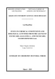
While studying the crosslinking of human replicative DNA polymerases with newly synthesized nucleic acids, we found that Pol e-crosslinked nascent RNA not rela- ted to DNA replication. This hinted at a possible association of Pol e with the transcription apparatus. To investigate this possibility, we performed reciprocal immunoprecipitations with antibodies against RNA pol II and the three replicative Pols (a, d and e). Preci- pitates were subjected to extensive washing at physiolo- gical salt concentration. The immunocomplexes were collected and analyzed by immunoblotting with anti- bodies against the cognate proteins (see Table 2, in Experimental procedures). A polyclonal antibody against RNA pol II coprecipitated Pol e, but not Pols a and d (Fig. 1). Reciprocal immunoprecipitation with antibodies against the replicative Pols a, d and e indicated that RNA pol II was co-immunoprecipitated only by the monoclonal antibody against Pol e. Immu- noprecipitations utilizing polyclonal antiserum K27, raised against a different region of Pol e, gave identical results (data not shown). We also excluded the possibil- ity that the RNA pol II–Pol e interaction was mediated by the presence of DNA, by performing the experi- ments also in the presence of ethidium bromide at concentrations known to disrupt protein–DNA interac- tions [28]. It is also noteworthy that no interaction between replicative pols could be observed under the moderately stringent employed in our experiments.
Pol e interacts with the hyperphosphorylated elongating isoform RNA pol IIO
Transcription factors may be involved in regulating DNA replication in eukaryotic cells indirectly, by modifying chromatin structure, or directly, by recruit- ing protein complexes [15,16]. Their effects are also reflected by a global co-ordination of cellular tran- scription and replication. In fact, the more transcrip- tionally active a chromosomal region, the greater the likelihood that replication initiates early in S phase within that domain [17]. Studies in yeast suggest that recruitment of the RNA pol II complex activates replication [18] and that the CTD is sufficient for this positive regulation of DNA replication initiation [19]. On the other hand, DNA replication and repair factors, such as MCM5, BRCA1 and Ku, have been implicated as positive or negative regulators of tran- scription [8,13,20]. Transcription has also been shown to induce homologous recombination [21].
RNA pol II exists in a dynamic equilibrium between the hypophosphorylated initiation isoform IIA and the hyperphosphorylated elongating isoform IIO that exhibits reduced electrophoretic mobility compared with the IIA isoform. Whereas the polyclonal anti- RNA pol II antibody precipitated both the fast and slowly migrating forms of RNA pol II, antibodies to Pol e coprecipitated only the slowly migrating form of RNA pol II (Fig. 1). This suggested that Pol e associ- ates with the RNA pol IIO isoform. To characterize further the phosphorylation status of the RNA pol II
Pol e is an essential replication protein involved in many cellular transactions [22]. Pol e, together with Pols a and d, is required for synthesizing the bulk of DNA during replication in mammalian cells [23,24], but its specific role in this process remains uncertain. In addition, Pol e is implicated in DNA repair and cell cycle regulation [22,25–27].
FEBS Journal 273 (2006) 5535–5549 ª 2006 The Authors Journal compilation ª 2006 FEBS
5536
DNA polymerase e associates with RNA polymerase II
A. K. Rytko¨ nen et al.
Ser2, but may or may not have phosphate at Ser5. H5 is specific for RNA pol II having phosphate at Ser2, and is considered a marker for elongating RNA pol II [31]. The form of RNA pol II coprecipitating with Pol e was recognized by all antibodies to be IIO, but not IIA (Fig. 2B). Conversely, all different RNA pol II antibodies always coprecipitated Pol e. The results indicate that the interaction does not depend on phos- phorylation of Ser2, because Pol e is coprecipitated by antibody 8WG16. Alternatively, antibody 8WG16 could have precipitated incompletely phosphorylated RNA pol II. Phosphorylation of Ser5 alone is suffi- cient for the interaction. The results do not exclude the possibility that phosphorylation of Ser2 alone would also be sufficient for the interaction.
Fig. 1. RNA polymerase (pol) II and DNA polymerase (Pol) e co-immunoprecipitate and copurify. Pol e, but not Pol a and Pol d, co-immunoprecipitates with RNA pol II, and RNA pol IIO co-immu- noprecipitates with Pol e. Immunoprecipitations were performed with antibody to RNA pol II (N20), antibody to Pol a (SJK 132–20), antibody to Pol d (K32), antibody to Pol e (GIA) or control antibody (mouse IgG). Immunopreciptitation (IP) and western blot analysis (WB) were performed as described in Experimental procedures. Where indicated, immunoprecipitations were performed in the presence of 50 lgÆmL)1 ethidium bromide (EtBr) to exclude interac- tions mediated by DNA. Hyperphosphorylated RNA pol IIO is indica- ted with a solid arrow and hypophosphorylated RNA pol IIA is indicated with an open arrow.
(DRB)
complexed with Pol e, we examined the effect of phosphatase on the precipitated RNA pol II. Immuno- precipitates were incubated at +37 (cid:2)C, in the presence intestinal phosphatase, prior to or absence of calf western analysis (Fig. 2A). Following phosphatase treatment, the cellular fraction of RNA pol II that associated with Pol e showed increased mobility, cor- responding to that of isoform IIA. The isoform IIO was also lost from RNA pol II precipitates after phos- phatase treatment. Thus, Pol e could interact specific- ally with hyperphosphorylated isoform RNA pol IIO, possibly during transcriptional elongation. Therefore, we studied this interaction more closely during the transcription cycle.
In order to study the interaction with RNA pol II and Pol e in more detail, we employed chemicals known to inhibit transcription. Whereas a-amanitin inhibits transcription both in the initiation and elonga- tion phases by preventing translocation of DNA and RNA through the enzyme [32,33], the cdk inhibitors 5,6-dichloro-1-beta-d-ribobenzimidazole and roscovitine inhibit phosphorylation of the CTD and thereby prevent transition from the initiation to the elongation complex [34,35]. RNA pol II complexes, already engaged in elongation, are not affected. As expected, treatment of the cells with DRB or roscovi- tine strongly decreased the hyperphosphorylation of RNA pol II in the whole-cell extract and in RNA pol II immunoprecipitate samples, because mainly the IIA form can be detected (Fig. 2C). Treatment with DRB or roscovitine also significantly decreased the amount of RNA pol II co-immunoprecipitating with Pol e (Fig. 2C). In contrast, a-amanitin did not affect the hyperphosphorylation of RNA pol II, and had no effect on the interaction between RNA pol II and Pol e (Fig. 2C). These results confirm that Pol e associ- specifically with elongation-competent RNA ates pol IIO. Moreover, this association persists, even when transcription is stalled.
Pol e associates with nascent RNA
RNA Pol II is sequentially phosphorylated during the transcription cycle, mainly by cyclin-dependent kinase activities. During transcription initiation, serine 5 residues of the CTD repeat become mainly phos- phorylated by Cdk7, a subunit of TFIIH [29,30]. Sub- sequent phosphorylation on serines at position 2 by the Cdk9 ⁄ pTEFb is required for transcription elonga- tion. We employed phospho-specific antibodies for immunoprecipitation and western blot analysis to study whether the Pol e interaction is specific in terms of CTD phosphorylation of RNA pol II. Polyclonal antibody (N20) recognizes both hyperphosphorylated IIO and hypophosphorylated IIA forms of RNA pol II. H14 antibody is specific for early stage RNA pol II phosphorylated at Ser5, and 8WG16 antibody recognizes RNA pol II that is not phosphorylated at
We employed a UV crosslinking technique to study the association of Pol e with nascent nucleic acids. This method is a modification of the polymerase trap tech- nique used to link newly synthesized DNA covalently to the synthesizing Pol or to various replication proteins intimately interacting with the nascent DNA [23,36,37]. We labelled permeabilized HeLa cells with either radio- active UTP or dATP in the presence of the photoreac- tive DNA precursor bromodeoxyuridine triphosphate (BrdUTP), and purified respective protein–nucleotide
FEBS Journal 273 (2006) 5535–5549 ª 2006 The Authors Journal compilation ª 2006 FEBS
5537
DNA polymerase e associates with RNA polymerase II
A. K. Rytko¨ nen et al.
A
B
C
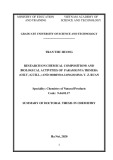
replication in mammalian cells [23]. Pol a and Pol e, but not Pol d, were labelled also with UTP. The label- ling of Pol a by UTP was expected because this enzyme synthesizes continuously RNA–DNA hybrid primers required for the initiation of the continuous strand and production of Okazaki fragments. Whether Pol e could also crosslink RNA-primed nascent DNA was not known. To find out, we compared the labelling of Pol a and Pol e by UTP using a panel of controls (Fig. 3B). In one control, BrdUTP, which facilitates the crosslink- ing to the DNA moiety of RNA-primed nascent DNA, was omitted. In a second control, DNA replication was inhibited by omitting dNTPs from the reaction mixture and including in it aphidicolin, an inhibitor of the repli- cative Pols [38]. In the last control, transcription was inhibited with a-amanitin. As expected, the labelling of Pol a by UTP was abolished when BrdUTP was omit- ted or replication inhibited, whereas a-amanitin had no effect. These results confirmed that the labelling of Pol a by UTP was linked to DNA replication, specific- ally, the synthesis of RNA–DNA primers. In contrast, Pol e labelling by radioactive UTP was not affected when the DNA crosslinker was omitted or DNA syn- thesis inhibited, but was abolished by the transcription inhibitor a-amanitin, which prevents incorporation of radioactive ATP in the polymerase trap reaction. It is noteworthy that the photolabelling of Pol e with incor- porated radioactive dATP absolutely depends on the presence of BrdUTP [23]. Thus, both Pols a and e could be UV crosslinked to newly synthesized RNA, but the RNA associated with Pol a primed DNA replication, whereas that associated with Pol e represented nascent RNA transcripts.
Pol e and RNA pol IIO colocalize in the nucleus
Fig. 2. DNA polymerase (Pol) e interacts with hyperphosphorylated RNA polymerase (pol) II. (A) Pol e co-immunoprecipitates the hyper- phosphorylated form RNA pol IIO. Immunoprecipitations were per- formed with antibody to RNA Pol II (N20), antibody to Pol e (GIA) or control antibody (mouse IgG). Precipitates were treated with calf intestinal phosphatase (CIP) where indicated and RNA pol II was detected as described in Experimental procedures. Whole-cell extract (WCE) represents 5–10% of the input. (B) Characterization of the carboxyterminal domain phosphorylation status of RNA pol II co-immunoprecipitated with Pol e. Immunoprecipitations and west- ern blot analyses were performed with the indicated phosphospe- cific RNA pol II antibodies, anti-Pol e antibody (GIA) or control antibody (mouse IgG), as described in Experimental procedures. The phosphorylation status of the heptapeptide repeat is indicated on the right. (C) Effects of the kinase inhibitors 5,6-dichloro-1-beta- D-ribobenzimidazole (DRB) and roscovitine, and of the transcription inhibitor, a-amanitin, on RNA pol II co-immunoprecipitation with Pol e. Cells were treated with the indicated inhibitors 1.5 h prior to preparation of cell extract, as described in Experimental proce- dures. Immunoprecipitations and western blot analyses were per- formed with antibody to RNA Pol II (N20), antibody to Pol e (GIA) or control antibody (mouse IgG), as in Fig. 1A. Whole-cell extract (WCE) represents 5–10% of the input. In all panels, hyperphosphor- ylated RNA pol IIO is indicated with a solid arrow and hypophos- phorylated RNA pol IIA with an open arrow.
complexes after UV crosslinking by immunoprecipita- tion with antibodies against Pol a, Pol d or Pol e. All three pols were labelled with dATP (Fig. 3A), as expec- three Pols are required for DNA ted, because all
Immunofluorescence and immunoelectron microscopy were next performed to study if the association of RNA pol IIO and Pol e is also reflected in their cellular local- ization. T98G cells were stained by indirect immunoflu- orescence, with antibody H5 recognizing RNA pol IIO and with Pol e antibody G1A. T98G cells showed quite even RNA pol IIO staining, with some cells showing very few distinct foci. Pol e localized to numerous foci (data not shown). These staining patterns correspond well to previous reports [39,40]. RNA pol II foci became more prominent after hypotonic permeabilization of cells and removal of the bulk DNA by restriction diges- tion prior to fixation (Fig. 4A). Most RNA pol IIO foci, detectable after this treatment, were found to colocalize or overlap with Pol e foci. As Pol e foci were by far more abundant, only few Pol e foci colocalized with RNA pol IIO.
FEBS Journal 273 (2006) 5535–5549 ª 2006 The Authors Journal compilation ª 2006 FEBS
5538
DNA polymerase e associates with RNA polymerase II
A. K. Rytko¨ nen et al.
A
(H3B)
B
Fig. 3. UV crosslinking of replicative DNA polymerases to nascent DNA or RNA chains. (A) DNA polymerases (Pols) a and e, but not Pol d, are photolabelled with radioactive UTP. HeLa cells were per- meabilized, incubated in the presence of radioactive UTP or dATP and the photoreactive DNA precursor BrdUTP, and subjected to UV crosslinking. The photolabelled derivatives of Pols a, d and e were then resolved by SDS ⁄ PAGE, as described in Experimental proce- dures. (B) The photolabelling of Pol a or Pol e with radioactive UTP depends on DNA replication or RNA transcription, respectively. UV crosslinking of the Pols to nascent RNA labelled from radioactive UTP in permeabilized HeLa cells was performed as described above (complete), without BrdUTP (no BrdUTP), without dNTP pre- cursors and with aphidicolin (+ aph, no dNTP) or in the presence of a-amanitin (a-amanitin). The photolabelled derivatives of Pols a and e were then resolved by SDS ⁄ PAGE and monitored as described in Experimental procedures.
ring-shaped aggregates of gold particles of (cid:2) 50– 100 nm diameter (Fig. 4B). Staining with antibody G1A is specific for Pol e, because it colocalizes exten- sively with antibody to Pol e in human fibroblasts (data not shown). The two antibodies recog- nize different epitopes of Pol e [41]. Quantitative analy- sis of staining series derived from two independent synchronizations indicate that, in general, 52% of Pol e and 66% of RNA pol IIO localizes to centres contain- ing at least three gold particles (Table 1). Pol e and RNA pol IIO demonstrated striking colocalization (Fig. 4B). Most of the centres identified contained both proteins, although centres containing only RNA pol IIO or Pol e were also present (see Fig. 4B, right). Micrographs from 64 nuclei were scored for the pres- ence of centres. Fifty-two per cent of all centres identi- fied contained both proteins, 23% contained only Pol e and 25% only RNA pol IIO. We did not detect any apparent differences in the localization pattern of the two proteins at the different time points investigated, indicating that colocalization is not limited to a specific cell cycle stage. We repeated immunoelectron microsco- py staining using RNA pol II antibody N20, which recognizes both the IIA and the IIO form. Although less RNA pol II appears to be recognized by the anti- body in this application, the focal colocalization of RNA pol II and Pol e remained apparent (Fig. 4C). Although only 45% of RNA pol II detected was in cen- tres, still 37% colocalized with Pol e (Table 1, Fig. 4C), confirming the authenticity of the observed colocaliza- tion. The reduced focal staining of RNA pol II by anti- body N20 compared with antibody H5 could reveal the functional difference between total RNA pol II and iso- form IIO, but may also simply reflect the apparently lower affinity of antibody N20 in the application. Taken together, the cell stainings indicate extensive, although not complete, colocalization of Pol e with RNA pol IIO at near-molecular level, as could be expected from the physical interaction studies.
The RNA pol IIO–Pol e interaction is not linked to nucleotide excision repair
For immunoelectron microscopy, T98G cells were serum deprived followed by serum stimulation, and cells were collected at different time points, ranging from 4 h (G0 ⁄ G1) to 22 h (late S ⁄ G2 phase) after serum stimulation, as determined by flow cytometry analysis of cultures (see supplementary Figs S1 and S2 for syn- chronization profiles). Cells were subjected to successive staining for Pol e (antibody G1A) and RNA pol IIO (antibody H5), using antibodies conjugated indirectly to gold particles of 5 and 10 nm, respectively. Both Pol e and RNA pol IIO localized predominantly to distinct sites or centres, represented by irregular or
TFIIH and Pol e are both components of an RNA pol II holoenzyme, and both have been implicated in nucleotide excision repair [25,26,42]. In addition, RNA Pol IIO has an important function in the transcription coupled repair pathway of nucleotide excision repair [43]. When the transcription apparatus encounters dam- age on the transcribed strand, nucleotide excision repair is performed, after which transcription can be resumed. Therefore, we studied whether the interaction between RNA pol IIO and Pol e could be linked to nucleotide
FEBS Journal 273 (2006) 5535–5549 ª 2006 The Authors Journal compilation ª 2006 FEBS
5539
DNA polymerase e associates with RNA polymerase II
A. K. Rytko¨ nen et al.
Fig. 4. Pol e and RNA pol II colocalize in the nucleus. (A) Immunofluorescent staining of T98G cell nuclei with a-RNA pol II H5 (red) and a-Pol e G1A (green). Yellow indicates colocalization in the merge image. Cells were permeabilized in hypotonic buffer in the presence of detergent, and DNA was digested to enhance visibility of RNA pol II foci. (B,C) A significant fraction of RNA pol II and Pol e colocalize in immunoelectron microscopy. Ultrathin cryosections of human T98G cells were subjected to immunostaining of Pol e (antibody G1A) followed by staining of RNA pol IIO (antibody H5) (B) or total RNA pol II (antibody N20) (C), as described in Experimental procedures. The antibodies were indirectly coupled to 5 nm and 10 nm gold particles, as indicated. Images represent serum-deprived cells 4 h (G1 phase; panel B left), 10 h (G1 ⁄ S phase; panel C left) 12 h (early S phase; panel B middle) and 16 h (mid S phase; panel B right) after serum stimulation or syn- chronization to the G1 ⁄ S boundary with mimosine (G1 ⁄ S phase; panel C right). No apparent differences were detected in the staining pattern at different time points. The scale bar is 100 nm.
excision repair. Staining patterns of Pol e and RNA pol IIO appeared unchanged after uniform UV expo- sure of T98G cells (data not shown). In order to evalu- ate changes in staining intensities caused by UV exposure, cells were UV irradiated through an 8 lm filter to cause local UV damage [44]. The sites of UV damage inside nuclei were visualized by indirect immu- nofluorescence staining, with antibody H3 recognizing cyclobutane thymidine dimers. As shown previously [45], nucleotide excision repair factor XP-B relocalized to damaged areas, where it gave a uniform staining
(Fig. 5, upper panel). Staining with an antibody against nucleotide excision repair factor XP-A gave a compar- able staining pattern (data not shown). Pol e detected with antibody G1A showed the punctuate staining typ- ical for nondamaged cells also at the damaged areas (Fig. 5, middle panel). We noticed a small, but very reproducible, increase in the staining intensity of Pol e in almost all UV irradiated areas compared to nonirra- diated areas of the same nuclei. The staining intensity of RNA pol IIO detected with antibody H5 in UV exposed decreased area
remained
appeared
constant
or
FEBS Journal 273 (2006) 5535–5549 ª 2006 The Authors Journal compilation ª 2006 FEBS
5540
DNA polymerase e associates with RNA polymerase II
A. K. Rytko¨ nen et al.
Table 1. Comparison of colocalization and focal staining of Pol e and RNA pol II by immunoelectron microscopy. The number of gold parti- cles representing Pol e (5 nm) and RNA pol II (10 nm), and the fraction of particles colocalizing and in centres, were determined in double stainings. Clusters of three or more gold particles were considered as centres. The table represents the summary of the analyses of sam- ples from G0 until late S phase (0–22 h after serum stimulation) derived from serum-deprived T98G cells stimulated to proliferate. Typically, 20 separate images, representing seven to nine nuclei, were scored for each sample. Eight samples from two independent synchronizations were stained for Pol e (antibody G1A) and transcriptionally active RNA pol II phosphorylated at Ser2 (antibody H5), and four samples derived from one synchronization were analysed for staining of Pol e (antibody G1A) and all forms of RNA pol II (antibody N20). No difference in focal staining and colocalization was detected at different time points.
Staining
Particles counted
Particles in centres
Particles colocalizing
Nuclei counted
Images counted
RNA Pol II Ser2-P
64
158
All RNA Pol II
33
80
Pol e RNA Pol II Pol e RNA Pol II
2519 1610 1133 177
1312 (52%) 1058 (66%) 680 (60%) 80 (45%)
976 (39%) 656 (41%) 192 (17%) 66 (37%)
Discussion
compared to the control area of the same nuclei (Fig. 5 lower panel). Overall, the staining intensities of RNA pol II and Pol e do not correlate, and there is only a very minor effect of local UV damage on the nuclear localization of RNA pol II and Pol e.
The RNA pol II–Pol e interaction does not depend on DNA replication
We describe here an extensive interaction of RNA pol II with Pol e. Our observation is based on detailed analysis of co-immunoprecipitation using multiple anti- bodies against the cognate proteins. This interaction appears to be specific for Pol e, because Pols a and d were absent from RNA pol II immunoprecipitates con- taining Pol e, RNA pol II was not precipitated with Pols a or d. What is more, the interaction is limited to the hyperphosphorylated RNA pol IIO, based on elec- trophoretic shift, treatment of immunoprecipitates with phosphatase and use of antibodies specific for differen- tially phosphorylated forms of the CTD.
(see
Maldonado et al. [5] reported the purification of an RNA pol II holoenzyme complex that also comprised several factors involved in DNA repair and recombina- including Pol e. The latter complex contained tion, included mainly hypophosphorylated RNA pol IIA, several general transcription factors and several sub- units of human Mediator and was capable of tran- scribing from a model promoter in vitro with minimal transcription factors, consistent addition of general with a transcription initiation complex [30]. The pres- ence of DNA repair or recombination factors in puri- fied RNA pol II has been found to depend strongly on the purification strategy employed. We see a striking results compared with those of difference in our Maldonado et al. [5], because the interaction described here correlated strictly with phosphorylation of the CTD. In support of this view, chemical kinase inhibi- tors that prevent phosphorylation-dependent transition from the initiation to the elongation of transcription strongly reduced the interaction observed by us. How- ever, the initiation and elongation inhibitor a-amanitin had no effect on this interaction, suggesting that it per- sists also when transcription stalls. What is more, we observed that part of the hyperphosphorylated RNA
Pol e is one of the three major replicases in the eukary- [22]. Therefore, we tested the effect of the otic cell replicative state of the cell on the interaction between Pol e and RNA pol II. A possible confinement of the interaction to late G1 and S phases (the cell cycle sta- ges dedicated to initiation and execution of DNA replication) would have strong implications on a func- tional interplay between transcription and DNA repli- cation. We therefore examined the interaction between RNA pol II and Pol e during the cell cycle. T98G cells were serum deprived, followed by stimulation to proliferate, to create samples at G0 ⁄ G1 (4 h), late G1 (10 h), early S (14 h), and late S ⁄ G2 (24 h) phases of the cell cycle, and T98G cells were blocked at the G1 ⁄ S boundary by mimosine supplementary Fig. S1 for flow cytometric cell cycle analysis). RNA Pol IIO and Pol e co-immunoprecipitated with com- parable efficiencies throughout the cell cycle (Fig. 6A). Although minor differences in the interaction may not be revealed by changes in immunoprecipitation, these results demonstrated that the interaction is not con- fined to a specific stage of the cell cycle. This is consis- tent with immunoelectron microscopy, revealing that RNA Pol II and Pol e colocalize throughout the cell cycle. What is more, the interaction prevailed in the presence of aphidicolin (Fig. 6B). Although our data do not address potential effects of the observed inter- action during DNA replication, they suggest that the interaction is not dependent on DNA replication.
FEBS Journal 273 (2006) 5535–5549 ª 2006 The Authors Journal compilation ª 2006 FEBS
5541
DNA polymerase e associates with RNA polymerase II
A. K. Rytko¨ nen et al.
Fig. 5. Pol e and RNA pol II do not relocalize to a nuclear site of UV exposure. T98G cells were UV irradiated at 60 JÆm)2 through an 8 lm pore polycarbonate filter. Cells were fixed 1 h after UV exposure and subjected to immunofluorescence staining. DNA staining (blue) shows positions of nuclei. The UV damage marker XP-B (S-19) localizes, as expected, to sites of UV damage visualized by an antibody specific to cyclobutane pyrimidine dimers (CPD) (upper panel), as opposed to Pol e (visualized by antibody G1A; middle panel) and RNA pol IIO (antibody H5; lower panel).
Pol e, it is tempting to speculate that the interaction may be mediated by the CTD, in particular when phosphorylated at the repeated serine 5 residues, although such an interaction could be mediated by another protein, or other regions of RNA pol II.
pol IIO form copurified during the three conventional column steps of the standard Pol e purification [46] (data not shown). Finally, Pol e could be UV cross- linked to newly synthesized a-amanitin-sensitive RNA, indicating close association of Pol e with nascent RNA pol II transcripts. All these results are consistent with the view that Pol e interacts with elongating RNA pol IIO.
excision repair
The CTD of RNA pol II has a central role in the co-ordination of the transcription cycle because it is capable of recruiting several factors involved in tran- scription and mRNA processing to the transcription apparatus [4]. The differential phosphorylation of the CTD provides means of discrimination during the transcription cycle. As we found that hyperphosphory- lation of the CTD correlates with the interaction with
Bulky DNA lesions block the passage of elonga- thereby stalling the transcription ting RNA pol II, [43]. To avoid consequent detrimental apparatus effects, nucleotide is preferentially directed to the impaired DNA template by the stalled transcription apparatus in a process called transcription-coupled repair. As Pol e, together with Pol d, are implicated in the DNA synthesis step of nucleotide excision repair [25,26], one could antici- pate that the interaction between Pol e and RNA pol IIO would facilitate transcription-coupled repair.
FEBS Journal 273 (2006) 5535–5549 ª 2006 The Authors Journal compilation ª 2006 FEBS
5542
DNA polymerase e associates with RNA polymerase II
A. K. Rytko¨ nen et al.
A
limited to between Pol e and RNA pol II is not S phase, indicating that it does not depend on DNA replication. This is also reflected by the fact that the replication inhibitor, aphidicolin, does not affect the interaction. What is more, the extent of interaction and ultrastructural colocalization observed between the two proteins is difficult to reconcile with such a specific function as the passing of colliding replica- tion and transcription apparatus.
B
the
Fig. 6. The interaction between RNA pol II and Pol e does not depend on DNA replication. (A) Pol e and RNA pol II co-immunopre- cipitate throughout the cell cycle. T98G cells were synchronized by serum deprivation and released, or blocked with mimosine, to obtain samples from different stages of the cell cycle. Immunoprec- ipitations were performed with antibody to RNA pol II (N20), anti- body to Pol e (GIA) or control antibody (mouse IgG), and analysed by western blot, as described in Experimental procedures. Whole- cell extract (WCE) represents 5–10% of the input. (B) Inhibition of DNA replication by aphidicolin does not affect the co-immunoprecip- itation of Pol e and RNA pol II. Cells were treated with aphidicolin for 1.5 h before preparation of the extract. Immunoprecipitations were performed and analysed as above. Hyperphosphorylated RNA Pol IIO is indicated with a solid arrow and hypophosphorylated RNA Pol IIA with an open arrow.
There are several direct links between transcription and DNA replication. Adolph et al. [50] showed that a-amanitin blocks cells specifically in the G1 phase of the cell cycle. This effect could be attributed to inhi- bition of transcription of cell cycle-regulated genes at this phase of the cell cycle [51]. Nevertheless, it was shown that cell cycle block by a-amanitin at the ori- gin decision point in G1 phase does not depend on protein synthesis of gene products [52]. It has been long known that actively transcribed genes are repli- cated early, and it has been proposed that the posit- ive regulation of DNA replication is a direct effect of the transcription apparatus, or is caused by remodel- ling of chromatin mediated by transcription factors [16,19,53–55]. It is also worth considering that the RNA pol IIO–Pol e interaction may help in load- replication origins within transcribed ing Pol e at domains. Links between transcription and DNA repli- cation have also been supported by ultrastructural studies. Sites of replication and transcription colocal- ized in human cells when studied by confocal micros- copy [56]. Using high-resolution electron microscopy, centres of DNA replication were found to contain also RNA processing components [57]. In our study, RNA pol IIO and Pol e displayed a partly dispersed staining with numerous foci in conventional immuno- fluorescence microscopy, but colocalization of the proteins became more prominent after hypotonic extraction and removal of part of the DNA. In con- trast, using cryo-electron microscopy without such manipulations, we found both proteins to colocalize extensively and to cluster into centres in the vicinity of small electron-dense domains of the nucleus. The RNA pol II staining corresponds well to previous ul- trastructural studies, where RNA pol II was concen- trated into clusters that overlapped with the sites of RNA synthesis [58,59].
Notwithstanding this expectation, the observed RNA pol IIO–Pol e interaction did not depend on, or was augmented in response to, DNA damage. This was most clearly demonstrated by the failure of the pro- tein to relocalize to a defined UV-irradiated nuclear area (Fig. 5). Although Sarker et al. [47] described recently an interaction between hyperphosphorylated RNA pol II and XP-G in undamaged cells, we were not able to detect XP-G, XP-A or XP-B in our Pol e or RNA pol II immunoprecipitates (data not shown), supporting the view of a sequential assembly of nucleotide excision repair factor at the sites of DNA damage [45]. Although we cannot exclude beneficial effects of the observed interaction on tran- scription-coupled repair, there is no indication that such repair represents its predominant function.
In theory, an interaction between RNA pol IIO and Pol e could be important for the nondisruptive bypass of a transcription bubble by a replication fork, as has been reported previously, for example, interaction in bacteria
[48,49]. Nevertheless,
the
Another example for prominent replication factors that interact with RNA pol II are the MCM proteins [10]. MCM proteins can be detected in RNA pol II holoenzyme affinity-purified against elongation factor TFIIS, but not in holoenzyme purified by anti-cdk7 [10,14]. Furthermore, the TFIIS holoenzyme form appears to be more abundant in the G1 ⁄ S phase of the
FEBS Journal 273 (2006) 5535–5549 ª 2006 The Authors Journal compilation ª 2006 FEBS
5543
DNA polymerase e associates with RNA polymerase II
A. K. Rytko¨ nen et al.
Experimental procedures
Antibodies
cell cycle. Antibodies against Mcm2 inhibited tran- scription, and, conversely, the yeast CTD mutants interacted genetically with mcm5 mutants and showed adverse effects on minichromosome maintenance and DNA replication [19].
297–542 (swissprot entry P28340)
secondary antibodies
Primary antibodies used are listed in Table 2. Rabbit poly- clonal antibody K32, against the human Pol d catalytic subunit, was raised against a peptide corresponding to amino acids and expressed as glutathione S-transferase fusion protein, as described previously [24]. Purified mouse IgG was pur- chased from Pierce (Rockford, IL, USA), rabbit anti-mouse IgG (Zymed, San Francisco, CA, USA) and rabbit anti-rat IgG (Jackson Immunoresearch, West Grove, PA, USA) in immunoelectron were used as microscopy, and horseradish peroxidase-conjugated anti- bodies (Jackson Immunoresearch or Chemicon, Chandlers Ford, UK) were used as secondary antibodies in western blotting.
Whereas other DNA repair and replication factors interacting with RNA pol II appear to regulate tran- scription positively [6,10,13], yeast Pol e has been implicated in transcriptional silencing of ribosomal DNA, the mating type locus and subtelomeric regions [60–62]. The B subunit of mouse Pol e interacts with SAP18, which is known to associate with the transcrip- tional corepressor, Sin3 [63]. Sin3 is a component of a protein complex possessing histone deacetylase activity, which interacts with several transcriptional corepres- sors and functions in transcriptional silencing [64]. Thus, a possible role of Pol e in transcription attenu- ation or silencing remains to be investigated.
Cell culture, synchronization and UV irradiation
HeLa CCL2 monolayer cells [American Type Culture Collection (ATCC), Manassas, VA, USA] were cultured DMEM (Invitrogen, Paisley, UK) containing 10% fetal bovine serum and antibiotics, at 37 (cid:2)C in a 5% carbon dioxide atmosphere. T89G cells (ATCC) were grown in
The physical and structural association between RNA pol II and Pol e presented here provide a direct link between central components of the transcription functional and DNA replication apparati. Further characterization of this interaction may provide valu- able insight into the crosstalk between transcription and DNA replication.
Table 2. Primary antibodies used in this study. IEM, immunoelectron microscopy; IF, immunofluorescence microscopy; IP, immunoprecipita- tion; Pol, DNA polymerase; pol, polymerase; WB, western blot; X-LINK, UV crosslinking.
Clone
Application
Reference
Target
Species ⁄ type
1Ct102, 2Ct25 SJK-132–20
Mouse monoclonal Mouse monoclonal
WB IP
Pol a catalytic subunit
Pol d catalytic subunit
Pol e catalytic subunit
p140 PDG-1E8 K30 K32 G1A H3B E24C K27 N20
Rabbit polyclonal Rat monoclonal Rabbit polyclonal Rabbit polyclonal Mouse monoclonal Mouse monoclonal Mouse monoclonal Rabbit polyclonal Rabbit polyclonal
X-LINK WB X-LINK, WB IP IP, X-LINK, WB, IEM IF, WB WB IP IP, WB, IF, IEM
RNA pol II
H5
Mouse monoclonal, IgM
IP, WB, IF, IEM
H14 8WG16 12F5 S-19
Mouse monoclonal IgM Mouse monoclonal IgG2a Mouse monoclonal Rabbit polyclonal
IP, WB IP, WB IP IF
XP-A XP-B
8H7
Mouse monoclonal
IP
XP-G
H3
mouse monoclonal
IF
[67] ATCC CRL-1640 [68] [69] This studya [70] This study [41] [41] [41] [71] Santa Cruz Biotechnologies Covance Research Products Nordic Biosite Nordic Biosite NeoMarkers Santa Cruz Biotechnologies Santa Cruz Biotechnologies Affitech
Cyclobutane pyrimidine dimers
a Chen S, Kremmer E, Weisshart K, Hubscher U & Nasheuer H-P, unpublished results.
FEBS Journal 273 (2006) 5535–5549 ª 2006 The Authors Journal compilation ª 2006 FEBS
5544
DNA polymerase e associates with RNA polymerase II
A. K. Rytko¨ nen et al.
UV crosslinking
amino glutamax, nonessential
(30 mm Hepes-KOH, pH 7.5,
following one the of
Eagle’s minimum essential medium (Sigma-Aldrich Fin- supplemented with 10% fetal land, Helsinki, Finland) bovine acids, serum, sodium pyruvate and antibiotics, at 37 (cid:2)C in 5% CO2. T98G cells were synchronized by arresting them at the G0 phase for 3–6 days using starvation medium (growth medium containing 0.5% serum). Cells were stimulated to grow by addition of equal volumes of complete and bal- anced growth medium. The efficacy of the synchroniza- tion was demonstrated by fluorescence cytometry analysis (Becton Dickinson, Helsinki, Finland) of propidium iod- ide-stained cells [65] in parallel cell cultures. Where indi- cated, cells were cultured in the presence of the indicated inhibitors: concentrations of 20 lgÆmL)1 a-amanitin, 40 lm roscovitine, 50 lm DRB or 5 lgÆmL)1 aphidicolin (all Sigma-Aldrich Finland). These inhibitors were added for 1.5 h before preparing the cell extracts.
Immunoprecipitation
(100 UÆmL)1)
times with washing buffer
UV crosslinking of proteins to nascent DNA ⁄ RNA in a monolayer of isolated nuclei was performed, as described previously [23], with minor modifications. Cells were washed with buffer KM (10 mm Mops-NaOH, pH 7.0, 10 mm NaCl, 1 mm MgCl2, 2 mm dithiothreitol, 1 · com- pleteTM protease inhibitor) and lysed by incubation for 30 min on ice with buffer KM containing 0.5% Nonidet P-40, resulting in a monolayer of isolated nuclei. The nuclear monolayer was then washed twice with buffer 5 mm K-acetate, KAc 0.5 mm MgCl2, 2 mm dithiothreitol, 1 · completeTM pro- tease inhibitor). The reaction mixture for UTP labelling contained 30 mm Hepes-KOH, pH 7.8, 50 mm K-acetate, 5 mm MgCl2, 2 mm dithiothreitol, 0.05% Nonidet P-40, 2 lm each of dATP, dGTP and dCTP, 20 lm BrdUTP, 1 lm [32P]UTP[aP] (specific activity 410 CiÆmmol)1), 2 mm ATP and 10 lm CTP and GTP. a-amanitin and aphidic- olin were used at 50 lgÆmL)1. Each 6 cm plate received 400 lL of reaction mixture and the reactions proceeded for 2.5 min at 30 (cid:2)C. The mixture was used for a total of three successive plates. After labelling, the nuclei were washed with buffer KAc and UV irradiated with a stand- ard UV transilluminator for 6 min. The irradiated nuclei were treated with HindIII and PstI for 30 min at 37 (cid:2)C to remove bulk DNA. Protein–nucleic acid complexes were extracted with phenol, precipitated with acetone, collected at 0 (cid:2)C and washed three times. The dried protein–nucleic acid pellet was resuspended by boiling in denaturation buffer (50 mm Tris ⁄ HCl, pH 7.5, 0.5% SDS, 70 mm b-mercaptoethanol) and renaturated (50 mm Tris ⁄ HCl, pH 7.5, 150 mm NaCl, 0.5% Nonidet P-40, 1 · CompleteTM protease inhibitors). Proteins were then ready for immunoprecipitation. For immunoprecipi- tations, 7 mg of protein A– or G–Sepharose and 3 lL of p140 serum, 4 lL of K30 serum and 8 lg of purified GIA antibody were used to precipitate, in parallel, Pols a, d and e, respectively. Immunoprecipitates were washed (50 mm Tris ⁄ HCl, three pH 7.5, 150 mm NaCl, 0.5% Nonidet P-40). Samples eluted with SDS loading buffer and separated were through 6% SDS ⁄ PAGE, transferred to poly(vinylidene difluoride) membrane and exposed to BioMax MS film (Kodak, Vantaa, Finland). After autoradiography, pro- teins were detected using enhanced chemiluminescence reagents (Pierce). Antibodies 1Ct102 and 2Ct25, PDG- 1E8, and a combination of G1A, H3B and E24C were used for detection of Pols a, d and e, respectively.
HeLa cells were washed with NaCl ⁄ Pi and scraped into lysis buffer [100 mm Tris ⁄ HCl, pH 7.5, 80 mm NaCl, 10% glycerol, 0.1% Nonidet P-40, CompleteTM protease inhibi- tors (Roche, Espo, Finland), 10 mm Na3VO4 and 10 mm NaF]. The cell extract from synchronized T98G cells used for analyzing protein–protein interactions during the cell cycle was prepared in lysis buffer II (50 mm Tris ⁄ HCl pH 7.5, 100 mm KCl, 5% glycerol, 0.1% Nonidet P-40, CompleteTM protease inhibitor, 1 mm EDTA, 10 mm NaF, 2 mm Na3VO4). Cells were broken by sonication (2 · 15 s) and the extract was further incubated on ice for 15 min and centrifuged in an Eppendorf 5415 table centrifuge (Eppendorf AG, Hamburg, Germany; 10 min, 20 000 g, 4 (cid:2)C). For immunoprecipitations, 0.2–3 mg of cell extract and 2–4 lg of antibody was used per sample. After pre- clearing, the antibody–protein complex was allowed to form overnight at 4 (cid:2)C. Rabbit anti-mouse IgM linkers were added in H14 and H5 immunoprecipitations to facili- tate collection of mouse IgM on protein G–Sepharose. Ten microlitres of GammaBind protein G–Sepharose (Amer- sham Biosciences, Helsinki, Finland) was used to collect the immunocomplex. Immunoprecipitates were washed five times with modified lysis buffer (without inhibitors, 100 mm NaCl). Samples were eluted with SDS loading buffer, separ- ated through 6% SDS ⁄ PAGE and transferred to poly(viny- lidene difluoride) membrane (Millipore, Espo, Finland). Proteins were detected using chemiluminescence reagents (Pierce). Antibodies GIA, E24C and H3B, or N20 were used for the detection of Pol e and RNA pol II, respect- ively.
Indirect immunofluorescence
For dephosphorylation,
FEBS Journal 273 (2006) 5535–5549 ª 2006 The Authors Journal compilation ª 2006 FEBS
5545
T98G cells were grown on glass slides to logarithmic phase and washed twice with hypotonic KM buffer (see above) on ice followed by permeabilization for 30 min in the same immunoprecipitates were first washed as usual and then treated with 15 U calf intestinal phosphatase (Amersham Biosciences) for 30 min at +37 (cid:2)C prior to elution to SDS ⁄ PAGE loading buffer.
DNA polymerase e associates with RNA polymerase II
A. K. Rytko¨ nen et al.
buffer containing 0.5% Nonidet P-40 and complete prote- ase inhibitors. The majority of DNA-bound proteins were removed by digesting DNA with 100 UÆmL)1 HindIII and PstI for 30 min at 37 (cid:2)C. Cells were fixed with 3% parafor- maldehyde in NaCl ⁄ Pi for 10 min, followed by quenching with 50 mm NH4Cl in NaCl ⁄ Pi.
protein A. The sections were then incubated with the sec- ond antibody for 60 min followed by antimouse for 30 min for mouse primary antibodies, and protein A–gold complex (size 10 nm) for 30 min, as described above. Antibodies G1A, N20 and H5 were used at 5 lgÆmL)1 for detection of Pol e and RNA pol II, respectively. The controls were prepared by carrying out the labelling procedure without primary antibody. The efficiency of blocking was controlled by performing the labelling procedure in the absence of the second primary antibody. The sections were embedded in methylcellulose and examined in a Philips CM100 transmis- sion electron microscope (FEI company, Hillsboro, OR, USA). Images were captured by charge-coupled device cam- era equipped with TCL-EM-Menu version 3 from Tietz Video and Image Processing Systems GmbH (Gaunting, Germany). For UV irradiation, T98G cells were grown to logarith- mic phase on glass slides, washed once with NaCl ⁄ Pi and partially exposed to 60 JÆm)2 UV light using an 8 lm micropore filter (Millipore). Cells were allowed to recover from the UV pulse for 1 h in balanced growth medium and were then permeabilized with 0Æ2% Triton X-100, fixed with 3% paraformaldehyde and quenched with 50 mm NH4Cl (all solutions prepared in NaCl ⁄ Pi), and applied to the cells for 10 min at room temperature. Samples to be stained with H3 antibody at a later stage were treated with 2 m HCl for 7 min at room temperature to denature the DNA.
Acknowledgements
This work was supported by grants from the Academy of Finland to JES and HP and a grant by the German- Isareli Foundation for Scientific Research and Develop- ment to GK. We are grateful to Tomi Ma¨ kela¨ for generous gifts of antibodies and a construct for recom- binant expression of the CTD. We would like to thank Sirpa Kellokumpu for excellent technical assistance.
References
Prior to antibody treatment, cells were treated with 0.2% coldwater fish skin gelatine (Sigma) in NaCl ⁄ Pi for 1 h. Pri- mary antibodies were diluted to 5 lgÆmL)1 and secondary antibodies to 4 lgÆmL)1 with 0.2% gelatine in NaCl ⁄ Pi. Cells were sequentially stained for 30 min at 37 (cid:2)C. DNA was stained with 1 lgÆmL)1 bisbenzimide (Hoechst 33258; Sigma) for 5 min at room temperature, followed by mount- ing to objective glass with Shandon Immu-Mount (Thermo Electron Cooperation, Vantaa, Finland). Images were taken with a charge-coupled device camera and Olympus BX-61 microscope (Olympus Finland, Vantaa, Finland) with a ·100 objective, followed by image processing using adobe photoshop (Adobe, San Jose, CA, USA). 1 Lee TI & Young RA (2000) Transcription of eukaryotic
Immunoelectron microscopy
protein-coding genes. Annu Rev Genet 34, 77–137. 2 Parvin JD & Young RA (1998) Regulatory targets in the RNA polymerase II holoenzyme. Curr Opin Genet Dev 8, 565–570.
3 Shilatifard A, Conaway RC & Conaway JW (2003) The RNA polymerase II elongation complex. Annu Rev Bio- chem 72, 693–715.
4 Howe KJ (2002) RNA polymerase II conducts a sym- phony of pre-mRNA processing activities. Biochim Biophys Acta 1577, 308–324. 5 Maldonado E, Shiekhattar R, Sheldon M, Cho H,
Drapkin R, Rickert P, Lees E, Anderson CW, Linn S & Reinberg D (1996) A human RNA polymerase II com- plex associated with SRB and DNA-repair proteins. Nature 381, 86–89.
6 Scully R, Anderson SF, Chao DM, Wei W, Ye W, Young RA, Livingston DM & Parvin JD (1997) BRCA1 is a component of the RNA polymerase II holoenzyme. Proc Natl Acad Sci USA 94, 5605– 5610.
FEBS Journal 273 (2006) 5535–5549 ª 2006 The Authors Journal compilation ª 2006 FEBS
5546
7 Neish AS, Anderson SF, Schlegel BP, Wei W & Parvin JD (1998) Factors associated with the mammalian RNA polymerase II holoenzyme. Nucleic Acids Res 26, 847– 853. T98G cells stimulated to proliferate were fixed for 30 min with 4% paraformaldehyde in 0.1 m phosphate buffer, pH 7.5, containing 2.5% sucrose, detached and centrifuged in an Eppendorf 5415 table centrifuge (2000 g for 3 min) to a tight pellet. The pellet was mixed to a small volume of 2% NuSieve agarose (FMC BioProducts, Philadelphia, PA, in NaCl ⁄ Pi at 37 (cid:2)C, cooled and immersed in USA) NaCl ⁄ Pi containing 2.3 m sucrose. The specimens were fro- zen in liquid nitrogen and thin cryosections were cut with a Leica Ultracut UCT microtome (Leica Microsystems, Wetz- lar, Germany). The sections were first incubated in NaCl ⁄ Pi containing 5% BSA and 0.1% gelatine. Antibodies and gold conjugates were diluted in NaCl ⁄ Pi containing 0.1% BSA-C (Aurion, Wageningen, the Netherlands). All wash- ing steps were performed in 0.1% BSA-C in NaCl ⁄ Pi. For the double-labelling experiments, after blocking, as des- cribed above, sections were exposed to the first primary antibody for 60 min followed by incubation with rabbit anti-mouse IgG at 1.9 lgÆmL)1 for 30 min for mouse pri- mary antibodies, and protein A–gold complex, size 5 nm [66] for 30 min. After washing, 1% glutaraldehyde in 0.1 m phosphate buffer was used to block free binding sites on
DNA polymerase e associates with RNA polymerase II
A. K. Rytko¨ nen et al.
8 Krum SA, Miranda GA, Lin C & Lane TF (2003)
(1996) DNA polymerase e may be dispensable for SV40- but not cellular-DNA replication. EMBO J 15, 2298–2305. BRCA1 associates with processive RNA polymerase II. J Biol Chem 278, 52012–52020.
9 Chiba N & Parvin JD (2002) The BRCA1 and BARD1 association with the RNA polymerase II holoenzyme. Cancer Res 62, 4222–4228.
24 Pospiech H, Kursula I, Abdel-Aziz W, Malkas L, Uitto L, Kastelli M, Vihinen-Ranta M, Eskelinen S & Syva¨ oja JE (1999) A neutralizing antibody against human DNA polymerase e inhibits cellular but not SV40 DNA repli- cation. Nucleic Acids Res 27, 3799–3804. 25 Aboussekhra A, Biggerstaff M, Shivji MK, Vilpo JA, 10 Yankulov K, Todorov I, Romanowski P, Ligatalosi D, Cilli K, McCracken S, Laskey R & Bentley DL (1999) MCM proteins are associated with RNA polymerase II holoenzyme. Mol Cell Biol 19, 6154–6163. 11 Holland L, Downey M, Song X, Gauthier L, Bell-
Moncollin V, Podust VN, Protic M, Hu¨ bscher U, Egly JM & Wood RD (1995) Mammalian DNA nucleotide excision repair reconstituted with purified protein com- ponents. Cell 80, 859–868.
Rogers P & Yankulov K (2002) Distinct parts of mini- chromosome maintenance protein 2 associate with his- tone H3 ⁄ H4 and RNA polymerase holoenzyme. Eur J Biochem 269, 5192–5202.
26 Araujo SJ, Tirode F, Coin F, Pospiech H, Syva¨ oja JE, Stucki M, Hu¨ bscher U, Egly JM & Wood RD (2000) Nucleotide excision repair of DNA with recombinant human proteins: definition of the minimal set of factors, active forms of TFIIH, and modulation by CAK. Genes Dev 14, 349–359. 12 Dziak R, Leishman D, Radovic M, Tye BK & Yanku- lov K (2003) Evidence for a role of MCM (mini-chro- mosome maintenance) 5 in transcriptional repression of sub-telomeric and Ty-proximal genes in Saccharomyces cerevisiae. J Biol Chem 278, 27372–27381.
27 Navas TA, Zhou Z & Elledge SJ (1995) DNA polymer- ase epsilon links the DNA replication machinery to the S phase checkpoint. Cell 80, 29–39.
28 Lai JS & Herr W (1992) Ethidium bromide provides a simple tool for identifying genuine DNA–independent protein associations. Proc Natl Acad Sci USA 89, 6958– 6962. 13 Mo X & Dynan WS (2002) Subcellular localization of Ku protein: functional association with RNA polymer- ase II elongation sites. Mol Cell Biol 22, 8088–8099. 14 Holland L & Yankulov K (2003) Two forms of RNA polymerase II holoenzyme display different abundance during the cell cycle. Biochem Biophys Res Commun 302, 484–488. 15 Murakami Y & Ito Y (1999) Transcription factors in 29 Palancade B & Bensaude O (2003) Investigating RNA polymerase II carboxyl-terminal domain (CTD) phos- phorylation. Eur J Biochem 270, 3859–3870. DNA replication. Front Biosci 4, D824–D833.
16 Kohzaki H & Murakami Y (2005) Transcription factors and DNA replication origin selection. Bioessays 27, 1107–1116. 30 Svejstrup JQ (2004) The RNA polymerase II transcrip- tion cycle: cycling through chromatin. Biochim Biophys Acta 1677, 64–73.
17 MacAlpine DM, Rodriguez HK & Bell SP (2004) Coor- dination of replication and transcription along a Droso- phila chromosome. Genes Dev 18, 3094–3105. 18 Stagljar I, Hu¨ bscher U & Barberis A (1999) Activation
of DNA replication in yeast by recruitment of the RNA polymerase II transcription complex. Biol Chem 380, 525–530.
31 Patturajan M, Schulte RJ, Bartholomew M, Sefton BM, Ronald Berezney R, Michel Vincent M, Olivier Bensa- ude O, Stephen L, Warren SL, Jeffry L et al. (1998) Growth-related changes in phosphorylation of yeast RNA polymerase II. J Biol Chem 273, 4689–4694. 32 Bushnell DA, Cramer P & Kornberg RD (2002) Struc- tural basis of transcription: a-Amanitin-RNA poly- merase II cocrystal at 2.8 A˚ resolution. Proc Natl Acad Sci USA 99, 1218–1222.
33 Gong XQ, Nedialkov YA & Burton ZF (2004) a-Aman- itin blocks translocation by human RNA polymerase II. J Biol Chem 279, 27422–27427. 34 Kim DK, Yamaguchi Y, Wada T & Handa H (2001) 19 Gauthier L, Dziak R, Kramer DJ, Leishman D, Song X, Ho J, Radovic M, Bentley D & Yankulov K (2002) The role of the carboxyterminal domain of RNA polymerase II in regulating origins of DNA replication in Saccharomyces cerevisiae. Genetics 162, 1117–1129.
The regulation of elongation by eukaryotic RNA poly- merase II: a recent view. Mol Cell 11, 267–274. 20 Haile DT & Parvin JD (1999) Activation of transcrip- tion in vitro by the BRCA1 carboxyl-terminal domain. J Biol Chem 274, 2113–2117. 21 Aguilera A (2002) The connection between transcription 35 Schang LM (2004) Effects of pharmacological cyclin- dependent kinase inhibitors on viral transcription and replication. Biochim Biophys Acta 1697, 197–209. and genomic instability. EMBO J 21, 195–201.
FEBS Journal 273 (2006) 5535–5549 ª 2006 The Authors Journal compilation ª 2006 FEBS
5547
36 Insdorf NF & Bogenhagen DF (1989) DNA polymer- ase c from Xenopus laevis. J Biol Chem 264, 21498– 21503. 22 Pospiech H & Syva¨ oja JE (2003) DNA polymerase epsi- lon – more than a polymerase. ScientificWorldJournal 3, 87–104. 37 Mass G, Nethanel T & Kaufmann G (1998) The middle subunit of replication protein A contacts growing 23 Zlotkin T, Kaufmann G, Jiang Y, Lee MYWT, Uitto L, Syva¨ oja J, Dornreiter I, Fanning E & Nethanel T
DNA polymerase e associates with RNA polymerase II
A. K. Rytko¨ nen et al.
RNA–DNA primers in replicating simian virus 40 chro- mosomes. Mol Cell Biol 18, 6399–6407. 51 Ohtani K (1999) Implication of transcription factor E2F in regulation of DNA replication. Front Biosci 4, D793– D804. 38 Mizushina Y, Kamisuki S, Mizuno T, Takemura M, 52 Keezer SM & Gilbert DM (2002) Sensitivity of the ori-
gin decision point to specific inhibitors of cellular signal- ing and metabolism. Exp Cell Res 273, 54–64. Asahara H, Linn S, Yamaguchi T, Matsukage A, Han- aoka F, Yoshida S et al. (2000) Dehydroaltenusin, a mammalian DNA polymerase a inhibitor. J Biol Chem 275, 33957–33961.
53 Hatton KS, Dhar V, Brown EH, Iqbal MA, Stuart S, Didamo VT & Schildkraut CL (1988) Replication pro- gram of active and inactive multigene families in mam- malian cells. Mol Cell Biol 8, 2149–2158. 39 Bregman DBL, van der Zee S & Warren SL (1995) Transcription-dependent redistribution of the large subunit of RNA polymerase II to discrete nuclear domains. J Cell Biol 129, 287–298.
54 Hassan AB & Cook PR (1994) Does transcription by RNA polymerase play a direct role in the initiation of replication? J Cell Sci 107, 1381–1387. 55 Bodmer-Glavas M, Edler K & Barberis A (2001) RNA 40 Fuss G & Linn S (2002) Human DNA polymerase e colocalizes with PCNA and DNA replication late, but not early in S phase. J Biol Chem 277, 8658– 8666.
polymerase II and III transcription factors can stimulate DNA replication by modifying origin chromatin struc- tures. Nucleic Acids Res 29, 4570–4580. 56 Hassan AB, Errington RJ, White NS, Jackson DA & 41 Uitto L, Halleen J, Hentunen T, Ho¨ yhtya¨ M & Syva¨ oja JE (1995) Structural relationship between DNA poly- merases e and e* and their occurrence in eukaryotic cells. Nucleic Acids Res 23, 244–247.
42 de Laat WL, Jaspers NG & Hoeijmakers JH (1999) Molecular mechanism of nucleotide excision repair. Genes Dev 13, 768–785. Cook PR (1994) Replication and transcription sites are colocalized in human cells. J Cell Sci 107, 425–434. 57 Philimonenko AA, Hodny Z, Jackson DA & Hozak P (2006) The microarchitecture of DNA replication domains. Histochem Cell Biol 125, 103–117.
58 Iborra FJ, Pombo A, Jackson DA & Cook PR (1996) Active RNA polymerases are localized within discrete transcription ‘factories’ in human nuclei. J Cell Sci 109, 1427–1436. 59 Cmarko D, Verschure PJ, Martin TE, Dahmus ME, 43 Sveistrup JQ (2002) Mechanisms of transcription-cou- pled DNA repair. Nat Rev Mol Cell Biol 3, 21–29. 44 Mone MJ, Volker M, Nikaido O, Mullenders LH, van Zeeland AA, Verschure PJ, Manders EM & van Driel R (2001) Local UV-induced DNA damage in cell nuclei results in local transcription inhibition. EMBO Report 2, 1013–1017. 45 Volker M, Mone MJ, Karmakar P, van Hoffen A,
Krause S, Fu XD, van Driel R & Fakan S (1999) Ultra- structural analysis of transcription and splicing in the cell nucleus after bromo-UTP microinjection. Mol Biol Cell 10, 211–223. 60 Ehrenhofer-Murray AE, Kamakaka RT & Rine J Schul W, Vermeulen W, Hoeijmakers JH, van Driel R, van Zeeland AA & Mullenders LH (2001) Sequential assembly of the nucleotide excision repair factors in vivo. Mol Cell 8, 213–224.
(1999) A role for the replication proteins PCNA, RF-C, polymerase e and Cdc45 in transcriptional silencing in Saccharomyces cerevisiae. Genetics 153, 1171–1182. 61 Smith JS, Caputo E & Boeke JD (1999) A genetic 46 Syva¨ oja J & Linn S (1989) Characterization of a large form of DNA polymerase d from HeLa cells that is insensitive to proliferating cell nuclear antigen. J Biol Chem 264, 2489–2497.
screen for ribosomal DNA silencing defects identifies multiple DNA replication and chromatin-modulating factors. Mol Cell Biol 19, 3184–3197.
47 Sarker AH, Tsutakawa SE, Kostek S, Ng C, Shin DS, Peris M, Campeau E, Tainer JA, Nogales E & Cooper PK (2005) Recognition of RNA polymerase II and tran- scription bubbles by XPG, CSB, and TFIIH: insights for transcription-coupled repair and Cockayne Syn- drome. Mol Cell 20, 187–198. 62 Iida T & Araki H (2004) Noncompetitive counterac- tions of DNA polymerase e and ISW2 ⁄ yCHRAC for epigenetic inheritance of telomere position effect in Saccharomyces cerevisiae. Mol Cell Biol 24, 217– 227. 48 Liu B, Wong ML, Tinker RL, Geiduschek EP &
Alberts BM (1993) The DNA replication fork can pass RNA polymerase without displacing the nascent tran- script. Nature 366, 33–39.
63 Wada M, Miyazawa H, Wang R-S, Mizuno T, Sato A, Asashima M & Hanaoka F (2002) The second largest subunit of mouse DNA polymerase e, DPE2, interacts with SAP18 and recruits the Sin3 Co-repressor protein to DNA. J Biochem (Tokyo) 131, 307–311. 64 Ng HH & Bird A (2000) Histone deacetylases: silencers 49 Liu B & Alberts BM (1995) Head-on collision between a DNA replication apparatus and RNA polymerase transcription complex. Science 267, 1131–1137. for hire. Trends Biochem Sci 25, 121–126. 50 Adolph S, Brusselbach S & Mu¨ ller R (1993) Inhibition
FEBS Journal 273 (2006) 5535–5549 ª 2006 The Authors Journal compilation ª 2006 FEBS
5548
of transcription blocks cell cycle progression of NIH3T3 fibroblasts specifically in G1. J Cell Sci 105, 113–122. 65 Vindelo¨ v LL, Christensen IJ & Nissen NI (1983) Stan- dardization of high-resolution flow cytometric DNA analysis by the simultaneous use of chicken and trout
DNA polymerase e associates with RNA polymerase II
A. K. Rytko¨ nen et al.
Supplementary material
red blood cells as internal reference standards. Cyto- metry 3, 323–327.
is available
66 Slot JW & Geuze HJ (1985) A novel method to make gold probes for multiple labeling cytochemistry. Eur J Cell Biol 38, 87–93.
67 Dornreiter I (1991) Generation of monoclonal antibodies against DNA polymerase a-primase and their use for investigating protein–protein interactions during viral DNA replication. PhD Thesis. Munich University, Germany.
68 Tanaka S, Hu SZ, Wang TS-F & Korn D (1982) Pre- paration and preliminary characterization of monoclo- nal antibodies against human DNA polymerase a. J Biol Chem 257, 8386–8390.
69 Stadlbauer F, Brueckner A, Rehfuess C, Eckerskorn C, Lottspeich F, Forster V, Tseng BY & Nasheuer HP (1994) DNA replication in vitro by recombinant DNA polymerase-a-primase. Eur J Biochem 222, 781–793. 70 Rytko¨ nen AK, Vaara M, Nethanel T, Kaufmann G,
Sormunen R, La¨ a¨ ra¨ E, Nasheuer HP, Rahmeh A, Lee MYWT, Syva¨ oja JE et al. (2006) Distinctive activities of DNA polymerases during human DNA replication. FEBS J 273, 2984–3001.
The following supplementary material online: Fig. S1. Cell cycle progression of human T98G glio- blastoma cells after serum stimulation. Human T98G cells were starved for 6 days in medium containing 0.25% serum and then stimulated to proliferate in complete medium, or were cultured in the presence of 1 mm mimosine in conditioned complete medium. The cells are from parallel cultures of cells cultured for immunoelectron microscopy and immunoprecipitation studies. The figure presents the flow cytometric analy- sis of cell cycle progression based on the DNA content of the cells at the indicated times. Fig. S2. Cell cycle progression of human T98G glio- blastoma cells after serum stimulation. Human T98G cells were starved for 6 days in medium containing 0.25% serum and then stimulated to proliferate in con- ditioned complete medium. The cells are from parallel cultures of cells cultured for immunoelectron microsco- py. The figure presents the flow cytometric analysis of cell cycle progression based on the DNA content of the cells at the indicated times.
This material is available as part of the online article
from http://www.blackwell-synergy.com
FEBS Journal 273 (2006) 5535–5549 ª 2006 The Authors Journal compilation ª 2006 FEBS
5549
71 Makiniemi M, Hillukkala T, Tuusa J, Reini K, Vaara M, Huang D, Pospiech H, Majuri I, Westerling T, Ma¨ kela¨ TP et al. (2001) BRCT domain-containing protein TopBP1 functions in DNA replication and damage response. J Biol Chem 276, 30399–30406.