doi:10.1111/j.1432-1033.2004.04430.x
Eur. J. Biochem. 271, 4582–4593 (2004) (cid:1) FEBS 2004
Inhibition of pea ferredoxin–NADP(H) reductase by Zn-ferrocyanide
Daniela L. Catalano Dupuy, Daniela V. Rial and Eduardo A. Ceccarelli
Molecular Biology Division, IBR (Instituto de Biologı´a Molecular y Celular de Rosario), Consejo Nacional de Investigaciones Cientı´ficas y Te´cnicas, Facultad de Ciencias Bioquı´micas y Farmace´uticas, Universidad Nacional de Rosario, Argentina
mainly affected. Zn-ferrocyanide interacts with the reduc- tase, probably increasing the accessibility of the prosthetic group to the solvent. Ferredoxin reduction was also inhib- ited by Zn-ferrocyanide in a noncompetitive manner, but the observed Ki was about nine times higher than those for the diaphorase reactions. The electron transfer to Anabaena flavodoxin was not affected by Zn-ferrocyanide. Binding of the apoflavodoxin to the reductase was sufficient to over- come the inhibition by Zn-ferrocyanide, suggesting that the interaction of FNRs with their proteinaceous electron partners may induce a conformational change in the reductase that alters or completely prevents the inhibitory effect.
ferredoxin; ferredoxin–NADP(H) reductase; Keywords: flavodoxin; flavoproteins; zinc. Ferredoxin–NADP(H) reductases (FNRs) represent a pro- totype of enzymes involved in numerous metabolic path- ways. We found that pea FNR ferricyanide diaphorase activity was inhibited by Zn2+ (Ki 1.57 lM). Dichloro- phenolindophenol diaphorase activity was also inhibited by Zn2+ (Ki 1.80 lM), but the addition of ferrocyanide was required, indicating that the inhibitor is an arrangement of both ions. Escherichia coli FNR was also inhibited by Zn-ferrocyanide, suggesting that inhibition is a consequence of common structural features of these flavoenzymes. The inhibitor behaves in a noncompetitive manner for NADPH and for artificial electron acceptors. Analysis of the oxida- tion state of the flavin during catalysis in the presence of the inhibitor suggests that the electron-transfer process between NADPH and the flavin is not significantly altered, and that the transfer between the flavin and the second substrate is
This reaction provides the NADPH necessary for CO2 assimilation in plants and cyanobacteria.
Some bacteria and algae possess an FMN-containing protein, flavodoxin (Fld), which is able to efficiently replace Fd as the electron partner of FNR in different metabolic processes, including photosynthesis. Fld expression is induced under conditions of iron deficit, when the [2Fe- 2S] cluster of Fd cannot be assembled [4–6].
Ferredoxin–NADP(H) reductases (FNRs) constitute a family of hydrophilic and monomeric enzymes that contain noncovanlently bound FAD [1,2]. One of the exceptional features of FNR is its ability to split electrons between obligatory one-electron and two-electron carriers, as a consequence of the biochemical properties of its prosthetic group. Flavoproteins with FNR activity have been found in phototrophic and heterotrophic bacteria, animal and yeast mitochondria, and apicoplasts of obligate intracellular parasites. They operate as general electronic switches at the bifurcation steps of many different electron-transfer pathways (for review see References [1–3]).
substituted phenols
In chloroplasts, they catalyze the final step of photosyn- thetic electron transport, which involves electron transfer from the iron-sulfur protein ferredoxin (Fd), reduced by photosystem I, to NADP+. At the molecular level, the reaction proceeds to the reduction of NADP+ via hydride transfer from the N5 atom of the flavin prosthetic group.
Plant FNRs
Correspondence to E. A. Ceccarelli, IBR, Facultad de Ciencias Bio- quı´ micas y Farmace´ uticas, Universidad Nacional de Rosario, Suipa- cha 531 (S2002LRK) Rosario, Argentina. Fax: +54 341 4390465, Tel.: +54 341 4351235, E-mail: cecca@arnet.com.ar Abbreviations: FNR, ferredoxin–NADP(H) reductase; Fd, ferredoxin; Fld, flavodoxin; DCPIP, 2,6-dichlorophenolindophenol; FPR, Escherichia coli FNR; GST, glutathione S-transferase; DNT, 2,4-dinitrotoluene. (Received 13 August 2004, revised 6 October 2004, accepted 11 October 2004)
FNR displays a strong preference for NADP(H) and is a very poor NAD(H) oxidoreductase. At variance, the reduced flavin can donate electrons to a remarkable variety of oxidants of very different structure and properties through a largely irreversible reaction named (cid:1)NADPH diaphorase(cid:2) [7]. The list of acceptors includes ferricyanide and other transition metal complexes, such as 2,6-dichlorophenolindophenol (DCPIP), nitroderivatives, tetrazolium salts, NAD+ (transhydrogenase activity), vio- logens, quinones, and cytochromes (reviewed in [8]). Some of these artificial reactions may have technological relevance for bioremediation and the pharmaceutical industry [9,10]. ((cid:1) 35 kDa) comprise two structural domains, each containing (cid:1) 150 amino acids [11]. The C-terminal region includes most of the residues involved in NADP(H) binding, and the large cleft between the two domains accommodates the FAD group. A large portion of the isoalloxazine moiety is shielded from the bulk solution, but the edge of the dimethyl benzyl ring that participates in electron transfer remains exposed to solvent in the native holoenzyme.
The structural determinants involved in the electron- transfer process, substrate recognition, and the likely
Inhibition of FNR by Zn-ferrocyanide (Eur. J. Biochem. 271) 4583
(cid:1) FEBS 2004
catalytic mechanism have been intensely analysed and debated but aspects of the subject remain to be revealed.
pH 8 (buffer A). The resin was extensively washed with the same buffer, and FNR eluted using a linear gradient from 0 to 0.3 M NaCl in buffer A. The fractions containing the enzyme were dialyzed against 50 mM Tris/HCl, pH 8, and concentrated on a DEAE Macroprep column (1 · 3 cm, equilibrated in 50 mM Tris/HCl) eluted with 250 mM NaCl in buffer A.
Here, we report the use of Zn2+, in conjunction with ferrocyanide, as a specific inhibitor to analyze the catalytic process and electron transfer in pea FNR. Zn2+ has catalytic, cocatalytic, and/or structural roles in a myriad of metalloenzymes [12]. In addition, it inhibits some enzymes that are not necessarily zinc ones [13–19]. Until now, no metal-binding site of high affinity has been identified in FNRs even though there is some evidence of metal-binding sites in FNR-like enzymes (e.g. NO synthase) [13,20], other flavoproteins [14,15,21,22] and a number of different enzymes [18,19,23,24]. Our results may also have some environmental significance in the light of the enormous amount of metal cyanides released as industrial waste and recent evidence of ferrocyanide transport by plants [25].
Recombinant pea Fd was obtained by expression in E. coli. Briefly, a pET28-Fd expression vector was con- structed by inserting the cDNA corresponding to the mature pea Fd into the pET28a vector (Novagen Inc., Madison, WI, USA). The coding sequence for the mature Fd was amplified by PCR using as primers the oligonucle- otides Fdup 5¢-GCAACACCATGGCTTCTTACAAAG TGAAA-3¢ and Fdlw 5¢-CCACAAGCTTGATATCATA TCATAGCATAGCAGT-3¢ and the full length pea Fd precursor cDNA as template. To facilitate the cloning process, NcoI and HindIII restriction sites were introduced in primers Fdup and Fdlw, respectively. After amplification, the product was digested with NcoI and HindIII, and the fragment ((cid:1)350 bp) was ligated to the pET28a vector digested with the same enzymes, obtaining the plasmid pET28-Fd. This vector allows the expression of pea Fd in E. coli as a soluble protein with high yield. Fd purification was performed essentially as described [31]. The E. coli Fd-NADP+ reductase was purified according to published procedures [32].
Fld from Anabaena was kindly provided by M. Medina (University of Zaragoza, Zaragoza, Spain). ApoFld from Anabaena Fld was obtained by treatment with trichloro- acetic acid [33].
Spectral analyses
FNRs can efficiently interact with and accommodate two completely different protein partners, i.e. Fd and Fld. Contacts between Fd and FNR occur through ionic interactions including acidic and basic residues present in each protein, respectively. These interactions determine the initial relative orientation between both proteins, which is finally tuned for electron exchange [3,26,27]. We found that pea FNR diaphorase activities were inhibited in a noncom- petitive manner by Zn2+ when equimolar concentrations of this metal and ferrocyanide were present. Escherichia coli FNR (FPR) behaves similarly to the pea enzyme with respect to the inhibitor. In contrast, to obtain a comparable inhibition of the Fd reduction catalyzed by pea FNR, (cid:2)9 times higher inhibitor concentration is needed. We observed that Fld from Anabaena is able to accept electrons from pea FNR, and that this reaction was not affected by Zn-ferrocyanide. Moreover, the addition of apoFld was sufficient to avoid enzyme inhibition by Zn-ferrocyanide, indicating that a conformational change is probably produced in the reductase upon binding of Fld. In addition, our data show that electron transfer from the reduced flavin to an oxidant can be inhibited without affecting the electron transfer between the NADPH and the prosthetic group. These results provide insights into enzyme catalysis and are discussed in the light of current knowledge.
Experimental procedures
Absorption spectra were recorded on a Shimadzu UV-2450 spectrophotometer. To study the inhibition by Zn-ferro- cyanide of the flavin reduction, the FNR samples were diluted in 50 mM HEPES, pH 7.5 (at 25 (cid:2)C) to a final concentration of (cid:1) 20 lM. Absorption spectra were recor- ded both before and after the addition of 2.5 mM NADPH (donor electron substrate), in either the absence or presence of 20 lM Zn-ferrocyanide. This procedure was also carried out with 1 mM potassium ferricyanide (electron acceptor substrate) in the solution.
Protein expression and purification
Protein and flavin fluorescence was monitored using a Kontron SFM 25A spectrofluorimeter (Zu¨ rich, Switzer- land) interfaced with a personal computer. Solution for fluorescence measurements contained (cid:1) 1 lM protein in 50 mM HEPES, pH 8. Assays were performed in either the absence or presence of 15 lM Zn-ferrocyanide, at 25 (cid:2)C.
Activity measurements
FNR-dependent diaphorase activity was determined by a published method [34]. The reaction mixture (1 mL) contained 50 mM HEPES, pH 7.5, 3 mM glucose 6-phos- phate, 0.3 mM NADP+, 1 U glucose-6-phosphate dehy- drogenase, and either 1 mM potassium ferricyanide or 0.033 mM DCPIP. After the addition of (cid:1) 20 nM pea FNR (or 150 nM E. coli FPR), reactions were monitored spec- trophotometrically by following ferricyanide reduction at Pea FNR, Y308 [28] and C266 FNR mutants were overexpressed in E. coli as reported [28] using vector pGF205+ [29]. Vector pGF205+ [29] was obtained by inserting an adapter formed from oligonucleotides 1 (TTGGTTCCGCGTGGATCCCGAGCT) and 2 (AGTT CCAGTTCCCAACATGATGATGACAGTAGC) at the SacI site of plasmid pGF105 [30]. The insertion generates a fusion protein GST-FNR+ containing the amino acid sequence LVPRGSRA, which includes a thrombin recog- nition site between the C-terminus of glutathione S-transferase (GST) and the first amino acid of the mature FNR. Purification of pea FNR from E. coli JM109 was carried out as described [30], except that the gel-filtration step was replaced by anion-exchange chromatography using a DEAE Macroprep column (1.5 · 15 cm; Bio-Rad, Hercules, CA, USA) equilibrated in 50 mM Tris/HCl,
4584 D. L. Catalano Dupuy et al. (Eur. J. Biochem. 271)
(cid:1) FEBS 2004
)1Æcm)1) or DCPIP reduction at )1Æcm)1).
420 nm (e420 ¼ 1 mM 600 nm (e600 ¼ 21 mM
Ferredoxin reductase activity of FNR was assayed in reaction medium (0.5 mL) containing 50 mM HEPES, pH 7.5, 0.3 mM NADPH and 25 lM Fd. After addition of (cid:1) 75 nM FNR, the reaction was monitored spectro- photometrically by following the decrease in A340 (e340 ¼ )1Æcm)1). Different FNR and Fd concentrations 6.22 mM were tested to ensure linearity of the reaction. (excitation at 270 nm; emission at 340 nm for the FNRÆFd complex) were monitored using a Kontron SFM 25A spectrofluorimeter. The fluorescence data were fitted to a theoretical equation as described in [35] for a 1 : 1 complex using the nonlinear regression program included in the SIGMAPLOT software package (Jandel Scientific) to optimize the value of Kd [36]. The effect of Zn-ferrocyanide on the Kd values of these complexes was also determined. The experimental setup was as above, except that 15 lM Zn-ferrocyanide was included in the solution.
Difference absorption spectroscopy was used to evaluate the dissociation constant of the FNRÆFld complex [37]. The experiment was performed essentially as described [37] on a solution containing 6.55 lM FNR in 50 mM HEPES, pH 8, at room temperature, to which aliquots of Anabaena Fld were added. The absorbance differences (DA) at 465 nm were registered and fitted to the theoretical equation [38]: Fld-dependent oxidase activity of FNR was determined using Anabaena Fld. The reaction mixture (0.6 mL) contained 50 mM HEPES, pH 7.5, 0.25 mM NADPH, 12.5 lM Fld and 100 nM FNR. The reaction was monitored by following NADPH oxidation at 340 nm. This activity was also assayed in the presence of 0.25 mM 2,4-dinitro- toluene (DNT). All kinetic experiments were performed at 30 (cid:2)C.
DA ¼ 0:5Defð½P(cid:3)t þ ½L(cid:3)t þ KdÞ (cid:4) ðð½P(cid:3)t þ ½L(cid:3)t þ KdÞ2 Inhibition assays (cid:4) 4½P(cid:3)t½L(cid:3)tÞ0:5g
for a 1 : 1 complex using a nonlinear regression, where [P]t and [L]t are the total concentration of FNR and Fld, respectively, and, De the molar absortivity of the complex [37]. This procedure does not require the determination of the titration endpoint [38]. Inhibition studies were performed by adding equimolar quantities of ZnSO4 and potassium ferrocyanide (0–20 lM) to the reaction medium, except for the ferricyanide diapho- in which only ZnSO4 was added. The rase reactions, inhibition reversibility was studied by adding 1 mM EDTA, pH 8.0, to the reaction medium.
Protection against inhibition by Fld
The inhibition by Zn-ferrocyanide of different FNR variants was studied by assaying ferricyanide diaphorase activity in the absence and presence of 5 lM ZnSO4 in the reaction medium after the addition of FNR as follows: wild- type, 0.021 lM; Y308G, 0.27 lM; Y308F, 0.026 lM; Y308S, 0.10 lM; Y308W, 0.06 lM; or C266A, 0.12 lM. The data obtained are presented as the percentage of the remaining activity observed in the presence of Zn-ferrocyanide in each case.
The inhibition of DCPIP diaphorase activity was assayed in the presence of Fld, apoFld or Fld previously treated with 1 : 100 (w/w) trypsin (15 h, at 24 (cid:2)C). After the addition of (cid:1) 20 nM FNR, the reaction was monitored spectrophoto- metrically by following DCPIP reduction at 600 nm. The inhibition protection profile was made in the presence of 20 lM Zn-ferrocyanide and varying either Fld or apoFld concentrations (0–8 lM). Determination of kinetic parameters
Inactivation of FNR by Zn-ferrocyanide
The DCPIP diaphorase activity of FNR was assayed using FNR samples previously treated with 3 lM Zn-ferrocyanide during different periods of time (0–80 min).
Results
To determine the kinetic parameters of the diaphorase and Fd reduction reactions, measurements were carried out at different NADPH, potassium ferricyanide, DCPIP or Fd concentrations, at a fixed saturating concentration of the in both the absence and presence of other substrate, inhibitor (0, 1.5, 3 and 5 lM Zn-ferrocyanide). Steady-state kinetic data were fitted to the theoretical curves using SIGMAPLOT software (Jandel Scientific, San Rafael, CA, USA). Inhibition constants (Ki) for the different substrates were determined by Dixon plots of 1/v plotted against inhibitor concentration (0, 1.5, 3 and 5 lM), at different concentrations of NADPH (5, 10, 25, 50, 100 and 300 lM), DCPIP (5, 10, 15, 25, 35 and 50 lM), ferricyanide (100, 250, 500, 750 and 1000 lM) and Fd (10, 15, 25, 40 and 50 lM).
Determination of the dissociation constants of the FNRÆNADP+, FNRÆFd and FNRÆFld complexes
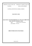
Inhibition of FNR activities by Zn-ferrocyanide The effect of the presence of Zn2+ during FNR catalysis is shown in Fig. 1A. Ferricyanide diaphorase activity was inhibited by the addition of increasing concentrations of ZnSO4, with an I0.5, the concentration that produces 50% inhibition, of about 1 lM Zn2+. In contrast, no effect was observed by Zn2+ addition on the DCPIP diaphorase activity (Fig. 1A). NADPH oxidation catalyzed by FNR in the presence of Fd or Fld was not affected by the presence of up to 1 mM ZnSO4.
The effect of equimolar concentrations of Zn2+ and ferrocyanide on diaphorase activities with different sub- strates and Fd reduction were then investigated. In all cases, the addition of metal ion and ferrocyanide to the reaction medium produced strong enzyme inhibition (Fig. 1B). To determine the Kd values of the complexes between FNR and NADP+ or Fd, 0.6 lM flavoprotein in 50 mM HEPES, pH 8, was titrated at 25 (cid:2)C with the corresponding substrate. After each addition, the flavin fluorescence (excitation at 456 nm; emission at 526 nm in the case of NADP+) or the flavoprotein fluorescence quenching
Inhibition of FNR by Zn-ferrocyanide (Eur. J. Biochem. 271) 4585
(cid:1) FEBS 2004
Fig. 1. Inhibition of FNR activities by Zn-fer- rocyanide. Residual FNR activity as a func- tion of ZnSO4 concentration (A) or ZnSO4 and potassium ferrocyanide equimolar con- centrations (B) using ferricyanide (d), DCPIP (s) and ferredoxin (m) as electron acceptors. (C) Inhibition of ferricyanide (d) and DCPIP (s) diaphorase activities of E. coli FPR as a function of Zn-ferrocyanide con- centration. In all cases, activity measurements were performed at pH 7.5. (D) A typical steady-state kinetics experiment of the FNR diaphorase activity for different DCPIP con- centrations at a fixed NADPH concentration of 300 lM, performed at increasing concen- trations of inhibitor [0 (s), 1.5 (d), 3 (m) and 5 (j) lM]. Inset: a typical Ki determination by Dixon plot of 1/v (B) vs. inhibitor concentra- tion (A) at different DCPIP concentrations.
100 lM for the ferricyanide diaphorase activity using any of the metals on pea reductase. Higher concentrations had some effect on the pea enzyme, but, in all cases, much lower inhibition was observed (not shown). In all cases, the addition of 1 mM EDTA final concentration after 2 min of reaction reversed the enzyme inhibition instantly and completely (not shown). This observation suggests that the Zn-ferrocyanide is accessible to the solvent. Incubation of the enzyme with Zn-ferrocyanide for longer periods of time resulted in inactivation of (cid:1) 57% in 60 min without the release of the prosthetic group (not shown). Zn-ferrocyanide was about six times more effective at inhibiting diaphorase activities than Fd reduction. However, in all cases total inhibition was obtained. At pH 7.5, a 50% inhibition was observed for the ferricyanide diaphorase activity with (cid:1) 1 lM Zn2+, meanwhile 6 lM Zn-ferrocyanide was necessary to obtain the same inhibition of the Fd reduction. Likewise, when the FPR from E. coli was investigated, inhibition of diaphorase activity was obtained with Zn-ferrocyanide (Fig. 1C). Neither FNR nor FPR activity was inhibited by sodium sulfate or ferrocyanide alone using DCPIP or ferricyanide as electron acceptors. Similarly, no enzyme inhibition was detected using ferrous sulfate.
Table 1. Kinetic, inhibition and binding parameters for various activities of FNR. The kinetics parameters were determined as describe in Experi- mental procedures. Each parameter value represents the average of three independent experiments. Ki values were calculated from Dixon plots of Zn-ferrocyanide noncompetitive inhibition with respect to the indicated substrate at a fixed saturating concentration of the other substrate. ND, Not determined.
Substrate
Km (lM) (no inhibitor)
kcat (s)1) (no inhibitor)
Kd (lM, NADP+) (no inhibitor)
Kd (lM, NADP+) (15 lM Zn-ferrocyanide)
Type of inhibition
Ki (lM)
NADPHa Ferricyanideb DCPIPc Ferredoxind
19.7 ± 2.3 106.0 ± 15.9 43.3 ± 4.9 42.1 ± 12.0
302.3 ± 7.8 321.0 ± 11.86 87.8 ± 4.3 3.3 ± 0.4
1.16 ± 0.1 1.57 ± 0.1 1.8 ± 0.2 13.8 ± 0.9
31.6 ± 2.3 ND ND 1.1 ± 0.1
33.0 ± 2.3 ND ND 1.4 ± 0.1
Noncompetitive Noncompetitive Noncompetitive Noncompetitive
a,b NADPH-ferricyanide diaphorase activity. c NADPH-DCPIP diaphorase activity. d NADPH-Fd reduction.
Co2+, which is able to replace Zn2+ in metaloenzymes, also inhibited the diaphorase reaction of pea FNR only if ferrocyanide was added with a I0.5 of 25 lM. Cu2+ and Ni2+ were also tested, and no inhibition was observed up to To evaluate this inhibitor in more detail, the steady-state kinetics of the FNR for the different substrates were examined at pH 7.5. Plots for the inhibitions of FNR diaphorase activities with increasing concentration of Zn-ferrocyanide (range 0–5 lM of inhibitor) showed that
4586 D. L. Catalano Dupuy et al. (Eur. J. Biochem. 271)
(cid:1) FEBS 2004
indicating that
the compound was a noncompetitive inhibitor of the enzyme for NADPH and ferricyanide. Similar results were obtained when DCPIP diaphorase activity and Fd reduc- tion were analysed for the substrates DCPIP and Fd, respectively (Table 1). In all cases a linear noncompetitive the inhibitor inhibition was observed, binding to the enzyme produces a nonproductive enzyme– substrate–inhibitor complex. Dixon plots were used to calculate the Ki values (Table 1), which were consistent with the I0,5 values extracted from Fig. 1. Calculation of enzyme activity at infinitive inhibitor concentration showed that total inhibition was obtained in all cases.
The dissociation constants of the FNRÆNADP+ and FNRÆFd complexes were estimated in the absence or presence of 15 lM inhibitor by measuring flavin fluores- cence and flavoprotein fluorescence quenching, respectively, after addition of each substrate. As shown in Table 1, the presence of the inhibitor did not change the enzyme affinity for its substrates. This is in agreement with the inhibition kinetic data presented above.
Zn-ferrocyanide inhibition of the reduction and oxidation of the flavin
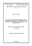
Fig. 2. Reduction and oxidation of the flavin. Optical spectra of FNR FAD reduction, 5 s after mixing, measured as the decrease in absorbance at 459 nm, and the increase at 550 nm to 650 nm range. All reactions were performed under aerobic conditions and contain 50 mM HEPES, pH 7.5, 20 lM FNR and the following additions. (A) In the absence of an electron acceptor: thick solid line, no addition; thick dashed line, 2.5 mM NADPH; thin solid line, 20 lM Zn-ferro- cyanide; thin dashed line, 20 lM Zn-ferrocyanide and 2.5 mM NADPH. (B) In the presence of an electron acceptor: thick solid line, no addition; thick dotted line, 1 mM potassium ferricyanide; thick dashed line, 1 mM potassium ferricyanide and 2.5 mM NADPH; thin solid line, 20 lM Zn-ferrocyanide; thin dotted line, 20 lM Zn-ferro- cyanide and 1 mM potassium ferricyanide; thin dashed line, 20 lM Zn-ferrocyanide, 1 mM potassium ferricyanide and 2.5 mM NADPH. Insets: amplified view of the region between 500 and 700 nm of the corresponding figures.
We have analyzed the spectral properties of FNR, and no differences were observed on addition of Zn-ferrocyanide (Fig. 2A, compare thick and thin solid lines). Then, we studied the spectral changes of the enzyme by addition of an excess amount of NADPH. The oxidation state of flavins can be distinguished by spectrophotometric means. They can exist in three different redox states: oxidized, one- electron reduced (semiquinone) radical, and fully reduced hydroquinone. The isolated FNR in solution contains mostly oxidized FAD. The neutral flavin radical absorbs light of long wavelength with a maximum at 570 nm, which is only detectable in FNR when the enzyme is anaerobically reduced. In aerobic conditions, when 2.5 mM NADPH was added to the enzyme solution and the spectral changes were recorded after 5 s, a decrease in absorbance was observed at 459 nm with a concomitant increase with a maximum at (cid:1) 590 nm. Similar results were obtained when reduction of the enzyme by NADPH was performed in the presence of Zn-ferrocyanide (Fig. 2A, thick and thin dashed lines). These results indicate that, in both cases under aerobic conditions and with an excess of NADPH, the neutral semiquinone of FAD appeared, with its typical absorption band which usually expands from 520 to 680 nm.
that the
Effect of Fld on the inhibition of FNR activities by Zn-ferrocyanide
The same experiment was then performed in the presence of the electron acceptor potassium ferricyanide in the absence of the inhibitor, recording the spectral changes 5 s after the addition of the substrates. Under these conditions, the enzyme containing a reduced flavin form was spectro- photometrically undetectable (Fig. 2B). The addition of 20 lM Zn-ferrocyanide in the reaction medium from the beginning of the measurement leads to the appearance of the reduced form of the enzyme, even in the presence of the electron acceptor (Fig. 2B). These results allow us to conclude electron-transfer process between NADPH and the flavin was not significantly altered by the presence of the inhibitor, and disrupting the electron transfer between the flavin and the second substrate mainly causes enzyme inhibition by Zn-ferrocyanide. In some photosynthetic systems, such as that of certain algae and cyanobacteria, the FMN-containing protein Fld
Inhibition of FNR by Zn-ferrocyanide (Eur. J. Biochem. 271) 4587
(cid:1) FEBS 2004
can efficiently replace Fd in the protein–protein electron- transfer process catalyzed by FNR. Fld and Fd bind to the same FNR site for catalysis, and, despite the difference in size, they seem to be equally oriented during binding to FNR and electron transfer [27]. Although there is no Fld in plants, Fld is able to efficiently accept electrons from plant FNR ([39] and this work).
Surprisingly, Zn-ferrocyanide was unable to inhibit the electron transfer from NADPH to Fld catalyzed by pea FNR. We tested concentrations up to 20 lM Zn-ferro- cyanide without any apparent loss of enzyme activity.
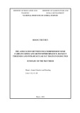
NADPH oxidation by FNR using Fld as electron acceptor proceeds at a low rate. This rate can be enhanced by the addition of the electron acceptor DNT. The interpretation of this observation is that Fld mediates the electron transfer between the reductase and DNT, as FNRs catalyze the reduction of DNT very slowly. This system can be used to better estimate the electron-transfer rate between the reductase and the Fld. Flavodoxin oxidase activity both in the absence and presence of DNT as artificial electron acceptor was insensitive to the addition of the metal ferrocyanide (Fig. 3A). Addition of Fld at saturating concentrations produced an increase of about 100% in the NADPH oxidation (Fig. 3A), which was not obtained by the addition of apoFld (not shown). Interestingly, Zn-ferrocyanide did not inhibit the reduction of DNT mediated by Fld, but completely prevented the direct transfer to DNT (Fig. 3A).
v ¼ 0.29 lmolÆmg)1Æmin)1 We also investigated the oxidase activity of FNR in the absence of added electron acceptors and, unexpectedly we found that it was completely insensitive to Zn-ferrocyanide (v ¼ 0.28 lmolÆmg)1Æmin)1 in the presence of 15 lM Zn-ferrocyanide vs in the absence of the inhibitor) (Fig. 3A).
Fig. 3. Inhibition by Zn-ferrocyanide in the presence of Fld. (A) The inhibition of flavodoxin oxidase activity was assayed in a DNT inde- pendent or dependent manner. The inhibition of DCPIP diaphorase activity was measured in the absence or presence of 12.5 lM Fld. Pure oxidase and DNT oxidase activities of FNR were assayed as controls. Activity was measured in the absence (hatched bars) or presence (solid bars) of Zn-ferrocyanide. Reactions were monitored by following NADPH oxidation at 340 nm. (B) The inhibition of DCPIP diapho- rase activity was measured in the absence or presence of 12.5 lM Fld, apoFld, or Fld digested with trypsin. DCPIP reduction was followed at 600 nm.
is inhibited by Zn-ferrocyanide,
spectroscopy. Figure 6 shows We then decided to investigate if Fld protects FNR against Zn-ferrocyanide inhibition. When the FNR DCPIP diaphorase activity was measured in the presence of 20 lM Zn-ferrocyanide and 12.5 lM Fld, no inhibition was observed (Fig. 3A,B). Under identical conditions, the reduc- tion of DCPIP was inhibited (cid:1) 98% by Zn-ferrocyanide. Two possible explanations for the unexpected protection displayed can be envisaged. Fld may bypass the pathway transferring the that electron to DCPIP or, the binding of the carrier protein to the FNR directly affects the interaction of the inhibitor with the enzyme. As shown in Fig. 3B the apoprotein protects FNR against Zn-ferrocyanide inhibition. As a control, a sample containing Fld previously treated with trypsin did not display any protection (Fig. 3B), indicating that the effect was a result of the presence of the polypeptide itself. Figure 4 shows a protection assay of the inhibition by Zn- ferrocyanide of the DCPIP diaphorase activity at different Fld concentrations. It can be observed that flavoprotein and its apoform displayed similar abilities to protect the enzyme. Moreover, the protection profile obtained can be correlated with the affinity of the FNRÆFld complex (13.4 lM) as obtained from the binding experiment depicted in Fig. 5.
Interaction of Zn-ferrocyanide with the FNR reductase
To further investigate the interaction of Zn-ferrocyanide with FNR, the prosthetic group environment was analyzed by fluorescence that Zn-ferrocyanide interacts with the enzyme in the absence of its substrates. Addition of the metal complex induces an increase in FAD fluorescence with a concomitant shift of emission maximum to a lower wavelength resembling the one obtained with FAD in solution. This observation can be considered to indicate that the prosthetic group undergoes a
4588 D. L. Catalano Dupuy et al. (Eur. J. Biochem. 271)
(cid:1) FEBS 2004
Fig. 4. Protection of DCPIP diaphorase activity by Fld. DCPIP diaphorase activity was assayed in the presence of 20 lM Zn-ferrocy- anide and different concentrations of either Anabaena Fld (solid bars) or apoFld (hatched bars).
rearrangement or that its exposure to the environment is increased.
Fig. 5. Determination of the dissociation constant of FNRÆFld complex. (A) Difference absorption spectra obtained during the titration of pea FNR (6.55 lM) with Anabaena Fld. (B) Absorbance difference data at 465 nm fitted to the theoretical equation for a 1 : 1 stoichiometric complex by means of nonlinear regression. The Kd value obtained was 13.4 lM.
A putative Zn2+-binding site within the FNR structure
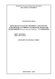
The crystal structure of pea FNR (PDB entry 1QG0 [40]), was analyzed searching for structures that could be able to bind Zn2+. All residues potentially able to co-ordinate Zn2+ were identified, and distances and geometries within the surrounding residues were determined using the SWISS- PDBVIEWER 3.7. The FAD was also took into account in the analysis because it has long been known that flavins interact specifically with metals [41,42]. We found a serine, a glutamic acid, a cysteine and a tyrosine residue near the isoalloxazine in a spatial orientation suitable for the interaction with metals (Fig. 7A). We also observed that the space available to accommodate Zn2+ is enough for appropriate binding of the metal ion, which remains accessible from the exterior (Fig. 7B). Distances between the N5 and O4 of the flavin, O of Ser90, S of Cys266, O of Glu306 and O of Tyr308 indicate that almost all of them are at bond distances between each other and nearly oriented correctly to participate in Zn2+ co-ordination (Fig. 7C). This amino acid arrangement around FAD is conserved in FPR and in the neuronal NO synthase (Fig. 7D,E).
affected by the inhibitor (Table 2). Similarly, replacing Tyr308 with other aromatic amino acids only slightly affects the inhibition by Zn-ferrocyanide on the enzyme. In contrast, replacing Tyr308 with glycine or serine consider- ably reduced the inhibition. Table 2 also shows the degree of nicotinamide ring occupancy of the binding site of Tyr mutants, as calculated by Piubelli et al. [43]. We found an inverse correlation between the extent of Zn2+ inhibition and nicotinamide ring occupancy in the FNR variants. These observations indicate that the binding of NADP+ to the enzyme either reduces the accessibility of the isoallox- azine itself to Zn2+ and/or ferrocyanide or partially impairs the entry of the inhibitor to the proposed binding site. Although a binding site for ferrocyanide, an octahedral To obtain supporting evidence for the proposed binding site and to investigate the participation of the above amino acids, several FNR mutants were analyzed. Table 2 shows the FNR inhibition by Zn-ferrocyanide obtained under identical experimental conditions with FNR mutants of Cys266 or Tyr308. Cysteine is one of the amino acids most commonly observed after histidine as a Zn2+ ligand in metalloproteins [12]. However, Cys266 does not appear to have a central role in the interaction with Zn-ferrocyanide as its replacement by alanine generates an enzyme that is still
Inhibition of FNR by Zn-ferrocyanide (Eur. J. Biochem. 271) 4589
(cid:1) FEBS 2004
Fig. 6. Interaction of Zn-ferrocyanide with FNR. Fluorescence emis- sion spectra of FNR (kexc. ¼ 459 nm) in the absence (thick solid line) or presence of 15 lM Zn-ferrocyanide (thin solid line). Fluorescence emission of free FAD (thin dashed line).
The kinetic analysis of all FNR activities inhibited by Zn-ferrocyanide revealed noncompetitive behavior for NADPH, for artificial electron acceptors and, for Fd. The FAD fluorescence of FNR showed a slight increase due to the addition of the inhibitor (10–15%), together with a shift of the maximum emission wavelength to 526 nm, closer to that of the free flavin. These rather small perturbations could be caused by changes in the microenvironment of the isoalloxazine ring, which is probably more exposed to the solvent after binding of the metal ferrocyanide. Changes may also be produced by the interaction of the metal itself with the isoalloxazine. However, the effect was only observed when Zn2+ and ferrocyanide were added, indica- ting that the combination of the two ions, and not each one separately, was responsible for the observed change.
complex anion with a diameter of about 6 A˚ , has never been mapped within the structure of FNR, it could be possible that this anion collides with the Zn2+-containing protein structure, interacting strongly and filling the open space near the isoalloxazine.
Discussion
Another hypothesis to explain the observed inhibition proposes that Zn2+ and ferrocyanide interact directly with the prosthetic group and/or with amino acid residues involved in the electron-transfer process. Searching for structures that may be able to bind Zn2+ on the crystal structure of FNR, we found a serine, a glutamic acid, a cysteine, and a tyrosine residue near the isoalloxazine in a spatial orientation suitable for the interaction with metals (Fig. 7A), although no definite sites were identified. It was also observed that the space available to accommodate Zn2+ is enough for appropriate binding of the metal ion. Interestingly, these amino acids are conserved in FPR and in the neuronal NO synthase (Fig. 7D,E). The residue homo- logous to FNR Tyr308 is a Phe in the NO synthase. Consequently, it may be suggested that Tyr308 may not be directly involved in the Zn-ferrocyanide inhibition of the reductase. Cysteine, histidine and glutamic acid are com- mon Zn2+ ligands in metalloproteins [12]. Although serine and tyrosine are less common Zn2+ ligands, they can interact with ions such as Zn2+ especially in proteins with more than one metal center such as alkaline phosphatase from E. coli [47].
The data presented in this work clearly show that the pea Fd-NADP(H) reductase is inhibited by Zn-ferrocyanide as a result of a specific combined interaction of both ions with the enzyme. The inhibition was also observed on the E. coli enzyme, a member of the same protein family, even though FPR is structurally distanced from the plant flavoprotein [44]. Inhibition by Zn2+ has been reported for other flavin- containing enzymes. Cu2+ and Zn2+ inhibit all NADPH- dependent reactions catalyzed by the neuronal NO synthase [13]. The authors of this work have concluded that inhibition is produced by the interaction of the metal with a unique site present in the reductase domain of the enzyme [13]. This domain binds one equivalent of FMN and one of FAD and, members of the Fd–NADP(H) reductase family share its structural features [45]. Similarly, it has been observed that Zn2+ inhibits the isolated a-oxoglutarate dehydrogenase mitochondrial complex [14]. A more de- tailed study has shown that the dihydrolipoyl dehydro- genase component of the complex is responsible for the observed Zn2+ inhibition [15]. This enzyme is a homo- dimeric molecule which contains FAD and belongs to the NAD-disulphide oxidoreductases class I group, which is led by the glutathione reductase as the model protein [46]. The latter group does not contain either the well-defined conserved sequences or displayed sequence similarity with the chloroplast-type FNRs. Taken together, these results may support the idea that the flavin itself may be involved in the interaction of the flavoproteins with the metal. We suggest that a partially or totally co-ordinated Zn2+ interacting with the bulky ferrocyanide, which can also interact with other amino acids, represents the true inhib- itor. The binding of ferricyanide and ferrocyanide has been detected in some enzymes [48,49]. Moreover, this interaction was proposed to occur via positively charged amino acids [49]. Indeed, the solubility of salts is a consequence of a large energy gain during hydration of ions that is surplus to the lattice energy. At the concentration of Zn-ferrocyanide used (0–30 lM), the salt is soluble but near its solubility product. Thus, a small change in the availability of water, as would occur with the inclusion of both ions in a protein hydrophobic pocket, may induce a stable ionic interaction between Zn2+ and ferrocyanide. We are not able to give an explanation for the participation of ferrocyanide as an obligate partner in the interaction of Zn2+ with the enzyme. However, it may be suggested that binding of ferrocyanide, an octahedral complex anion with a diameter of about 6 A˚ , throughout the interaction with the bound Zn2+ near the isoalloxazine could produce the observed inhibitory effect on FNR activity. This hypothesis is also supported by the finding that mutants with a catalytic site greatly occupied by the NADP+ nicotinamide displayed a reduced susceptibility to Zn-ferrocyanide inhibition (Table 2). It is worth men- tioning that the crystal structures of these mutants have
4590 D. L. Catalano Dupuy et al. (Eur. J. Biochem. 271)
(cid:1) FEBS 2004
Fig. 7. Putative Zn2+-binding site in FNR-like enzymes. Detail view of the spatial distribution of residues putatively involved in the interaction with Zn2+ in (A) pea FNR, (D) E. coli FPR and (E) rat neuronal NO synthase. Nitrogen 5 (N5) and oxygen 4 (O4) from FAD isoalloxazine are indicated. (B) Ribbon diagram of the putative Zn2+-binding site in pea FNR. (C) Distances between the atoms of pea FNR probably involved in the interaction with Zn2+ were measured in A˚ . The schemes were drawn using SWISS-PDBVIEWER 3.7 and rendered with POV-RAY from the tridimensional structures as determined by X-ray diffraction (Protein Data Bank entries 1QG0, 1FDR, 1TLL) [40].
Table 2. Inhibition of wild-type and mutant FNR diaphorase activity by Zn-ferrocyanide. ND, Not determined.
Nicotinamide ring occupancy of the binding site (%)b
FNR variant
Remaining activity (% of control)a
been obtained and that the overall conformations are equivalent to those of wild-type spinach and pea leaf FNRs, with no significant changes in the relative orientation of amino acids, the FAD or the conformation and binding of the 2¢-P-AMP portion of NADP+ [40].
WT C266A Y308S Y308G Y308F Y308W
1.8 15.0 79.5 41.0 23.2 5.9
14 ND 100 84 85 40
a Remaining ferricyanide diaphorase activity in the presence of 5 lM Zn-ferrocyanide with respect to the control. b Taken from Ref [43], calculated from the absorption coefficients at the peak near 510 nm of the difference spectra elicited by nicotinamide nucleotide binding to the various pea FNR forms; 100% refers to NADP+ occupancy of FNR-Y308S.
It has been observed that A-type monoamine oxidase is inhibited by the zinc benzoate salt [16]. Similarly, a-chymotrypsin may be inhibited by a substrate analog that interacts with a Zn2+ ion that is partially co-ordinated at the active site [17].
The Ki for the inhibition of Fd reduction is 8.8 times higher than those for diaphorase activity inhibition (Table 1). These results may be explained by the observa- tion that binding of Fd to FNR leads to structural changes in the reductase. After complex formation, the entire NADP(H) domain is displaced slightly as a single unit, and Glu306, which is located near the isoalloxazine, moves to within hydrogen-bonding distance of the hydroxy group of Ser90, as observed by Kurisu et al. [50] in crystals of the
Inhibition of FNR by Zn-ferrocyanide (Eur. J. Biochem. 271) 4591
(cid:1) FEBS 2004
FNRÆFd complex from maize. This glutamic acid is sufficiently exposed and readily available for the interaction with Zn2+ (Fig. 7A,B). In the Anabaena FdÆFNR crystal- lographic association resolved by Morales et al. [26] the carboxy group of the homologous Glu301 is no more exposed to solvent but is hydrogen-bonded to the hydroxy group of Fd Ser64. electron partners, Fd and Fld [27]. Moreover, it has been shown that Fd and Fld could be completely overlapped on the basis of their surface electrostatic potentials [56], but the interaction with Fld has been proposed to involve a larger FNR surface [57]. Although the interaction of FNR with its substrates exhibits co-operativity [58,59], modifications of the structure that should lead to the observed effects have remained elusive or hard to detect [11,40,60].
The role of these residues has been thoroughly inves- tigated by site-directed mutagenesis. When the homolog Glu301 from Anabaena FNR was mutated to Ala, the altered enzyme obtained was only 1% as active as the wild-type enzyme in electron transfer to Fd [51]. As the photoreduction of NADP+ was not affected to the same degree as the Fd reduction, the authors suggested that the rate-determining step during catalysis involves other processes in addition to the electron-transfer process between the two prosthetic groups [51]. The semiquinone state of FAD was significantly destabilized in the FNR mutant in which Glu301 was changed to Ala, and this was probably the main reason for the electron-transfer alter- ation observed in this mutant. Similarly, four different spinach FNR mutants of the equivalent Glu312 were obtained and analyzed [52]. The authors concluded that this residue is directly involved in the electron transfer between FNR and Fd. They also hypothesized that the residue may contribute to the tuning of the redox potential of the flavin semiquinone to enhance efficient electron transfer and/or may be acting as a proton donor/acceptor to FAD [51,52].
Changes in hydrophobic patches of Anabaena FNR influenced the rates of electron transfer to and from Fld and Fd. However, the observed effects were more dramatic in the processes involving Fld than those involving Fd, suggesting that these Anabaena FNR residues do not participate to the same extent in the processes for the two proteins [61]. Recently, electron transfer was obtained with the hybrid system bovine adrenodoxin reductase/Anabaena Fld, indicating that a highly specific interaction is not essential and that the process may proceed through multiple weak interactions. So far no residue on the Fld surface has been identified to be critical for the interaction and the electron-transfer processes between Fld and FNR [62]. It has therefore been suggested that there is a lower specificity for the FNR–Fld interaction than for the FNR–Fd one [62]. Therefore a dynamic assembly of the former complex in which multiple orientations may exist can be proposed. The fact that Zn-ferrocyanide was unable to inhibit the pea FNR electron transfer to Fld may not only be related to the protein size or a specific residue but also to the mechanisms of interaction between the reductase and Fld. The very short distance predicted between the two redox centers [62] may also account for our observations.
On the other hand, interaction of FNR with Fd does lead to structural changes in both electron carriers relative to the free protein conformations [26,50]. The protein–protein interaction also affects the microenvironments of the two prosthetic groups. In the case of the Fd and FNR, their redox potentials (Em) were shifted by )25 mV and +20 mV, respectively, reflecting theses changes [63].
The O of Ser90 and the S of Cys266 of pea FNR are close to N5 and O4 of the isoalloxazine, which are involved in hydride transfer. The hydroxy group of Ser90 could accept a hydrogen bond and thus help to stabilize the reduced flavin. Meanwhile its interaction probably affects the transition state of hydride transfer. The Ser96Val mutant of FNR displayed a kcat nearly 2000 times lower than that of the wild-type enzyme [53]. Analysis of the crystal structure of wild-type pea FNR shows that the Zn2+ ion can easily access Ser90. Moreover, serine is among the amino acids that could, although infrequently, co-ordinate Zn2+ [12]. Thus, it may be one of the amino acid residues involved in the inhibition of FNR by Zn-ferrocyanide.
We have observed that both Fld and its apoprotein are able to impede the inhibition of FNR by Zn-ferrocyanide. More remarkable, the solely polypeptide interaction between FNR and apoFld is sufficient to prevent the indicating that no participation of the Fld inhibition, electronic transfer is involved in the observed protection. In addition, we observed that the Fld concentration needed to protect FNR from Zn-ferrocyanide inhibition is similar to the Kd for the pea FNRÆFld complex (13.4 lM). It is worth mentioning that it has been determined that the structure of apoFld is virtually equivalent to that of the holoprotein, the only exception being that the isoalloxazine- binding site closed [64].
Our results (Fig. 2) allow us to suggest that Zn-ferro- cyanide mainly causes an interruption of the oxidative half reaction in the diaphorase activity and the electron transfer between FNRred and Fd. It has been observed that electrophilic metal ions such as Zn2+ prefer co-ordination with the one-electron reduced semiquinone state of flavin [42]. Thus, Zn-ferrocyanide may interact after reduction of the enzyme by NADPH with the semiquinone state of the flavoprotein, producing the observed inactive form or altering the proton exchange between the flavin and the surrounding amino acid residues, in particular Glu306 and Ser90. Both residues have been proposed to participate in the proton-transfer pathway between the exterior and isoalloxazine [26,45,51–55].
It is interesting that Zn-ferrocyanide was unable to inhibit the electron transfer from NADPH to Fld catalyzed by pea FNR. At present, no crystal structures of the complex between FNR and Fld have been obtained. Using several charge-reversal mutants, it has been possible to infer that FNR uses the same site for the interaction with both In summary, our results indicate that determinants on the FNR polypeptide are essential for electron transfer between the reduced flavin and the substrate and, that this process can be completely inhibited by Zn2+ in the presence of ferrocyanide. We have obtained evidence that isoalloxazine and the surrounding amino acids are the binding site of the inhibitor. Clearly, the observation that binding of Fld or apoFld to the reductase was sufficient to overcome the inhibition may be taken as evidence for a conformational change produced in the reductase on interaction with this electron partner, modifying either the FAD environment or
4592 D. L. Catalano Dupuy et al. (Eur. J. Biochem. 271)
(cid:1) FEBS 2004
14. Brown, A.M., Kristal, B.S., Effron, M.S., Shestopalov, A.I., Ullucci, P.A., Sheu, K.F., Blass, J.P. & Cooper, A.J. (2000) Zn2+ inhibits alpha-ketoglutarate-stimulated mitochondrial respiration and the isolated alpha-ketoglutarate dehydrogenase complex. J. Biol. Chem. 275, 13441–13447.
the amino acids involved in the electron-transfer process. These findings are significant for understanding the changes in the reductase on binding of Fd and Fld that were not detected by other approaches and should help in further studies of the enzyme.
Acknowledgements
15. Gazaryan, I.G., Krasnikov, B.F., Ashby, G.A., Thorneley, R.N., Kristal, B.S. & Brown, A.M. (2002) Zinc is a potent inhibitor of thiol oxidoreductase activity and stimulates reactive oxygen spe- cies production by lipoamide dehydrogenase. J. Biol. Chem. 277, 10064–10072.
16. Egashira, T., Sakai, K., Takayama, F., Sakurai, M. & Yoshida, S. (2003) Zinc benzoate, a contaminating environmental compound derived from polystyrene resin inhibits A-type monoamine oxi- dase. Toxicol. Lett. 145, 161–165.
E.A.C. is a staff member of the Consejo Nacional de Investigaciones Cientı´ ficas y Te´ cnicas (CONICET, Argentina). D.L.C.D. and D.V.R. are fellows of the same institution. This study was supported by grants from CONICET and Agencia de Promocio´n Cientı´fica y Tecnolo´gica (ANPCyT, Argentina). We thank Dr M. Medina (University of Zaragoza, Spain) for the gift of flavodoxin from Anabaena, and Matı´ as Musumeci for providing the C266A FNR mutant.
17. Han, M.S., Oh, D.J. & Kim, D.H. (2004) Inhibition of alpha- chymotrypsin with thiol-bearing substrate analogues in the pre- sence of zinc ion. Bioorg. Med. Chem. Lett. 14, 701–705.
References
18. Maret, W., Jacob, C., Vallee, B.L. & Fischer, E.H. (1999) Inhibitory sites in enzymes: zinc removal and reactivation by thionein. Proc. Natl Acad. Sci. USA 96, 1936–1940.
1. Carrillo, N. & Ceccarelli, E.A. (2003) Open questions in ferre- doxin-NADP+ reductase catalytic mechanism. Eur. J. Biochem. 270, 1900–1915.
19. Maret, W., Yetman, C.A. & Jiang, L. (2001) Enzyme regulation by reversible zinc inhibition: glycerol phosphate dehydrogenase as an example. Chem. Biol. Interact. 130–132, 891–901.
2. Ceccarelli, E.A., Arakaki, A.K., Cortez, N. & Carrillo, N. (2004) Functional plasticity and catalytic efficiency in plant and bacterial ferredoxin-NADP (H) reductases. Biochim. Biophys. Acta 1698, 155–165.
20. Luna, C.M., Casano, L.M. & Trippi, V.S. (2000) Inhibition of wheat nitrate reductase activity by zinc. Biol. Plant. 43, 257–262. 21. Egashira, T., Takayama, F. & Sakai, K. (2003) Inhibition by Zn (2+) of A-form monoamine oxidase in monkey brain mitochon- dria. J. Pharmacol. Sci. 91, 239–245.
22. Brohawn, S.G., Miksa, I.R. & Thorpe, C. (2003) Avian sulfhydryl oxidase is not a metalloenzyme: adventitious binding of divalent metal ions to the enzyme. Biochemistry 42, 11074–11082.
3. Hurley, J.K., Morales, R., Martinez-Julvez, M., Brodie, T.B., Medina, M., Gomez-Moreno, C. & Tollin, G. (2002) Structure- function relationships ferredoxin/ferredoxin: in Anabaena NADP(+) reductase electron transfer: insights from site-directed mutagenesis, transient absorption spectroscopy and X-ray crys- tallography. Biochim. Biophys. Acta 1554, 5–21.
4. Fillat, M.F., Sandmann, G. & Gomez-Moreno, C. (1988) Flavo- doxin from the nitrogen-fixing cyanobacterium Anabaena PCC 7119. Arch. Microbiol. 150, 160–164.
23. Kannt, A., Ostermann, T., Muller, H. & Ruitenberg, M. (2001) Zn (2+) binding to the cytoplasmic side of Paracoccus denitrificans cytochrome c oxidase selectively uncouples electron transfer and proton translocation. FEBS Lett. 503, 142–146.
24. Mills, D.A., Schmidt, B., Hiser, C., Westley, E. & Ferguson- Miller, S. (2002) Membrane potential-controlled inhibition of cytochrome c oxidase by zinc. J. Biol. Chem. 277, 14894–14901.
5. Razquin, P., Fillat, M.F., Schmitz, S., Stricker, O., Bohme, H., Gomez-Moreno, C. & Peleato, M.L. (1996) Expression of ferre- doxin-NADP+ reductase in heterocysts from Anabaena sp. Bio- chem. J. 316 (1), 157–160.
25. Samiotakis, M. & Ebbs, S.D. (2004) Possible evidence for trans- port of an iron cyanide complex by plants. Environ. Pollut. 127, 169–173.
6. Ragsdale, S.W. & Ljungdahl, L.G. (1984) Characterization of ferredoxin, flavodoxin, and rubredoxin from Clostridium for- micoaceticum grown in media with high and low iron contents. J. Bacteriol. 157, 1–6.
7. Avron, M. & Jagendorf, A.T. (1956) A TPNH diaphorase from
chloroplast. Arch. Biochem. Biophys. 65, 475–490.
26. Morales, R., Charon, M.H., Kachalova, G., Serre, L., Medina, M., Gomez-Moreno, C. & Frey, M. (2000) A redox-dependent interaction between two electron-transfer partners involved in photosynthesis. EMBO Rep. 1, 271–276.
8. Carrillo, N. & Vallejos, R.H. (1987) Ferredoxin-NADP+ oxido- reductase. In Topics in Photosynthesis (Barber, J., ed.), pp. 527– 560. Elsevier, Amsterdam, New York, Oxford.
27. Martinez-Julvez, M., Medina, M. & Gomez-Moreno, C. (1999) Ferredoxin-NADP (+) reductase uses the same site for the interaction with ferredoxin and flavodoxin. J. Biol. Inorg. Chem. 4, 568–578.
9. Shah, M.M. & Spain, J.C. (1996) Elimination of nitrite from the explosive 2,4,6-trinitrophenylmethylnitramine (tetryl) catalyzed by ferredoxin NADP oxidoreductase from spinach. Biochem. Bio- phys. Res. Commun. 220, 563–568.
28. Orellano, E.G., Calcaterra, N.B., Carrillo, N. & Ceccarelli, E.A. (1993) Probing the role of the carboxyl-terminal region of ferre- doxin-NADP+ reductase by site-directed mutagenesis and dele- tion analysis. J. Biol. Chem. 268, 19267–19273.
29. Rial, D.V., Arakaki, A.K. & Ceccarelli, E.A. (2000) Interaction of the targeting sequence of chloroplast precursors with Hsp70 molecular chaperones. Eur. J. Biochem. 267, 6239–6248.
10. Kranendonk, M., Commandeur, J.N., Laires, A., Rueff, J. & Vermeulen, N.P. (1997) Characterization of enzyme activities and cofactors involved in bioactivation and bioinactivation of chemi- cal carcinogens in the tester strains Escherichia coli K12 MX100 and Salmonella typhimurium LT2 TA100. Mutagenesis 12, 245– 254.
30. Serra, E.C., Carrillo, N., Krapp, A.R. & Ceccarelli, E.A. (1993) One-step purification of plant ferredoxin-NADP+ oxidoreduc- tase expressed in Escherichia coli as fusion with glutathione S-transferase. Protein Expr. Purif. 4, 539–546.
11. Bruns, C.M. & Karplus, P.A. (1995) Refined crystal structure of spinach ferredoxin reductase at 1.7 A resolution: oxidized, reduced and 2¢-phospho-5¢-AMP bound states. J. Mol. Biol. 247, 125–145. 12. McCall, K.A., Huang, C. & Fierke, C.A. (2000) Function and
mechanism of zinc metalloenzymes. J. Nutr. 130, 1437S–1446S. 13. Perry, J.M., Zhao, Y. & Marletta, M.A. (2000) Cu2+ and Zn2+ inhibit nitric-oxide synthase through an interaction with the reductase domain. J. Biol. Chem. 275, 14070–14076.
31. Hurley, J.K., Salamon, Z., Meyer, T.E., Fitch, J.C., Cusanovich, M.A., Markley, J.L., Cheng, H., Xia, B., Chae, Y.K. & Medina, M. (1993) Amino acid residues in Anabaena ferredoxin crucial to interaction with ferredoxin–NADP+ reductase: site-directed mutagenesis and laser flash photolysis. Biochemistry 32, 9346– 9354.
Inhibition of FNR by Zn-ferrocyanide (Eur. J. Biochem. 271) 4593
(cid:1) FEBS 2004
32. Jenkins, C.M. & Waterman, M.R. (1998) NADPH–flavodoxin reductase and flavodoxin from Escherichia coli: characteristics as a soluble microsomal P450 reductase. Biochemistry 37, 6106– 6113.
50. Kurisu, G., Kusunoki, M., Katoh, E., Yamazaki, T., Teshima, K., Onda, Y., Kimata-Ariga, Y. & Hase, T. (2001) Structure of the electron transfer complex between ferredoxin and ferredoxin- NADP (+) reductase. Nat. Struct. Biol. 8, 117–121.
33. Genzor, C.G., Beldarrain, A., Gomez-Moreno, C., Lopez- Lacomba, J.L., Cortijo, M. & Sancho, J. (1996) Conformational stability of apoflavodoxin. Protein Sci. 5, 1376–1388.
51. Medina, M., Martinez-Julvez, M., Hurley, J.K., Tollin, G. & Gomez-Moreno, C. (1998) Involvement of glutamic acid 301 in the catalytic mechanism of ferredoxin-NADP+ reductase from Anabaena PCC 7119. Biochemistry 37, 2715–2728.
34. Zanetti, G. (1976) A lysyl residue at the NADP binding site of ferredoxin-NADP reductase. Biochim. Biophys. Acta 445, 14–24. 35. Geren, L.M. & Millett, F. (1981) Fluorescence energy transfer studies of the interaction between adrenodoxin and cytochrome c. J. Biol. Chem. 256, 10485–10489.
52. Aliverti, A., Deng, Z., Ravasi, D., Piubelli, L., Karplus, P.A. & Zanetti, G. (1998) Probing the function of the invariant glutamyl residue 312 in spinach ferredoxin-NADP+ reductase. J. Biol. Chem. 273, 34008–34015.
36. Aliverti, A., Lubberstedt, T., Zanetti, G., Herrmann, R.G. & Curti, B. (1991) Probing the role of lysine 116 and lysine 244 in the spinach ferredoxin–NADP+ reductase by site-directed mutagen- esis. J. Biol. Chem. 266, 17760–17763.
53. Aliverti, A., Bruns, C.M., Pandini, V.E., Karplus, P.A., Vanoni, M.A., Curti, B. & Zanetti, G. (1995) Involvement of serine 96 in the catalytic mechanism of ferredoxin-NADP+ reductase: struc- ture–function relationship as studied by site-directed mutagenesis and X-ray crystallography. Biochemistry 34, 8371–8379.
37. Sancho, J. & Gomez-Moreno, C. (1991) Interaction of ferredoxin– NADP+ reductase from Anabaena with its substrates. Arch. Biochem. Biophys. 288, 231–238.
54. Aliverti, A., Piubelli, L., Zanetti, G., Lubberstedt, T., Herrmann, R.G. & Curti, B. (1993) The role of cysteine residues of spinach ferredoxin-NADP+ reductase as assessed by site-directed muta- genesis. Biochemistry 32, 6374–6380.
38. Wang, Z.X., Kumar, N.R. & Srivastava, D.K. (1992) A novel spectroscopic titration method for determining the dissociation constant and stoichiometry of protein-ligand complex. Anal. Biochem. 206, 376–381.
55. Mayoral, T., Medina, M., Sanz-Aparicio, J., Gomez-Moreno, C. & Hermoso, J.A. (2000) Structural basis of the catalytic role of Glu301 in Anabaena PCC 7119 ferredoxin-NADP+ reductase revealed by x-ray crystallography. Proteins 38, 60–69.
39. Pirola, M.C., Monti, F., Aliverti, A. & Zanetti, G. (1994) A functional heterologous electron-transfer protein complex: Desulfovibrio vulgaris flavodoxin covalently linked to spinach ferredoxin-NADP+ reductase. Arch. Biochem. Biophys. 311, 480–486.
56. Ullmann, G.M., Hauswald, M., Jensen, A. & Knapp, E.W. (2000) Structural alignment of ferredoxin and flavodoxin based on electrostatic potentials: implications for their interactions with photosystem I and ferredoxin–NADP reductase. Proteins 38, 301–309.
40. Deng, Z., Aliverti, A., Zanetti, G., Arakaki, A.K., Ottado, J., Orellano, E.G., Calcaterra, N.B., Ceccarelli, E.A., Carrillo, N. & Karplus, P.A. (1999) A productive NADP+ binding mode of ferredoxin-NADP+ reductase revealed by protein engineering and crystallographic studies. Nat. Struct. Biol. 6, 847–853.
57. Martinez-Julvez, M., Medina, M. & Gomez-Moreno, C. (1999) Ferredoxin–NADP (+) reductase uses the same site for the interaction with ferredoxin and flavodoxin. J. Biol. Inorg. Chem. 4, 568–578.
41. Yu, M.W. & Fritchie, C.J. Jr (1975) Interaction of flavins with
electron-rich metals. J. Biol. Chem. 250, 946–951.
58. Batie, C.J. & Kamin, H. (1984) Electron transfer by ferredoxin: NADP+ reductase. Rapid-reaction evidence for participation of a ternary complex. J. Biol. Chem. 259, 11976–11985.
42. Kaim, W., Schwederski, B., Heilmann, O. & Hornung, F.M. (1999) Coordination compounds of pteridine, alloxazine and fla- vin ligands: structures and properties. Coord. Chem. Rev. 182, 323–342.
59. Batie, C.J. & Kamin, H. (1986) Association of ferredoxin- NADP+ reductase with NADP (H) specificity and oxidation– reduction properties. J. Biol. Chem. 261, 11214–11223.
43. Piubelli, L., Aliverti, A., Arakaki, A.K., Carrillo, N., Ceccarelli, E.A., Karplus, P.A. & Zanetti, G. (2000) Competition between C-terminal tyrosine and nicotinamide modulates pyridine nu- cleotide affinity and specificity in plant ferredoxin- NADP (+) reductase. J. Biol. Chem. 275, 10472–10476.
44. Arakaki, A.K., Ceccarelli, E.A. & Carrillo, N. (1997) Plant-type ferredoxin–NADP+ reductases: a basal structural framework and a multiplicity of functions. FASEB J. 11, 133–140.
60. Serre, L., Vellieux, F.M., Medina, M., Gomez-Moreno, C., Fontecilla-Camps, J.C. & Frey, M. (1996) X-ray structure of the ferredoxin: NADP+ reductase from the cyanobacterium Anab- aena PCC 7119 at 1.8 A resolution, and crystallographic studies of NADP+ binding at 2.25 A resolution. J. Mol. Biol. 263, 20–39. 61. Nogues, I., Martinez-Julvez, M., Navarro, J.A., Hervas, M., Armenteros, L., de la Rosa, M.A., Brodie, T.B., Hurley, J.K., Tollin, G., Gomez-Moreno, C. & Medina, M. (2003) Role of hydrophobic interactions in the flavodoxin mediated electron transfer from photosystem I to ferredoxin-NADP+ reductase in Anabaena PCC 7119. Biochemistry 42, 2036–2045.
45. Karplus, P.A. & Bruns, C.M. (1994) Structure–function relations for ferredoxin reductase. J. Bioenerg. Biomembr. 26, 89–99. 46. Kuriyan, J., Krishna, T.S., Wong, L., Guenther, B., Pahler, A., Williams, C.H. Jr & Model, P. (1991) Convergent evolution of similar function in two structurally divergent enzymes. Nature (London) 352, 172–174.
62. Medina, M. & Gomez-Moreno, C. (2004) Interaction of ferre- doxin–NADP+ reductase with its substrates. Optimal interact for efficient electron transfer. Photosynth. Res. 79, 113–131.
47. Kim, E.E. & Wyckoff, H.W. (1991) Reaction mechanism of alkaline phosphatase based on crystal structures. Two-metal ion catalysis. J. Mol. Biol. 218, 449–464.
63. Smith, J.M., Smith, W.H. & Knaff, D.B. (1981) Electrochemical titrations of a ferredoxin-ferredoxin: NADP+ oxidoreductase complex. Biochim. Biophys. Acta 635, 405–411.
48. Eley, C.G., Ragg, E. & Moore, G.R. (1984) Kinetics of electron transfer between mitochondrial cytochrome c and iron hexa- cyanides. J. Inorg. Biochem. 21, 295–310.
64. Maldonado, S., Lostao, A., Irun, M.P., Fernandez-Recio, J., Gustavo, G.C., Begona, G.E., Rubio, J.A., Luquita, A., Daoudi, F. & Sancho, J. (1998) Apoflavodoxin: structure, stability, and FMN binding. Biochimie 80, 813–820.
49. Colotti, G., Verzili, D., Boffi, A. & Chiancone, E. (1994) Identi- fication of the site of ferrocyanide binding involved in the intra- molecular electron transfer process to oxidized heme in Scapharca dimeric hemoglobin. Arch. Biochem. Biophys. 311, 103–106.