doi:10.1046/j.1432-1033.2003.03354.x
Eur. J. Biochem. 270, 20–27 (2003) (cid:3) FEBS 2003
Structure of the O polysaccharides and serological classification of Pseudomonassyringaepv. porri from genomospecies 4
Evelina L. Zdorovenko1, George V. Zatonskii1, Nina A. Kocharova1, Aleksander S. Shashkov1, Yuriy A. Knirel1 and Vladimir V. Ovod2 1N. D. Zelinsky Institute of Organic Chemistry, Russian Academy of Sciences, Moscow, Russia; 2Institute of Medical Technology, University of Tampere, Tampere, Finland
attachment of GlcNAc, were found in the two strains stud- ied, O repeat 1 being major in strain NCPPB 3365 and 2 in strain NCPPB 3364T.
Strains of Pseudomonas syringae pv. porri are characterized by a number of pathovar-specific phenotypic and genomic characters and constitute a highly homogeneous group. Using monoclonal antibodies, they all were classified in a
fi3)-a-L-Rhap-(1fi2)-a-L-Rhap-(1fi3)-a-L-Rhap-(1fi3)-a-L-Rhap-(1fi (1)
2 › 1 b-D-GlcpNAc
fi2)-a-L-Rhap-(1fi2)-a-L-Rhap-(1fi3)-a-L-Rhap-(1fi3)-a-L-Rhap-(1fi (2)
2 › 1 b-D-GlcpNAc
The relationship between OPS chemotype and serotype on one hand and the genomic characters of P. syringae pv. porri and other pathovars delineated in genomospecies 4 on the other hand is discussed.
Keywords: lipopolysaccharide; O polysaccharide structure; serological classification; monoclonal antibody; Pseudo- monas syringae.
novel P. syringae serogroup O9. The O polysaccharides (OPS) isolated from the lipopolysaccharides (LPS) of P. syringae pv. porri NCPPB 3365 and NCPPB 3364T possess multiple oligosaccharide O repeats, some of which are linear and composed of L-rhamnose (L-Rha), whereas the major O repeats are branched with L-rhamnose in the main chain and GlcNAc in side chains (structures 1 and 2). Both branched O repeats, which differ in the position of substitution of one of the Rha residues and in the site of
(epiphytes) and pathogenic strains, all of which are able to induce the hypersensitive reaction to tobacco [2,3]. There- fore, pathovars have no taxonomic impact [2,4,5].
Strains of the phytopathogenic bacterium Pseudomonas syringae are characterized by a high degree of heterogeneity in respect to phenotypic and genotypic characters. More than 50 infraspecific taxa, so-called pathovars, of P. syrin- gae and related species have been described based on the distinctive pathogenicity of strains to one or more host plants [1]. However, P. syringae is known to be an opportunistic pathogen that includes both nonpathogenic
P. syringae strains of different pathovars also reveal heterogeneity of their genomic characters [6–11]. Recently, pathotype strains of 48 pathovars of P. syringae and eight related phytopathogenic pseudomonads have been delinea- ted in nine genomospecies [4]. However, the genomospecies cannot be properly discriminated based on the nutritional characters of strains [2,4,7,12,13]. Therefore, new pheno- typic characters are necessary for discrimination between pathovars/genomospecies and identification of the bacteria. Recently, it has been suggested that chemotype of the lipopolysaccharide (LPS) and the corresponding O serotype of P. syringae are conserved phenotypic characters, which may correlate with pathovars and genomospecies [14]. Previously, we have elucidated the structures of the O polysaccharide chains (OPS) of LPS of a number of P. syringae strains belonging to different pathovars
Correspondence to Yuriy A. Knirel, N. D. Zelinsky Institute of Organic Chemistry, Russian Academy of Sciences, Leninsky Prospekt 47, 119991 Moscow, GSP-1, Russia. Fax: +7 095 1355328, Tel.: +7 095 9383613, E-mail: knirel@ioc.ac.ru Abbreviations: HSQC, heteronuclear single-quantum coherence; LPS, lipopolysaccharide; OPS, O polysaccharide; Rha, rhamnose. (Received 30 August 2002, revised 24 October 2002, accepted 7 November 2002)
O polysaccharides of Pseudomonas syringae pv. porri (Eur. J. Biochem. 270) 21
(cid:3) FEBS 2003
Chemical analyses
[14–23]. Here we report on structural and serological studies of LPS of strains of P. syringae pv. porri, the causative agent of the bacterial blight of leek (Allium porrum) [24,25], which together with P. syringae pvs. garcae, atropurpurea, oryzae, striafaciens, zizaniae, and Pseudomonas coronafac- iens have been delineated in genomospecies 4 [4].
Materials and methods
For sugar analysis, the OPS was hydrolyzed with 2 M CF3CO2H (120 (cid:4)C, 2 h), monosaccharides were identified by GLC as the alditol acetates [26] on a Hewlett-Packard 5880 chromatograph (USA) equipped with a DB-5 capillary column using a temperature gradient of 160 (cid:4)C (1 min) to 250 (cid:4)C at 3 (cid:4)C min)1. The absolute configurations of the monosaccharides were determined by GLC of the acetyl- ated glycosides with (S)-octan-2-ol [27].
Cultivation of bacteria, isolation of lipopolysaccharides and polysaccharides
Methylation was carried out with CH3I in dimethyl sulfoxide in the presence of solid NaOH [28]. Hydrolysis of the methylated polysaccharides was performed as in sugar analysis, partially methylated monosaccharides were con- verted into the alditol acetates and analyzed by GLC/MS on a Hewlett Packard 5890 chromatograph (USA) equipped with a DB-5 capillary column and a NERMAG R10–10 L mass spectrometer (France) in the same chromatographic conditions as above.
NMR spectroscopy
Bacterial strains of pathovars delineated in genomospecies 4 (Table 1) were cultivated on potato agar at 22 (cid:4)C for 24 h. LPS were isolated by extraction with Tris/EDTA buffer as described [23]. The LPS of P. syringae pv. porri NCPPB 3364T (GSPB 2654) and NCPPB 3365 (GSPB 2655) were degraded by hydrolysis with 2% (v/v) HOAc for 1.5 h at 100 (cid:4)C. The OPS were isolated by gel- permeation chromatography on a column (70 · 2.6 cm) of Sephadex G-50 using pyridinium acetate buffer pH 4.5 (4 mL pyridine and 10 mL HOAc in 1 L water) and monitoring of elution with a differential refractometer (Knauer, Germany).
For NMR spectroscopy, samples were deuterium- exchanged by freeze-drying from 99.9% D2O and dissolved
Table 1. LPS-based serological and chemical classification of strains of Pseudomonas syringae pathovars and Pseudomonas coronafaciens from genomospecies 4 [4]. CFBP, French Collection of Phytopathogenic Bacteria (INRA, Angers, France); ICMP, International Collection of Micro- organisms from Plants (Auckland, New Zealand); NCPPB, National Collection of Plant Pathogenic Bacteria (Harpenden, UK).
Strain Host plant Serotype Chemotype Geographical origin Year of isolation P. syringae pathovar or Pseudomonas species
atropurpurea
P. coronafaciens 3C 3C 4E0 3C 3C
garcae
Lolium multiflorum Lotium sp. Agrostis sp. Avena sativa Avena sativa Avena sativa Avena sativa Avena sativa Avena sativa Avena sativa Avena sativa Avena sativa Coffea arabica Coffea arabica Coffea arabica Coffea arabica Coffea arabica Coffea arabica oryzae
porri
4E1-I 4E1-I 4E1-I 4E1-I 4E1-I 4E1-I 4E1-I 4E1-I 4E1-I 4 A 4 A 4E2 8C 4E1-I 9C 9C 9C
9C striafaciens Japan Japan UK UK UK Canada Canada Canada Germany Kenya New Zealand Canada Brazil Brazil Brazil Kenya Kenya Kenya Japan Japan France France France France Netherlands unknown Zimbabwe O3(3c,3c1) O3(3c,3c1) O4(4a,4e) O3(3c) O3(3c) Rough O4(4a1,4e) O4(4a1,4e) O4(4a1,4e) O4(4a1,4e) O4(4a1,4e) O4(4a1,4e) O4(4a1,4e) O4(4a1,4e) O4(4a1,4e) O4(4a,4a1,4a2) O4(4a,4a1,4a2) O4(4a1,4e2) O8(8c) O4(4a1,4e) O9(9c,9c1) O9(9c) O9(9c) Rough O9(9c,9c1) Rough O3(3c) O1[(1–2)a,(1–2)a1,1a,1b]
zizaniae NCPPB 2397T NCPPB 2396 NCPPB 1768 NCPPB 600T NCPPB 1253 NCPPB 1356 NCPPB 1357 NCPPB 1327 NCPPB 874 NCPPB 2481 NCPPB 2680 NCPPB 2816 NCPPB 588T NCPPB 512 ICMP 5802 NCPPB 2708 NCPPB 2710 ICMP 8047 NCPPB 3683T Oryza sativa Oryza sativa CFBP 4363 NCPPB 3364T Allium porrum Allium porrum NCPPB 3365 Allium porrum NCPPB 3366 Allium porrum NCPPB 3367 Allium porrum NCPPB 3545 NCPPB 1898T Avena sativa Avena sativa NCPPB 2480 Secale & Triticum sp. Mexico NCPPB 2713 Avena sativa ICMP 4483 Avena sativa ICMP 8815 NCPPB 3690T Zizania aquatica 1967 1967 1965 1958 1962 1954 1962 1962 1959 1970 1969 1933 1956 1956 1976 1972 1973 1974 1983 1983 1978 1964 1975 1979 1984 1966 1971 1973 New Zealand Unknown O4(4a,4e) 1973 Mexico 1983 USA 3C 1B 4E0 1B 4E1-I O1[(1–2)a,(1–2)a1,1a,1b] O4(4a1,4e)
22 E. L. Zdorovenko et al. (Eur. J. Biochem. 270)
(cid:3) FEBS 2003
in 99.96% D2O. The 1H and 13C NMR spectra were recorded on Bruker DRX-500 and DRX-600 spectrometers (Germany) at 60 (cid:4)C. Chemical shifts were determined with acetone as internal standard (dH 2.225, dC 31.45). Spectra were run using standard Bruker software, and the XWINNMR 2.1 program was used to acquire and process the data. A mixing time of 100 and 200 ms was used in TOCSY and NOESY experiments, respectively.
Remarkably, MAb Pscor1 reacted with the LPS P. syr- ingae pv. porri rough strain NCPPB 3367 but with none of the other, smooth strains of P. syringae pv. porri. This MAb is known to be specific to the outer core region of P. syringae LPS and reactive in Western immunoblotting with R- and SR (semirough)-form LPS, which are coex- pressed with S-form LPS in smooth strains of most P. syringae pathovars [23,31]. Other epitopes related to the LPS core, which are common for all P. syringae strains, were recognized by the corresponding core-specific MAbs in P. syringae pv. porri strains too (data not shown).
Production of monoclonal antibodies and serological tests
Structural studies of the OPS of P.syringae pv. porri NCPPB 3365
A high-molecular-mass OPS was isolated by mild acid degradation of the LPS from P. syringae pv. porri NCPPB 3365 followed by gel-permeation chromatography on Sephadex G-50. Sugar analysis of the OPS, including determination of the absolute configurations of monosac- charides, demonstrated the presence of L-rhamnose (L-Rha) and 2-amino-2-deoxy-D-glucose (D-GlcN). Methylation analysis of the OPS revealed 2-substituted, 3-substituted, and 3,4-disubstituted Rha in the ratios 3 : 3 : 2 as well as terminal GlcNAc.
Murine MAbs Ps3c, Ps4a, Ps4e, and Ps8c have been produced and characterized previously [17,19,29–31]. New O polysaccharide-specific MAbs Ps4a1 (IgM) and Ps4e2 (IgG3) were generated against P. syringae pv. garcae ICMP 8047, MAbs Ps9c (IgG2a) and Ps9c1 (IgG2a) against P. syringae pv. porri NCPPB 3364T, and MAb Ps4a2 (IgM) was produced against P. syringae pv. delphinii NCPPB 1879T. Immunization protocol, hybridomas gen- eration, selection of specific clones and determination of MAb isotypes were performed as described earlier [14,23,29,31]. ELISA, SDS/PAGE and Western immuno- blotting were performed essentially as described [23,29,31]. Crude and proteinase K-digested LPS and isolated OPS were used as antigens to coat Nunc-Immuno MaxiSorp Surface ELISA plates (Nunc, Roskilde, Denmark).
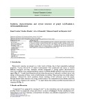
The 1H and 13C NMR spectra of the OPS (Fig. 1A) showed signals of different intensities, thus indicating a structural heterogeneity. The 13C NMR spectrum contained
Results
Serological characterization and classification of strains of P.syringaepv. porri in serogroup O9
Two MAbs, Ps9c and Ps9c1, were produced against type strain of P. syringae pv. porri, NCPPB 3364T. In ELISA, both MAbs strongly reacted with the homologous LPS whether it was crude or digested with proteinase K. In Western immunoblotting, only MAb Ps9c was reactive (data not shown). MAb Ps9c cross-reacted with all strains of P. syringae pv. porri (Table 1), except for strain NCPPB 3367, whereas MAb Ps9c1 recognized only strains NCPPB 3364T and NCPPB 3545. Strains of none of the other pathovars delineated in genomospecies 4 (Table 1) reacted with these MAbs.
Based on the reactivity with MAbs Ps9c and Ps9c1, strains of P. syringae pv. porri were classified in a new serogroup O9 as two serotypes designated correspondingly as O9(9c) and O9(9c,9c1). A stable epitope 9c is present in all strains of pathovar porri studied that have an S-form LPS, whereas only a few strains coexpose epitope 9c1 (Table 1). The inability of MAbs Ps9c and Ps9c1 to recognize P. syringae pv. porri NCPPB 3367 was accounted for by the R-form of LPS of this strain revealed by SDS/PAGE (data not shown). The crude LPS from strains P. syringae pv. porri NCPPB 3364T and NCPPB 3545 cross-reacted in ELISA with MAb Ps4a1, which is specific to LPS of strains from P. syringae serogroup O4 [17,29]. However, the reaction was only weak, epitope 4a1 was not stably expressed by OPS of this strain and was absent from LPS of P. syringae pv. porri NCPPB 3365. Therefore, the observed cross-reactivity is not suffi- cient for classification of strains of P. syringae pv. porri in serogroup O4 rather than in a new serogroup O9.
Fig. 1. 13C NMR spectra of the O polysaccharides of P. syringae pv. porri NCPPB 3365 (A) and NCPPB 3364T (B). Signals for anomeric carbons of the major O repeats are designated in the expansions as follows: G, GlcNAc; RI, RhaI; R2, RhaII; RIII, RhaIII; RIV, RhaIV; other major anomeric signals are superpositions of signals from minor O repeats (data of the two-dimensional 1H,13C HSQC spectra).
O polysaccharides of Pseudomonas syringae pv. porri (Eur. J. Biochem. 270) 23
(cid:3) FEBS 2003
signals for anomeric carbons at d 101.8–103.9, CH3-C groups (C6 of Rha residues) at d 17.9, one HOCH2-C group (C6 of GlcN) at d 61.9, one nitrogen-bearing carbon (C2 of GlcN) at d 57.1, sugar ring carbons linked to oxygen at d 70.5–79.1 and one N-acetyl group (CH3 at d 23.6, CO at d 175.4).
is the site of attachment of
The linkage and sequence analyses of the OPS were performed using a NOESY experiment. The NOESY spectrum contained the following correlations between the anomeric protons and the protons at the linkage carbons: RhaI H1/RhaIV H3, RhaII H1/RhaI H3, RhaIII H1/RhaII H3, RhaIV H1/RhaIII H2 and GlcNAcI H1/ RhaII H2 at d 5.05/3.85, 5.25/3.91, 5.25/3.99, 4.96/4.04 and 4.63/4.15, respectively. These data defined the sequence of rhamnose residues in the main chain and showed that RhaII the GlcNAc side chain. The NOESY data were in agreement with the methylation analysis data, and the glycosylation pattern was further confirmed by the 13C NMR chemical shift data (Table 3). Particularly, the positions of substi- tution of the rhamnose residues followed from downfield displacements of the signals for C3 of RhaI and RhaIV, C2 of RhaIII, C2 and C3 of RhaII to d 77.3–79.1, i.e. by 6–8 p.p.m. as compared with their positions in the
The assignment of the 1H and 13C NMR spectra of the OPS was performed using two-dimensional 1H,1H COSY, TOCSY and 1H,13C HSQC experiments, and spin systems for four major residues of Rha and one residue of GlcNAc were identified (Tables 2 and 3). A relatively large J1,2 coupling constant value of 8 Hz showed that the GlcNAc residue is b-linked. The a configuration of all rhamnosidic linkage followed from the comparison of the H5 and C5 NMR chemical shifts (Tables 2 and 3) with published data for a- and b-rhamnopyranose [32]. Therefore, the major O repeat of the OPS is a pentasaccharide consisting of four residues of a-L-Rha and one residue of b-D-GlcNAc.
Table 2. 1H NMR data of O polysaccharides of P. syringae pv. porri (d, p.p.m.). Assignment of the signals for H6 of rhamnose residues could be interchanged.
Chemical shift for
Monosaccharide residue H1 H2 H3 H4 H5 H6a H6b CH3CON
P. syringae pv. porri NCPPB 3365 O repeat 1 3.89 2.09
4.63 5.05 5.25 5.25 4.96 3.71 4.15 4.15 4.04 4.16 3.62 3.91 3.99 3.86 3.85 3.49 3.59 3.51 3.54 3.58 3.43 3.87 3.72 3.77 3.75 3.77 1.32 1.34 1.34 1.27 b-D-GlcpNAc-(1fi fi3)-a-L-RhapI-(1fi fi2,3)-a-L-RhapII-(1fi fi2)-a-L-RhapIII-(1fi fi3)-a-L-RhapIV-(1fi
P. syringae pv. porri NCPPB 3364T O repeat 2 3.89 2.09
4.61 5.18 5.00 5.17 5.12 3.72 4.19 4.12 4.08 4.09 3.59 3.89 3.76 3.96 3.91 3.47 3.48 3.62 3.49 3.47 4.42 3.71 3.78 3.83 3.70 3.76 1.27 1.34 1.30 1.29 b-D-GlcpNAc-(1fi fi2,3)-a-L-RhapI-(1fi fi3)-a-L-RhapII-(1fi fi2)-a-L-RhapIII-(1fi fi2)-a-L-RhapIV-(1fi
Table 3. 13C NMR data of O polysaccharides of P. syringae pv. porri (d, p.p.m). Assignment of the signals for N˜ 5 e` N˜ 6 of rhamnose residues could be interchanged.
Chemical shift for
Monosaccharide residue C1 C2 C3 C4 C5 C6 CH3CON CH3CON
P. syringae pv. porri NCPPB 3365 O repeat 1 23.6 175.4
103.9 103.4 102.5 101.8 103.1 57.1 71.2 79.1 79.1 71.1 74.4 79.1 77.3 71.4 79.1 71.3 72.6 73.5 73.5 72.7 76.8 70.5 70.6 70.6 70.6 61.9 17.9 17.9 17.9 17.9 b-D-GlcpNAc-(1fi fi3)-a-L-RhapI-(1fi fi2,3)-a-L-RhapII-(1fi fi2)-a-L-RhapIII-(1fi fi3)-a-L-RhapIV-(1fi
P. syringae pv. porri NCPPB 3364T O repeat 2 23.9 175.6
103.2 101.9 103.7 101.9 101.7 56.9 78.6 71.0 78.5 79.1 74.4 78.9 78.8 71.3 71.0 71.1 72.9 72.8 73.4 73.4 76.8 70.4 70.2 70.2 71.0 61.8 17.9 17.9 18.1 17.9 b-D-GlcpNAc-(1fi fi2,3)-a-L-RhapI-(1fi fi3)-a-L-RhapII-(1fi fi2)-a-L-RhapIII-(1fi fi2)-a-L-RhapIV-(1fi
24 E. L. Zdorovenko et al. (Eur. J. Biochem. 270)
(cid:3) FEBS 2003
spectrum of nonsubstituted a-rhamnopyranose [32]. The C2–C6 chemical shifts of the GlcNAc residue were close to those of nonsubstituted b-GlcNAc [32].
These data together showed that the major O repeat of the OPS of P. syringae pv. porri NCPPB 3365 has structure 1.
The O repeat 3 has been previously found as one of two linear O repeats in the OPS of P. syringae pv. garcae NCPPB 2708 [33]. Similar NMR spectroscopic studies of the OPS of P. syringae pv. atrofaciens IMV 948 showed that, in addition to the branched O repeat 4, whose structure was determined by us earlier [20] (Table 4), it also contains the minor O repeat 3.
fi3)-a-L-RhapIV-(1fi2)-a-L-RhapIII-(1fi3)-a-L-RhapII-(1fi3)-a-L-RhapI-(1fi (1)
2 › 1 b-D-GlcpNAc
Structural studies of the OPS of P.syringae pv. porri NCPPB 3364T
Sugar analysis of the OPS isolated by mild acid degradation of the LPS from P. syringae pv. porri NCPPB 3364T showed the presence of L-rhamnose (L-Rha) and 2-amino-2- deoxy-D-glucose (D-GlcN). Methylation analysis of the OPS
Studies of minor series in the NMR spectra of this OPS, including tracing connectivities in the two-dimensional spectra, showed that, in addition to the major O repeat 1, there is another branched O repeat, which is identical to the major O repeat in the OPS of P. syringae pv. porri NCPPB 3364T (structure 2, see below), and a linear O repeat 3 having the following structure:
fi2)-a-L-Rhap-(1fi2)-a-L-Rhap-(1fi3)-a-L-Rhap-(1fi3)-a-L-Rhap-(1fi (3)
Table 4. Structures of the O polysaccharides of P. syringae having a main chain of L-rhamnose tetrasaccharide O repeats and side chains of single D-GlcNAc residues.
O repeat structure Chemotype Serotype Reference Pathovar and strain
9C This work fi3)-a-L-Rhap-(1fi2)-a-L-Rhap-(1fi3)-a-L-Rhap-(1fi3)-a-L-Rhap- (1fi 1a O9(9c) O9(9c,9c1)
Porri NCPPB 3365, porri NCPPB 3364T 2 › 1 b-D-GlcpNAc fi2)-a-L-Rhap-(1fi2)-a-L-Rhap-(1fi3)-a-L-Rhap-(1fi3)-a-L-Rhap-(1fi 2a
2 › 1 b-D-GlcpNAc 3C O3(3c) [20] fi2)-a-L-Rhap-(1fi2)-a-L-Rhap-(1fi3)-a-L-Rhap-(1fi3)-a-L-Rhap-(1fi 4
Atrofaciens IMV 948
2 › 1 b-D-GlcpNAc 8C O8(8c) [19] fi2)-a-L-Rhap-(1fi2)-a-L-Rhap-(1fi3)-a-L-Rhap-(1fi3)-a-L-Rhap-(1fi 6 Ribicola NCPPB 1010
3 › 1 b-D-GlcpNAc fi3)-a-L-Rhap-(1fi2)-a-L-Rhap-(1fi3)-a-L-Rhap-(1fi3)-a-L-Rhap-(1fi 7
a The O repeat 1 is major in strain NCPPB 3365 and minor in strain NCPPB 3364T, and the O repeat 2 is major in strain NCPPB 3364T and minor in strain NCPPB 3365.
3 › 1 b-D-GlcpNAc
O polysaccharides of Pseudomonas syringae pv. porri (Eur. J. Biochem. 270) 25
(cid:3) FEBS 2003
revealed 2- and 3-substituted, and 3,4-disubstituted Rha in the ratios 10 : 1 : 3 as well as terminal GlcNAc.
each other in the position of substitution of one of the rhamnose residues (RhaIV) in the main chain and the site of attachment of the GlcNAc side chain (at RhaII or RhaI). Remarkably, both O repeats are present in each strain of P. syringae pv. porri studied, the O repeat 1 being major in strain NCPPB 3365 and 2 in strain NCPPB 3364T (Table 4).
The 1H and 13C NMR spectra of the OPS (Fig. 1B) showed signals of different intensities, thus indicating a structural heterogeneity. The 13C NMR spectrum contained signals for anomeric carbons at d 101.7–103.7, CH3-C groups (C6 of Rha residues) at d 17.9–18.1, one HOCH2- C group (C6 of GlcN) at d 61.8, one nitrogen-bearing carbon (C2 of GlcN) at d 56.9, sugar ring carbons linked to oxygen at d 70.2–79.1 and one N-acetyl group (CH3 at d 23.9, CO at d 175.6).
The assignment of the 1H and 13C NMR spectra of the OPS was performed as described above and the results are given in Tables 2 and 3. Again, the major pentasac- the OPS was identified, which charide O repeat of consists of four residues of L-Rha and one residue of D-GlcNAc. A relatively large J1,2 coupling constant value of 8 Hz for the H1 signal of the GlcNAc residue and the NMR chemical shifts of H5 and C5 of the rhamnose residues showed that the former is b-linked and the latter are a-linked.
In previous studies of structurally heterogeneous OPS of P. syringae having an L-rhamnan backbone, it has been demonstrated that both major and minor O repeats enter into the same polysaccharide chain, where they form blocks of structurally identical oligosaccharides [19,21,34,35]. This could be determined making use of a different behavior of the O repeats towards Smith degradation, from which only one was oxidized, whereas the other was stable. In the OPS of P. syringae pv. porri both major and minor O repeats are oxidizable by periodate, and therefore this approach could not be used to solve the problem. Assuming that biosyn- thesis of all L-rhamnan-based OPS of P. syringae proceeds by the same mechanism, it can be concluded that the O repeats of both types occur in the same polysaccharide chain in P. syringae pv. porri strains too.
The NOESY experiment revealed the following correla- tions between the anomeric protons and the protons at the linkage carbons: RhaI H1/RhaIV H2, RhaII H1/RhaI H3, RhaIII H1/RhaII H3, RhaIV H1/RhaIII H2 and GlcNAcI H1/RhaI H2 at d 5.18/4.09, 5.00/3.89, 5.17/3.76, 5.12/4.08 and 4.61/4.19, respectively. The glycosylation pattern was confirmed by downfield displacements of the signals for the linkage carbons, namely C3 of RhaII, C2 of RhaIII and RhaIV, and C2 and C3 of RhaI to d 78.5–79.1 (by 6–8 p.p.m.), and the similarity of the C2-C6 chemical shifts of the GlcNAc residue to those of nonsubstituted b-GlcNAc [32].
These data showed that the major O repeat of the OPS
The structural data of the OPS revealed the molecular basis for strong serological cross-reactivity of these strains and their classification in the same serogroup O9. Serolog- ical studies using MAbs Ps9c and Ps9c1 produced against P. syringae pv. porri NCPPB 3364T showed that all and only smooth strains of P. syringae pv. porri fell in the novel serogroup O9, which can be divided into two serotypes, O9(9c) or O9(9c,9c1) (Table 1). From the two correspond- ing epitopes on the LPS, only epitope 9c, which is common for all strains, was stable, whereas epitope 9c1, present only in a few strains, could be revealed only in ELISA and therefore can be considered as a conformational epitope. Epitope Ps9c, which is restricted to strains of P. syringae pv.
has structure 2:
fi2)-a-L-RhapIV-(1fi2)-a-L-RhapIII-(1fi3)-a-L-RhapII-(1fi3)-a-L-RhapI-(1fi (2)
2 › 1 b-D-GlcpNAc
porri, is evidently associated with the lateral b-GlcNAc residue but it remains unknown which O repeat, 1, 2 or both, carries this epitope.
Analysis of minor series in the NMR spectra of the OPS of P. syringae pv. porri strain NCPPB 3364T demonstrated that, in addition to the major O repeat 2, there are two minor O repeats: the branched O repeat 1 and the linear O repeat 3.
Discussion
Two major branched O repeats 1 and 2 present in the OPS of P. syringae pv. porri have the same monosaccha- rides composition and similar structures differing from
A weak cross-reactivity of the crude LPS from P. syrin- gae pv. porri NCPPB 3364T and NCPPB 3545 was observed in ELISA with MAb Ps4a1. This MAb has been produced against P. syringae pv. garcae ICMP 8047 and is specific to the L-rhamnan backbone. The cross-reactivity could be accounted for by the presence of the same of L-rhamnan main chain in OPS of P. syringae pv. porri (O repeat 1) and P. syringae pv. garcae ICMP 8047 [18] (O repeat 5).
fi2)-a-L-RhapIV-(1fi2)-a-L-RhapIII-(1fi3)-a-L-RhapII-(1fi3)-a-L-RhapI-(1fi (5)
4 › 1 a-D-Fucp3NAc
26 E. L. Zdorovenko et al. (Eur. J. Biochem. 270)
(cid:3) FEBS 2003
Acknowledgment
This work was supported by the Russian Foundation for Basic Research (grant 02-04-48721) and INTAS (grant YS 00–12).
References
LPS of smooth strains of P. syringae pv. porri did not react with MAb Pscor1, which is specific to the core oligosaccharide and recognizes LPS of most other P. syringae strains studied [23,31]. This suggests a difference in either the LPS core structure or/and in the mode of the attachment of the OPS to the core. Therefore, strains of pathovar porri are clearly distinct from other P. syringae pathovars in serology of both OPS moiety and LPS core. Strains of this pathovar are also distinguished in a number of other phenotypic and genotypic characters [4,24,25]. These data together suggest that P. syringae pv. porri is a separate ancestral line that can be identified on the basis of distinctive chemical characters.
1. Young, J.M., Saddler, G.S., Takikawa, Y., DeBoer, S.H., Vau- terin, L., Gardan, L., Gvozdyak, R.I. & Stead, D.E. (1996) Names of plant pathogenic bacteria 1864–1995. ISPP Subcommittee on Taxonomy of Plant Pathogenic Bacteria. Rev. Plant Pathol. 75, 721–763.
2. Hirano, S.S. & Upper, C.D. (1990) Population biology and epi- demiology of Pseudomonas syringae. Annu. Rev. Phytopathol. 28, 155–177.
3. Lattore, B.A. & Jones, A.L. (1979) Evaluation of weeds and plant refuse as potential sources of inoculum of Pseudomonas syringae in bacterial canker of cherry. Phytopathology 69, 1122–1125.
4. Gardan, L., Shafik, H., Belouin, S., Brosch, R., Grimont, F. & Grimont, P.A.D. (1999) DNA relatedness among the pathovars of P. syringae and description of Pseudomonas tremae sp. nov. & Pseudomonas cannabina sp. nov. (ex. Sutic and Dowson 1959). Int. J. Syst. Bacteriol. 49, 469–478.
5. Schaad, N.W., Vidaver, A.K., Lacy, G.L., Rudolph, K. & Jones, J.B. (2000) Evaluation of proposed amended names of several Pseudomonads and Xanthomonads and recommendations. Phyto- pathology 90, 208–213.
6. Pecknold, P.C. & Grogan, R.C. (1973) Deoxyribonucleic acid homology groups among phytopathogenic Pseudomonas species. Int. J. Syst. Bacteriol. 23, 111–121.
The pathotype strain of P. syringae pv. porri, NCPPB 3364T, was delineated in genomospecies 4 [4]. It showed as much as 78–95% DNA–DNA homology with the patho- type strains of the other pathovars delineated in genomo- species 4, namely P. syringae pvs. garcae, atropurpurea, oryzae, porri, striafaciens, zizaniae, and Pseudomonas coronafaciens, which altogether constitute a distinct ribo- group F [4]. Studies of the representative strains of these pathovars using ELISA and Western immunoblotting with MAbs specific to the OPS and LPS core showed their serological heterogeneity (Table 1). Most strains from genomospecies 4 belong to three serotypes: O3(3c) [29], O4(4a1,4e) (authors’ unpublished data), and O9(9c) (this work), which correspond to OPS chemotypes 3C, 4E1-I, and 9C, respectively. The less common serotype O8(8c) and the corresponding chemotype 8C, which has been described earlier for P. syringae pv. ribicola NCPPB 1010 [17], is characteristic of only one strain from genomospecies 4, namely the pathotype strain of P. syringae pv. oryzae, NCPPB 3683T.
7. Palleroni, N.J. (1984) Genus I. Pseudomonas Migula 1894. 237AL. In Bergey’s Manual of Systematic Bacteriology, 1 (Krieg, N.R. & Holt, G.D., eds), pp. 141–199. William & Wilkins, Baltimore, London.
8. Denny, T.P., Gilmour, M.N. & Selander, R.K. (1988) Genetic diversity and relationships of two pathovars of Pseudomonas syringae. J. Gen. Microbiol. 134, 1949–1960.
9. Janse, J.D., Rossi, P., Angelucci, L., Scortichini, M., Derks, J.H., Akkermans, A.D.L., De Vrijer, R. & Psallidas, P.G. (1996) Reclassification of Pseudomonas syringae pv. avellanae as Pseu- domonas avellanae (spec. nov.), the bacterium causing cancer of hazelnut (Corylus avellana L.). System. Appl. Microbiol. 19, 598– 595.
10. Manceau, C. & Horvais, A. (1997) Assessment of genetic diversity among strains of Pseudomonas syringae by PCR-restriction frag- ment length polymorphism analysis of rRNA operons with special emphasis on P. syringae pv. tomato. Appl. Environ. Microbiol. 63, 498–505.
11. Clerc, A., Manceau, C. & Nesme, X. (1998) Comparison of ran- domly amplified polymorphic DNA with amplified fragment length polymorphism to assess genetic diversity and genetic relatedness within genospecies III of Pseudomonas syringae. Appl. Environ. Microbiol. 64, 1180–1187.
12. Young, J.M., Takikawa, Y., Gardan, L. & Stead, D.E. (1992) Changing concepts in the taxonomy of plant pathogenic bacteria. Annu. Rev. Phytopathol. 30, 67–105.
OPS of strains from genomospecies 4 have marked compositional and structural similarities. Particularly, they all have a backbone of a-(1fi2)- and a-(1fi3)- linked L-rhamnose residues and lack a strict regularity owing to the occurrence of several types of O repeats in the main chain. The OPS are either linear (chemotype 4A) or branched with side chains of single a-D-Fuc3NAc residues (chemotypes 4E0, 4E1-I, and 4E2) or b-D- GlcNAc residues (chemotypes 3C, 8C, and 9C) (Table 1). The OPS of chemotypes 3C, 8C and 9C differ from each other in the site of the attachment of b-D-GlcNAc residues to the main L-rhamnan chain (Table 4). The OPS of P. syringae pv. atrofaciens IMV 948 [20] resembles most closely that of P. syringae pv. porri NCPPB 3364T: both OPS have similar major branched O repeats 4 and 2, respectively (Table 4), and the same minor linear O repeat 3. In spite of this similarity, neither P. syringae pv. atrofaciens IMV 948 (chemotype 3C) nor P. syringae pv. ribicola NCPPB 1010 [19] (chemotype 8C, O repeats 6 and 7, Table 4) is serologically related to P. syringae pv. porri (chemotype 9C), and, accordingly, they were classified into different serogroups O3 and O8, respectively.
Genetic and antigenic
the same ancestral origin of
13. Young, J.M. & Triggs, C.M. (1994) Evaluation of determinative tests for pathovars of Pseudomonas syringae van Hall 1902. J. Appl. Bacteriol. 77, 195–207.
(chemical and serological) similarities suggest the strains from pathovars delineated in genomospecies 4, and their antigenic diversity may result from a divergent evolution of the bacteria during a relatively short period of time.
14. Ovod, V., Knirel, Y.A., Samson, R. & Krohn, K. (1999) Immunochemical characterization and taxonomic evaluation of the O polysaccharides of the lipopolysaccharides of Pseudomonas syringae serogroup O1 strains. J. Bacteriol. 181, 6937–6947. 15. Knirel, Y.A. & Zdorovenko, G.M. (1997) Structures of O-poly- saccharide chains of lipopolysaccharides as the basis for classifi- cation of Pseudomonas In syringae and related strains.
O polysaccharides of Pseudomonas syringae pv. porri (Eur. J. Biochem. 270) 27
(cid:3) FEBS 2003
26. Sawardeker, J.S., Sloneker, J.H. & Jeanes, A. (1965) Quantitative determination of monosaccharides as their alditol acetates by gas liquid chromatography. Anal. Chem. 37, 1602–1603. and Related Pathogens Pseudomonas Syringae Pathovars (Rudolph, K., Burr, T.J., Mansfield, J.W., Stead, D., Vivian, A. & von Kietzell, J., eds), pp. 475–480. Kluwer Academic Publishers, Dordrecht, Boston, London.
27. Leontein, K., Lindberg, B. & Lo¨ nngren, J. (1978) Assignment of absolute configuration of sugars by g.1.c. of their acetylated glycosides formed from chiral alcohols. Carbohydr. Res. 62, 359–362. 16. Knirel, Y.A., Ovod, V.V., Paramonov, N.A. & Krohn, K. (1998) Heterogeneity in the O polysaccharide structure of Pseudomonas syringae pv. coriandricola GSPB 2028 (W-43). Eur. J. Biochem. 258, 716–721.
28. Ciucanu, I. & Kerek, F. (1984) A simple and rapid method for the permethylation of carbohydrates. Carbohydr. Res. 131, 209–217. (1998) Structure of
17. Knirel, Y.A., Ovod, V.V., Zdorovenko, G.M., Gvozdyak, R.I. & Krohn, K. the O polysaccharide and immunochemical relationships between the lipopolysaccharides of Pseudomonas syringae pathovar tomato and pathovar maculicola. Eur. J. Biochem. 258, 657–661.
29. Ovod, V., Rudolph, K. & Krohn, K. (1997) Serological classifi- cation of Pseudomonas syringae pathovars based on monoclonal antibodies towards the lipopolysaccharide O-chains. In Pseudo- monas Syringae Pathovars and Related Pathogens (Rudolph, K., Burr, T.J., Mansfield, J.W., Stead, D., Vivian, A. & von Kietzell, J., eds), pp. 526–531. Kluwer Academic Publishers, Dordrecht, Boston, London. 18. Zdorovenko, E.L., Ovod, V., Shashkov, A.S., Kocharova, N.A., Knirel, Y.A. & Krohn, K. (1999) Structure of the O-poly- saccharide of the lipopolysaccharide of Pseudomonas syringae pv. garcae ICMP 8047. Biochemistry (Moscow) 64, 765–773.
19. Ovod, V., Zdorovenko, E.L., Shashkov, A.S., Kocharova, N.A. & Knirel, Y.A. (2000) Structure of the O polysaccharide and ser- ological classification of Pseudomonas syringae pv. ribicola NCPPB 1010. Eur. J. Biochem. 267, 2372–2379.
30. Ovod, V., Knirel, Y. & Krohn, K. (1997) Demonstration of the immunochemical diversity of O-chains of lipopolysaccharide of Pseudomonas syringae and inferring of the serogroup- and sero- type-specific epitopes with monoclonal antibodies. In Pseudo- monas Syringae Pathovars and Related Pathogens (Rudolph, K., Burr, T.J., Mansfield, J.W., Stead, D., Vivian, A. & von Kietzell, J., eds), pp. 532–537. Kluwer Academic Publishers, Dordrecht, Boston, London.
31. Ovod, V., Ashorn, P., Yakovleva, L. & Krohn, K. (1995) Classi- fication of Pseudomonas syringae with monoclonal antibodies against the core and O-side chains of the lipopolysaccharide. Phytopathology 85, 226–232.
20. Zdorovenko, G.M., Shashkov, A.S., Zdorovenko, E.L., Kochar- ova, N.A., Yakovleva, L.M., Knirel, Y.A. & Rudolph, K. (2001) Characterization of the lipopolysaccharide and structure of the O-specific polysaccharide of the bacterium Pseudomonas syringae pv. atrofaciens IMV 948. Biochemistry (Moscow) 66, 369–377. 21. Zdorovenko, E.L., Zatonsky, G.V., Zdorovenko, G.M., Pasich- nik, L.A., Shashkov, A.S. & Knirel, Y.A. (2001) Structural het- erogeneity in the lipopolysaccharides of Pseudomonas syringae with O-polysaccharide chains having different repeating units. Carbohydr. Res. 336, 329–336.
32. Jansson, P.-E., Kenne, L. & Widmalm, G. (1989) Computer- assisted structural analysis of polysaccharides with an extended version of CASPER using 1H- and 13C-n.m.r. data. Carbohydr. Res. 188, 169–191.
22. Zdorovenko, E.L., Ovod, V., Zatonsky, G.V., Shashkov, A.S., Kocharova, N.A. & Knirel, Y.A. (2002) Structure of the O polysaccharide of Pseudomonas syringae pv. delphinii NCPPB 1879T having side chains of multiple 3-acetamido-3,6-dideoxy- D-galactose residues. Biochemistry (Moscow) 67, 558–565.
33. Zdorovenko, E.L., Knirel, Y.A. & Ovod, V.V. (1999) Structures of O-polysaccharide chains of Pseudomonas syringae pv. garcae LPS. In Abstract. 13th International Congress of the Hungarian Society for Microbiology, Budapest, Hungary, 29 August)1 Sep- tember 1999, p. 112.
23. Ovod, V., Rudolph, K., Knirel, Y.A. & Krohn, K. (1996) Immunochemical characterization of O polysaccharides compos- ing the a-D-rhamnose backbone of lipopolysaccharide of Pseu- domonas syringae and classification of bacteria into serogroups O1 and O2 with monoclonal antibodies. J. Bacteriol. 178, 6459–6465. 24. Koike, S.T., Barak, J.D., Henderson, D.M. & Gilbertson, R.L. (1999) Bacterial blight of leek: a new disease in California caused by Pseudomonas syringae. Plant Dis. 83, 165–170.
34. Knirel, Y.A., Zdorovenko, G.M., Shashkov, A.S., Mamyan, S.S., Gubanova, N.Y., Yakovleva, L.M. & Solyanik, L.P. (1988) the Antigenic polysaccharides of bacteria. 30. Structure of polysaccharide chain of the Pseudomonas syringae pv. syringae 281 (serogroup I) lipopolysaccharide. Bioorg. Khim. 14, 180–186. 35. Knirel, Y.A., Zdorovenko, G.M., Shashkov, A.S., Yakovleva, L.M., Gubanova, N.Y. & Gvozdyak, R.I. (1988) Antigenic polysaccharides of bacteria. 29. Structure of the O-specific poly- saccharide chain of the Pseudomonas holci 8300 (serogroup I) lipopolysaccharide. Bioorg. Khim. 14, 172–179. 25. Samson, R., Shafik, H., Benjama, A. & Gardan, L. (1998) Description of the bacterium causing blight of leek as Pseudo- monas syringae pathovar porri (pathovar nov.). Phytopathology 88, 844–850.