Suppression of microtubule dynamics by benomyl decreases tension across kinetochore pairs and induces apoptosis in cancer cells K. Rathinasamy and D. Panda
School of Biosciences and Bioengineering, Indian Institute of Technology Bombay, Mumbai, India
Keywords apoptosis; benomyl; bcl2; centrosomes; microtubule dynamics
Correspondence D. Panda, School of Biosciences and Bioengineering, Indian Institute of Technology Bombay, Powai, Mumbai, 400 076, India Fax: +91 22 257 23480 Tel: +91 22 257 67838 E-mail: panda@iitb.ac.in
(Received 9 May 2006, revised 27 June 2006, accepted 11 July 2006)
doi:10.1111/j.1742-4658.2006.05413.x
We found that benomyl, a benzimidazole fungicide, strongly suppressed the reassembly of cold-depolymerized spindle microtubules in HeLa cells. Benomyl perturbed microtubule-kinetochore attachment and chromosome alignment at the metaphase plate. Benomyl also significantly decreased the distance between the sister kinetochore pairs in metaphase cells and increased the level of the checkpoint protein BubR1 at the kinetochore region, indicating that benomyl caused loss of tension across the kineto- chores. In addition, benomyl decreased the intercentrosomal distance in mitotic HeLa cells and blocked the cells at mitosis. Further, we analyzed the effects of benomyl on the signal transduction pathways in relation to mitotic block, bcl2 phosphorylation and induction of apoptosis. The results suggest that benomyl causes loss of tension across the kinetochores, blocks the cell cycle progression at mitosis and subsequently, induces apoptosis through the bcl2–bax pathway in a manner qualitatively similar to the powerful microtubule targeted anticancer drugs like the vinca alkaloids and paclitaxel. Considering the very high toxicity of the potent anticancer drugs and the low toxicity of benomyl in humans, we suggest that benomyl could be useful as an adjuvant in combination with the powerful anticancer drugs in cancer therapy.
shown to cause impaired liver
Benomyl is a benzimidazole fungicide that is widely used in agriculture against a range of fungal diseases of field crops, fruit trees and ornamentals. It is a broad spectrum systemic fungicide that is selectively toxic to microorganisms [1]. The acute toxicity of benomyl is reported to be very low [median lethal dose (LD50) of (cid:2) 10 gÆkg)1] in rats [1]. However, other studies sugges- larger than ted that benomyl at a single dose of 100 mgÆkg)1 is capable of inducing testicular toxicity in experimental animals [2,3]. Chronic administration of benomyl at doses higher than 500 mgÆkg)1 in mice function and was increased liver weights [4]. Most of the toxicity studies are performed using very high dosages of benomyl, which are unlikely to be used at the therapeutic level
in humans. Benomyl and its major metabolite carbend- azim have been shown to exhibit diffential sensitivity against fungal tubulin and mammalian brain tubulin [5,6]. It is believed that the selective toxicity of beno- is due to its higher affinity for fungal myl to fungi tubulin than for mammalian tubulin [5]. Benomyl is extensively used as a research tool in fungal genetics and cell biology [7–9]. Recently, it has been shown that benomyl inhibits mitosis in mammalian cells, inhibits polymerization of purified mammalian tubulin into microtubules and suppresses dynamic instability of reconstituted bovine brain microtubules in vitro [10]. Benomyl binds to mammalian tubulin with a moderate affinity and the binding of benomyl to tubulin is shown to induce conformational changes in tubulin
Abbreviations CI, congression index; DAPI, 4¢,6-diamidino-2-phenylindole; FITC, fluorescein isothiocyanate; IC50, half-maximal inhibitory concentration; PARP, poly (ADP ribose) polymerase; SRB, sulforhodamine B.
FEBS Journal 273 (2006) 4114–4128 ª 2006 The Authors Journal compilation ª 2006 FEBS
4114
K. Rathinasamy and D. Panda
Antiproliferative mechanism of action of benomyl
the propensity to form homodimers or heterodimers. It is believed that the heterodimerization of bcl2 and bax prevents the cell from undergoing bax mediated apop- tosis [27]. Hence, the balance between bcl2–bax het- erodimer and bax–bax homodimer determines the fate of a cell [27,28].
[10]. The binding site of benomyl in tubulin is yet to be determined. However, it has been suggested that benomyl binds to tubulin at a site that is distinct from the colchicine and vinbastine binding sites in tubulin [10,11]. Although benomyl inhibits mitosis in mamma- lian cells, its antimitotic mechanism of action is not clearly understood.
In this study we found that benomyl suppressed the regrowth of spindle microtubules in HeLa cells and per- turbed the attachment of microtubules to kinetochores, leading to mitotic irregularities in the cells arrested at mitosis. The relatively nontoxic benomyl was, thus far, considered to be a systemic fungicide targeting fungal microtubules. Here, we show that benomyl also targets mammalian microtubule assembly dynamics, disrupts the microtubule–kinetochore interactions, decreases the tension across the sister kinetochores, and activates the spindle checkpoint protein BubR1 in the mitotically arrested HeLa cells. We also present evidence indicating that the cells blocked at mitosis were eliminated by apoptosis through the bcl2–bax pathway. The results suggest that the suppression of microtubule dynamics by benomyl was the cause for the loss of tension across the kinetochores and activation of the checkpoint pro- teins and induction of apoptosis.
Results
Benomyl suppressed the reassembly of spindle microtubules in HeLa cells
Microtubules are dynamic cytoskeletal polymers in all eukaryotic cells that play which are present important roles in various cellular processes such as cell signaling, cell motility, organelle transport and maintenance of cell polarity, separation of the duplica- ted centrosomes, and in cell division and mitosis [12– 19]. At the onset of mitosis, the interphase microtubule network rapidly disassembles and reorganizes to form bipolar mitotic spindles [13]. The interactions of spin- dle microtubules and kinetochores play an important role in the congression of chromosomes at the meta- phase plate [14]. Kinetochores are specialized pairs of disc shaped structures that are located on either side of the centromere, through which the chromosomes are [15,16]. The attached to the spindle microtubules microtubules attached to the kinetochores are called kinetochore fibres. Several lines of evidence indicate that the checkpoint proteins like Mad2, Bub1 and BubR1 can sense the attachment of microtubules and kinetochores, and the tension across the sister chrom- atids [17]. Kinetochores that are not attached to the microtubules acquire increased concentration of the motor proteins and the spindle checkpoint proteins [18]. The interaction of kinetochores with the micro- tubules, resulting in the formation of the kinetochore fibre, leads to a reduction in the concentration of the motor proteins and spindle checkpoint proteins [19]. This reduction of checkpoint proteins is required for inactivating the spindle checkpoint signal and progres- sion in the cell cycle [20].
The functions of microtubules are thought to be highly dependent on the assembly dynamics of micro- It tubules is well established that minor [12,13]. perturbation of the microtubule dynamics by the microtubule targeted drugs like the vinca alkaloids, taxanes, noscopaine, and griseofulvin, arrest the cell cycle progression at mitosis [13,21–24]. Hence, micro- tubule dynamics acts as a potential target for most of the successful anticancer drugs. Cells arrested in the cell cycle will be eliminated by apoptosis, executed either through the p53 pathway or bcl2 pathway depending upon the inducer of apoptosis. The bcl2 pathway involves the bcl2 family of proteins consisting of the proapoptotic proteins like bax, bad, bid, bak and the antiapoptotic proteins like bcl2, bcl-XL, bcl- W, and Bfl1 [25,26]. The bcl2 family of proteins have
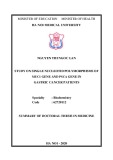
HeLa cells were incubated at 2 (cid:2)C for 1 h. Then, the cold media was replaced with warm media containing different concentrations of benomyl. Subsequently, the cells were incubated at 37 (cid:2)C in a CO2 incubator. The cold treatment caused complete depolymerization of mitotic spindle microtubules as observed by fluores- cence microscopy after immunostaining the HeLa cells with antia-tubulin IgG (Fig. 1). In the absence of spin- dle microtubules, centrosomes were located near the spindle equator and the chromosomes were compactly aligned at the metaphase plate (Fig. 1). In control HeLa cells, mitotic spindle microtubules were reassem- incubation at 37 (cid:2)C (Fig. 1), bled within 5 min of while benomyl caused a significant delay in the reas- sembly of the spindle microtubules. In the presence of 5 lm benomyl, the spindles were only partially reas- sembled after 5 min of incubation at 37 (cid:2)C (Fig. 1). They appeared to be in the initial stage of recovery near the centrosomes. After 10 min of incubation, mitotic spindles had considerable amount of micro- tubules, although their spindle lengths (6.9 ± 1.0 lm*) were found to be significantly shorter than that of the control cells (10.1 ± 1.3 lm*). Mitotic cells treated
FEBS Journal 273 (2006) 4114–4128 ª 2006 The Authors Journal compilation ª 2006 FEBS
4115
K. Rathinasamy and D. Panda
Antiproliferative mechanism of action of benomyl
Fig. 1. Benomyl suppressed the reassembly of cold depolymerized spindle microtubules in HeLa cells. Twenty-four hours after seeding, the cells were cold treated (2 (cid:2)C) for 1 h to disassemble the spindles. Then, the cells were incubated in a CO2 incubator at 37 (cid:2)C in the absence and presence of different concentrations of benomyl. The cells were fixed at the desired time points and stained with antic-tubulin (green), antia-tubulin (red) IgG and DAPI (blue) to visualize the centrosomes, microtubules and DNA, respectively. Bars, 5 lm.
with 20 lm benomyl did not recover their spindle microtubules even after 10 min at 37 (cid:2)C. After 20 min of incubation, microtubules appeared near the centro- somes and the chromosomes were found to be in condensed structures (Fig. 1). However, the average spindle length was measured (3.65 ± 0.64 lm*) and shown to be considerably smaller than that of the con- trol cells (10.1 ± 1.3 lm*) (*P < 0.01, n ¼ 15 cells).
Benomyl caused disruption of chromosomal alignment at the metaphase plate
HeLa cells treated with vehicle alone displayed normal spindle morphology and the mitotic chromosomes were properly aligned at the metaphase plate (Fig. 2A). As documented recently [10], the spindle microtubules
in the cells treated with 5 lm benomyl appeared nearly normal, but a few chromosomes were unable to con- gress at the metaphase plate (Fig. 2A). The effects of microtubule perturbation by benomyl on the chromo- somal alignment were assessed by determining the chromosomal congression index (CI), which is a measure of the ratio of the width to height of the chromosomal masses of cells with metaphase type chromosome alignment [29]. Consistent with the previ- ous report [29], the CI for the control HeLa cells was found to be 0.31 ± 0.05. Benomyl treatment strongly increased the CI (Fig. 2B). For example, in the pres- ence of 5 lm benomyl, the CI was increased by 58% from 0.31 ± 0.05 to 0.49 ± 0.11, and the CI was increased by 74% in the presence of 10 lm benomyl (Fig. 2B). Finally, higher concentrations of benomyl
FEBS Journal 273 (2006) 4114–4128 ª 2006 The Authors Journal compilation ª 2006 FEBS
4116
K. Rathinasamy and D. Panda
Antiproliferative mechanism of action of benomyl
A
B
0.8
) I C
0.6
0.4
0.2
( x e d n I n o i s s e r g n o C
0
0
10.0
5.0 Benomyl (μM)
Fig. 2. Effects of benomyl on the spindle microtubules and chromosome organization. (A) HeLa cells were incubated with different concen- trations of benomyl for 20 h. Spindle microtubules (green) and chromosomes (blue) were analyzed as described in Experimental procedures. Bars, 5 lm. (B) The chromosomal congression index (CI) of the control and drug treated HeLa cells. The CI was calculated as described in Experimental procedures. Error bars represent SD.
(20 lm) produced clear abnormalities in chromosomal alignment with nearly 70% of all the mitotic cells hav- ing multiple poles with short spindles and the chromo- somes were rounded to a ball shaped structure (data not shown).
The
1.74 ± 0.3 lm* (n ¼ 75). This distance was signifi- cantly reduced by 29% and 34% in the presence of 5 and 10 lm benomyl, being equal to 1.25 ± 0.4 lm* (n ¼ 96) and 1.15 ± 0.4 lm* (n ¼ 98) respectively (*P < 0.01) (Fig. 3B). results
Benomyl perturbed microtubule–kinetochore attachment and caused loss of tension across the sister kinetochores
attachment of microtubules
(Fig. 4), a significant decrease
cells
All the kinetochores of vehicle treated HeLa cells were found to be attached to the spindle microtubules and the chromosomes were properly aligned at the meta- phase plate (Fig. 3A). Benomyl treatment perturbed the to kinetochores (Fig. 3A). In benomyl treated cells, some kinetochore pairs were found to be attached to kinetochore fibres from both the poles whereas others were attached to the kinetochore fibre from one side of the pole, result- ing in misalignment of chromosomes at the metaphase plate.
It has been shown that
(Fig. 4). Benomyl
the tension generated between kinetochore pairs is finely regulated by the combined action of microtubule dynamics and motor proteins [30,31]. The tension across the paired kineto- chores is thought to be proportional to the distance between the sister kinetochore pairs [31–33]. In control cells the distance between the sister kinetochores was
suggested that benomyl perturbed microtubule–kinetochore interactions and caused a reduction in the tension exerted by microtubules on kinetochores. This was further investigated by limited cold treatment of the mitotic spindles and calcium induced depolymerization of nonkinetochore micro- tubules. It has been shown that limited cold treat- ment disassembles most of the spindle microtubules but keeps the kinetochore fibres intact [34]. HeLa cells were thus incubated on ice for 10 min in the absence and presence of benomyl. While the stable kinetochore microtubules were present in the con- in the trol amount of cold stable microtubules was observed in the presence of 5 lm benomyl (Fig. 4). At 20 lm benomyl, almost all the spindle microtubules were disassembled indicating a concentration dependent perturbation of microtubule–kinetochore interactions by benomyl caused a similar concentration dependent decrease in the quantity of spindle microtubules in the presence of 1 mm calcium chloride in a microtubule stabilizing buffer (data not shown).
FEBS Journal 273 (2006) 4114–4128 ª 2006 The Authors Journal compilation ª 2006 FEBS
4117
K. Rathinasamy and D. Panda
Antiproliferative mechanism of action of benomyl
A
B
24
24
Control
20
20
5 μM Benomyl
16
16
12
12
d e t n u o C s r i a P
d e t n u o C s r i a P
8
8
4
4
l a t o T %
l a t o T %
0.8
1.2
1.6
2.0
2.4
0 0.4
0 0.4
0.8
1.2
1.6
2.0
2.4
Sister Kinetochore Distance (μm)
Sister Kinetochore Distance (μm)
24
20
10 μM Benomyl
16
12
d e t n u o C s r i a P
8
4
l a t o T %
0 0.4
0.8
1.2
1.6
2.0
2.4
Sister Kinetochore Distance (μm)
Fig. 3. Benomyl reduced the tension at the kinetochores. (A) Immunofluorescent ima- ges of mitotic spindle of HeLa cells, cen- tromeres (green), DNA (blue) and merged images of microtubules (red) and centrom- eres are shown. HeLa cells were treated with 5 and 10 lM benomyl and compared with those of the vehicle treated (control) cells. Bars, 5 lm; inset bars, 1 lm. (B) Benomyl decreased the distance between the sister kinetochores. Sister kinetochore distances were measured as described in Experimental procedures.
FEBS Journal 273 (2006) 4114–4128 ª 2006 The Authors Journal compilation ª 2006 FEBS
4118
K. Rathinasamy and D. Panda
Antiproliferative mechanism of action of benomyl
Fig. 4. Effects of benomyl on the kinetochore–microtubule attachment: Cells were first incubated with different concentrations (0, 5, and 20 lM) of benomyl for 10 min at 37 (cid:2)C. Then, the warm media was replaced with ice-cold media containing the same concentration of beno- myl and incubated on ice for 10 min and subsequently fixed and processed for immunofluorescence using antitubulin IgG (red) and DAPI (blue). Bars, 5 lm.
Activation of the spindle checkpoint protein BubR1 by benomyl treatment
treatment, BubR1 was
benomyl (5 and 10 lm), BubR1 was localized in large quantities at the kinetochores on both the chromo- somes that are aligned at the metaphase plate and those that are not aligned at the metaphase plate (Fig. 5). The results indicated that the sister kineto- chores are not under tension in the presence of beno- myl, resulting in the inhibition of degradation of the checkpoint protein BubR1 at the kinetochore region.
Benomyl treatment decreased the spindle length and intercentrosomal distance
The movement of centrosomes towards the opposite poles is thought to be microtubule dependent and it is
To investigate the status of the checkpoint protein treatment, we examined BubR1 following benomyl the cellular the checkpoint protein localization of BubR1 in the benomyl arrested mitotic cells. In the control localized to the kinetochore region in the prometaphase cells and upon chromosomal alignment the concentration of BubR1 was decreased at the kinetochore region. In some control cells a very small amount was detectable in the kinetochore region of the metaphase chromo- somes and in other cells BubR1 was undetectable after the chromosomal alignment. In the presence of
FEBS Journal 273 (2006) 4114–4128 ª 2006 The Authors Journal compilation ª 2006 FEBS
4119
K. Rathinasamy and D. Panda
Antiproliferative mechanism of action of benomyl
Fig. 5. Localization patterns of the checkpoint protein BubR1. Cells treated with 5 lM benomyl for 20 h were compared with the control metaphase cells. BubR1 (green) and DNA (blue) were stained and visualized as described in Experimental procedures. Bars, 5 lm.
carried out in association with motor proteins and other cytoskeleton network like actin filaments [35]. In order to check the effect of benomyl on centrosomes and the spindle length, the cells were stained to visual- and microtubules. The distance ize
centrosomes
between the two centrosomes in the mitotic cells that had achieved a metaphase type of chromosomal align- ment was 11.1 ± 1.5 lm* (n ¼ 60) (Fig. 6). Centro- some separation in the mitotic cells was affected significantly in the presence of benomyl, with an
FEBS Journal 273 (2006) 4114–4128 ª 2006 The Authors Journal compilation ª 2006 FEBS
4120
K. Rathinasamy and D. Panda
Antiproliferative mechanism of action of benomyl
A
B
16
12
8
4
s e l o P e h t n e e w t e B e c n a t s i D
0
0
5.0
10.0
Benomyl (μM)
Fig. 6. Benomyl reduced the intercentrosomal distance. (A) Immunofluorescent images showing centrosomes (green), merged images of microtubules (red) and centrosomes, and merged images of microtubules, centrosomes and DNA (blue) in metaphase HeLa cells. HeLa cells were treated with 5 and 10 lM benomyl and compared with those of the vehicle treated (control) cells. (B) Benomyl treatment caused a reduction of intercentrosomal distance. The distance between the intercentrosomes was measured as described in Experimental proce- dures. Error bars represent SD.
intercentrosomal distance of 7.9 ± 1.5 lm* at 5 lm (n ¼ 87) (Fig. 6) (29% reduction compared to control), and 6.9 ± 2.2 lm* at 10 lm (n ¼ 75) (38% reduction compared to control) (Fig. 6) (*P < 0.01). The interc- entrosomal distance was calculated only for those cells having bipolar centrosomal organization. Cells treated with 20 lm benomyl showed huge abnormalities in the centrosomal organization; nearly 40% of all the pro- phase cells showed three to four centrosomal struc- tures, while the control cells had one centrosome in interphase and two in prophase, which were separated by 2–10 lm in different cells.
Benomyl arrests the cells at mitosis, inhibits the proliferation of HeLa cells and induces apoptosis
Consistent with a previous report [10], we found that benomyl inhibited HeLa cell proliferation with an IC50 of 5 ± 1 lm (Fig. 7A) and arrested the cell cycle
progression at mitosis in a concentration dependent fashion. In the present work, we found that the mitotic block caused by benomyl paralleled its ability to inhibit cell proliferation. For example, 5 and 20 lm benomyl blocked 26% and 59% of cells at mitosis and inhibited cell proliferation by 50% and 70%, respect- ively (Fig. 7A). In order to find the fate of the cells arrested at mitosis, cells were incubated with different concentrations (0–40 lm) of benomyl for 20 or 40 h and the live and dead ⁄ apoptotic cells were counted using the combination of hoechst 33342 and propidium iodide staining. Total cells were counted by hoechst fluorescence and the dead or apoptotic cells by propi- dium iodide fluorescence. After 20 h of drug treatment 8% and 23% of dead ⁄ apoptotic cells were detected at 5 and 20 lm benomyl, respectively, whereas, at the same concentrations 19% and 42% of dead ⁄ apoptotic cells were detected after 40 h of benomyl treatment (Fig. 7B). The number of cells undergoing death or
FEBS Journal 273 (2006) 4114–4128 ª 2006 The Authors Journal compilation ª 2006 FEBS
4121
K. Rathinasamy and D. Panda
Antiproliferative mechanism of action of benomyl
A
100
100
benomyl from the culture media indicating that the continued presence of benomyl is required to inhibit cell proliferation (data not shown).
75
75
n o i t a r e f i l o r P
50
50
l l e C
Benomyl induced apoptosis in HeLa cells was confirmed by the cleavage of poly (ADP) ribose polymerase and the fragmentation of DNA
s l l e C c i t o t i
M %
25
25
f o n o i t i b i h n I
%
0
0
0
40
60
We found that the enzyme poly (ADP ribose) poly- merase (PARP) (specifically cleaved in many forms of programmed cell death [36,37]) was cleaved in HeLa cells upon benomyl treatment for 40 h (Fig. 8A). The apoptosis induced by benomyl was also confirmed by the presence of a DNA laddering pattern (Fig. 8B).
20 Benomyl (μM)
B
80
Benomyl caused an increase in the hyperphosphorylation of bcl2
Dead cells (20 h)
Dead cells (40 h)
60
40
s l l e C d a e D %
20
0
0
2
20
40
unphosphorylated
phosphorylated
5 Benomyl (μM)
After 20 h treatment of HeLa cells with benomyl, the level of phosphorylation of bcl2 increased in a concen- tration dependent manner as evidenced by the decreased mobility of the protein (Fig. 9A). At 5 lm concentration of benomyl, which showed 50% inhibi- tion of proliferation and 26% mitotic block, nearly 45% of the total bcl2 was hyperphosphorylated, while at 20 lm benomyl, which caused 70% inhibition of proliferation and 59% mitotic block, nearly 80% of bcl2 became hyperphosphorylated. Benomyl did not alter the overall expression of bcl2 as the total amount bcl2 and of remained the same as that of the control (Fig. 9A).
Dissociation of bax from bcl2 correlated with the phosphorylation of bcl2 caused by benomyl treatment
Fig. 7. Benomyl inhibited HeLa cell proliferation by arresting the cells at mitosis and induced cell death. (A) Inhibition of proliferation (s) and mitotic progression (m) in HeLa cells by benomyl. The inhi- bition of cell proliferation was determined by sulforhodamine B (SRB) assay after incubating the cells with various concentrations (0–60 lM) of benomyl for 20 h. The mitotic index was calculated by DAPI staining method after incubating the cells with different con- centrations (0–60 lM) of benomyl for 20 h. The experiment was performed four times. Data represent mean ± SD. (B) Percentage of cell death after 20 (light gray) and 40 h (dark gray) of drug incu- bation. Live and dead ⁄ apoptotic cells were counted after incubating the HeLa cells with different concentrations (0–40 lM) of benomyl for 20 h and 40 h as described in Experimental procedures. Error bars are SD.
It has been shown that bcl2 protects cancer cells from its dimeric partner undergoing apoptosis, whereas bax induces apoptosis [27,38,39]. Benomyl caused an increase in the expression of bax minimally (Fig. 9B). The efficiency of the binding of phosphorylated bcl2 to bax was tested by coimmunoprecipitation with antibcl2 IgG and subsequent immunoblot analysis with antibax IgG. In the presence of benomyl, the binding of bcl2 to bax was inhibited considerably (Fig. 9C). For exam- ple, cells treated with 20 lm benomyl showed nearly 40% reduction of bax protein in the immunocomplex precipitated by the antibcl2 IgG.
Discussion
apoptosis increased after one cell cycle as shown in Fig. 7B. The effects of benomyl on the inhibition of proliferation were found to be reversible. HeLa cells were incubated with benomyl for 4 h, the cells were then washed with fresh media, and incubated for an additional 20 h. Neither mitotic arrest nor inhibition of cell proliferation was detected after the removal of
In this study, we found that benomyl at its lower effective concentration range (at IC50, 5 lm), strongly
FEBS Journal 273 (2006) 4114–4128 ª 2006 The Authors Journal compilation ª 2006 FEBS
4122
K. Rathinasamy and D. Panda
Antiproliferative mechanism of action of benomyl
116 KD
A PARP
85 KD
β-Actin
Control
5 μM
20 μM 100 nM Taxol
Benomyl
B
Fig. 9. Benomyl induced hyperphosphorylation of bcl2 and dissoci- ation of bax from bcl2. HeLa cells were treated with the vehicle control (lane 1), 5 and 20 lM benomyl (lane 2 and 3), and 100 nM Taxol (lane 4) for 20 h. Equal amounts of cell lysates were resolved by SDS ⁄ PAGE followed by immunoblotting with (A) antibcl2 IgG or (B) antibax IgG. (C) Cell lysates equivalent to 150 lg of total protein was immunoprecipitated (IP) with antibcl2 IgG, resolved by SDS ⁄ PAGE (12% gel), and immunoblotted with antibax IgG.
move both toward and away from the poles [30,40,41]. Finally, the chromosomes are positioned at the equa- torial center because opposing forces are balanced at the center of the equator [40]. In this study, we found that benomyl suppressed the assembly dynamics of spindle microtubules in HeLa cells (Fig. 1) and per- turbed the organization of the chromosomes at the metaphase plate (Fig. 2A). Benomyl has also been shown to suppress dynamic instability of purified microtubules in vitro [10]. Therefore, it is reasonable to propose that the suppression of microtubule dynamics by benomyl inhibits spindle microtubules to capture the chromosomes and align them at the metaphase plate.
Fig. 8. Benomyl induced apoptosis in HeLa cells. (A) Cleavage of PARP by benomyl treatment. HeLa cells were treated with the indi- cated concentrations of drugs for 40 h and equal amounts of cell lysates were resolved by SDS ⁄ PAGE followed by immunoblotting with antiPARP IgG, which detects the 116 kDa protein and the 85 kDa fragments. (B) Benomyl caused fragmentation of DNA. The DNA ladder assay was performed as described in Experimental pro- cedures. Shown are, control after 40 h (lane 1); 20 h after 5 and 20 lM of benomyl treatment (lanes 2 and 3); and 40 h after 5 and 20 lM of benomyl treatment (lanes 4 and 5).
tension at kinetochores
suppressed the reassembly of cold depolymerized spin- dle microtubules of HeLa cells, suggesting that beno- myl perturbs spindle microtubule assembly dynamics. At higher concentration (20 lm), benomyl suppressed microtubule nucleation from the centrosomes. Beno- myl treatment decreased the interpolar distance in the mitotic HeLa cells, reduced the distance between sister kinetochores, caused loss of tension across the kineto- chores and activated the checkpoint protein BubR1. Further, benomyl efficiently arrested the cells at mito- sis and induced apoptosis.
The functions of microtubules are shown to be lar- gely determined by their polymerization dynamics. The frequent transitions between microtubule growth and shortening are required for the congression of the chromosomes. During congression the chromosomes
The spindle assembly checkpoint acts as a surveil- lance mechanism to prevent errors during cell division. The checkpoints can detect the attachment of microtu- bules and kinetochores [42,43] and they can also sense the absence of that are attached to microtubules [44]. The checkpoints block the metaphase ⁄ anaphase transition until all the kineto- chores have successfully attached to the spindle [42] and sufficient tension is generated across the sister kinetochores [43]. In the present work, we found that benomyl decreased the distance between sister kineto- chore pairs in the chromosomes aligned at the meta- phase plate and those that are not aligned at the metaphase plate. The checkpoint BubR1 was present on both the kinetochores of the chromosomes that are aligned and not aligned at the metaphase plate, indica- ting that in both cases the chromosomes were not under tension in the presence of benomyl. The loss tension across the kinetochore pairs caused by of
FEBS Journal 273 (2006) 4114–4128 ª 2006 The Authors Journal compilation ª 2006 FEBS
4123
K. Rathinasamy and D. Panda
Antiproliferative mechanism of action of benomyl
benomyl treatment has caused the sustained activation of the mitotic spindle checkpoints to induce metaphase block. The reduction in the distance between sister kinetochores observed in the presence of benomyl was found to be comparable with the effects of well known microtubule targeted agents [23].
indicating that
It was shown by Ostergren that the poleward force on a chromosome is proportional to the kinetochore fibre length [45]. Hays and Salmon have demonstrated that the net poleward force on a chromosome depends both on the number of kinetochore microtubules as well as the distance from the pole [46]. Kinetochore fibres exhi- bit cold stability [34] and show resistance to calcium induced depolymerization [47]. We found that benomyl decreased the quantity of kinetochore microtubules as observed by the limited cold treatment of HeLa cells and the calcium induced depolymerization of spindles, which depolymerized all the microtubules except those microtubules that are attached to the kinetochores. At higher concentration of benomyl, the spindle microtubules are completely depolymerized indicating reduced the microtubule–kinetochore that benomyl interactions. Benomyl treatment decreased the spindle length and the interpolar distance. The decreased inter- polar distance together with the reduced number of sta- ble kinetochore microtubules might have decreased the poleward force on the chromosomes required to segre- gate the sister chromatids during mitosis.
has been shown in many cells types that bax undergoes homodimerization in response to a death signal, and integrates into the mitochondrial membrane, triggering the release of cytochrome c which causes activation of the caspases. Caspases initiate the apoptotic DNA fragmentation by activating certain nucleases [50]. The 116 kDa enzyme PARP is cleaved into a 85 kDa frag- ment and a 25 kDa fragment in many forms of apop- totic cell death [36]. We observed that benomyl induced mitotic arrest in HeLa cells was accompanied by the hyperphosphorylation of bcl2 (Fig. 9A). Beno- myl induced phosphorylation of bcl2 released bax from the the bcl2–bax complex (Fig. 9C) physical association of bcl2 and bax was disrupted sig- nificantly in the presence of benomyl. The increased ratio of free bax might have triggered the apoptosis. The cleavage of PARP and the fragmentation of DNA confirm the apoptosis induced by benomyl. The results demonstrate that benomyl induces apoptosis through the bcl2–bax pathway, in a way similar to that of paclitaxel induced apoptosis [48]. While benomyl was used extensively to study the role of checkpoints in yeast, its actions on mammalian checkpoints and mechanism of cell death was not studied; we show that the antitubulin fungicide can activate mammalian checkpoint and induce apoptosis through the bcl2 pathway by perturbing the dynamics of microtubules in HeLa cells. One of the major obstacles in the treat- ment of cancer is the toxicity of anticancer drugs. Combination of two inducers of apoptosis that work synergistically at the nontoxic concentrations could be beneficial in chemotherapy. Similar to taxol and vin- blastine, benomyl also induces apoptosis in HeLa cells, indicating that benomyl may be useful in combination with other potent anticancer drugs for the treatment of cancer.
Experimental procedures
Materials
Cells treated with higher benomyl concentration (20 lm) caused fragmentation of the centrosomes resulting in three to four centrosomes in the prophase cells leading to multipolar mitosis. Following the sup- pression of microtubule dynamics and abnormalities in the spindle structure the cells are arrested at mitosis. We could not detect any anaphase or telophase type cells in the drug treated (5, 10 and 20 lm) cases. How- ever, there were significant numbers of multinucleate cells after two cycles of cell division, indicating that a fraction of the cells might have had an aberrant exit from the mitotic block without the completion of ana- phase. It was observed by the hoechst and propidium iodide staining that the number of cells undergoing apoptosis increased after one cell cycle (Fig. 7B) and paralleled its ability to cause the mitotic block (Fig. 7A). Thus, the block of cell cycle progression at mitosis in HeLa cells by benomyl correlates well with its ability to inhibit the proliferation and induction of apoptosis ⁄ cell death.
It has been proposed that prolonged mitotic arrest stimulates the phosphorylation of bcl2, thereby making it inactive [39,48,49]. Bcl2 in the unphosphorylated form complexes with bax, and phosphorylation of bcl2 releases bax from the bcl2–bax complex [27,38,39]. It
FEBS Journal 273 (2006) 4114–4128 ª 2006 The Authors Journal compilation ª 2006 FEBS
4124
Benomyl was purchased from Sigma-Aldrich (Milwaukee, sulforhodamine B (SRB), mouse WI, USA), paclitaxel, monoclonal antibcl2 IgG, mouse monoclonal antia-tubulin IgG, affinity isolated rabbit antic-tubulin IgG, alkaline phosphatase-conjugated antimouse IgG, alkaline phospha- tase-conjugated antirabbit IgG, fluorescein isothiocyanate (FITC)-conjugated antirabbit IgG, propidium iodide, hoechst 33342, fetal bovine serum, BSA, and dithiothreitol were purchased from Sigma (St. Louis, MO, USA). Mouse monoclonal antip53 IgG and rabbit polyclonal antiPARP IgG were purchased from Santa Cruz Biotechnology (Santa Cruz, CA, USA). Antimouse IgG-alexa 568 conjugate and
K. Rathinasamy and D. Panda
Antiproliferative mechanism of action of benomyl
Cell culture, cell proliferation and mitotic index assays
in an Eclipse TE-2000U microscope (Nikon). The images were analyzed by using image-pro plus software (Media Cybernetics, Silver Spring, MD, USA). 4¢,6-diamidino-2-phenylindole (DAPI) were purchased from Molecular Probes (Eugene, OR, USA). All other reagents are of analytical grade.
Reassembly of spindle microtubules after cold treatment
The effect of benomyl on chromosome congression was calculated by measuring the ratio of the width to the height of the chromosomal mass (congression index) of HeLa cells with metaphase type chromosome alignment in the absence and presence of different concentrations of the drug [29]. Chromosomes located outside the metaphase plate were not included in the calculation.
Cultured cells were grown on glass coverslips for 24 h and then incubated at 2 (cid:2)C for 1 h. After cold treatment, the cold medium was replaced with warm medium containing different concentrations (0, 5 and 20 lm) of benomyl and incubated at 37 (cid:2)C. Cells were then fixed at different time points (0, 5, 10 and 20 min) with 3.7% (v ⁄ v) formaldehyde at room temperature for 20 min. The fixed cells were then processed with rabbit antic-tubulin IgG, mouse monoclonal antia-tubulin IgG and DAPI to visualize the centrosomes, spindle microtubules and DNA, respectively. The secondary antibodies used were FITC-conjugated antirabbit IgG and Alexa 568-conjugated antimouse IgG. The average length of the spindles at different time points was calculated by measuring the distance between the poles using the image- pro plus software.
Measurement of sister kinetochore distance
Immunofluorescence microscopy
HeLa cells were grown in minimal essential medium (Hi media) supplemented with 10% (v ⁄ v) fetal bovine serum and sodium bicarbonate (1.5 mgÆmL)1) in the presence of antibiotics (100 units of penicillin, 0.1 mg of streptomycin, 0.25 lg of amphotericin B per mL) at 37 (cid:2)C in a humid- ified atmosphere of 5% CO2 and 95% air [10]. Cells were grown in tissue culture flasks or on poly l-lysine coated coverslips and treated with benomyl or paclitaxel suspen- ded in 100% dimethylsulfoxide. The final dimethylsulfox- ide concentration in all the experiments was 0.1% (v ⁄ v). The effects of benomyl on cell proliferation were deter- mined in 96-well tissue culture plates by SRB assay [10,51]. Dimethylsulfoxide alone was used as a vehicle control. To calculate mitotic index, cells were grown on poly l-lysine coated coverslips and treated with benomyl for 20 h and then centrifuged in a Labofuge 400R cyto- spin (Heraeus, Hanau, Germany) for 10 min (1200 g at 30 (cid:2)C), fixed with 3.7% (v.v) formaldehyde for 20 min at room temperature and washed with NaCl ⁄ Pi. Cells were then stained with DAPI and the mitotic and interphase cells were counted using the Eclipse TE-2000U microscope (Nikon, Kanagawa, Japan). At least 600 cells were scored for each concentration of benomyl and the experiment was repeated four times.
Visualization of kinetochore microtubules and BubR1
Cells grown on glass coverslips were treated with vehicle or benomyl (5 and 10 lm) for 20 h and then fixed with 3.7% (v ⁄ v) formaldehyde, permeabilized with 0.2% (v ⁄ v) Triton X-100 ⁄ NaCl ⁄ Pi [23], and processed with primary and sec- ondary antibodies as described above. Antibodies raised against the centromere were kindly provided by KF Sulli- van (Scripps Research Institute, La Jola, CA, USA) and were used at 1 : 1500 dilution, monoclonal antia-tubulin IgG was used to identify the mitotic cells and to visualize the attachment of spindle microtubules at the kinetochores. The secondary antibodies used were goat antihuman-FITC conjugate and antimouse IgG-Alexa 568 conjugate. The sis- ter kinetochores that lie in the same focal plane were selec- ted and the distance between them was measured as the distance between the centre of one sister kinetochore to the other using the image-pro plus software.
FEBS Journal 273 (2006) 4114–4128 ª 2006 The Authors Journal compilation ª 2006 FEBS
4125
Kinetochore microtubules were detected as described previ- ously [34]. Briefly, cells grown on coverslips were first Immunofluorescence staining of microtubules and chromo- somes was performed as described previously [10]. Cells were grown on poly l-lysine coated coverslips at a density of 1 · 105 cellsÆmL)1 in 24-well tissue culture plates. After 20 h of the drug treatment, cells were fixed in 3.7% (v ⁄ v) formaldehyde for 30 min at 37 (cid:2)C and then transferred to cold ()20 (cid:2)C) methanol for 20 min, and washed in NaCl ⁄ Pi for 5 min. Nonspecific binding sites were blocked by incubating with 2% (v ⁄ v) BSA ⁄ NaCl ⁄ Pi at 37 (cid:2)C for 30 min. Cells were then incubated at 37 (cid:2)C with mouse monoclonal antia-tubulin IgG (Sigma) at 1 : 300 dilution for 2 h, washed with 2% (v ⁄ v) BSA ⁄ NaCl ⁄ Pi at room tem- perature before incubating with a 1 : 200 dilution of anti- mouse IgG labelled with Alexa 568 (Molecular Probes) for 1 h. Coverslips were washed with 2% (v ⁄ v) BSA ⁄ NaCl ⁄ Pi and incubated in DAPI (1 lgÆmL)1) solution for 20 s at room temperature. Finally, the coverslips were mounted in 80% glycerol in NaCl ⁄ Pi containing 5 mm DABCO (1,4- diazabicyclo[2,2,2]octane). The coverslips were observed by using a 60· water immersion objective in a FV 500 laser scanning confocal microscope (Olympus, Tokyo, Japan), or
K. Rathinasamy and D. Panda
Antiproliferative mechanism of action of benomyl
Western analysis
incubation,
Measurement of intercentrosomal distance
incubated with different concentrations (0, 5, and 20 lm) of benomyl for 10 min at 37 (cid:2)C. Then, the warm media was replaced with ice-cold media containing the same concen- trations of benomyl and incubated on ice for 10 min and subsequently fixed and processed for immunofluorescence microscopy using antibodies raised against tubulin, and DAPI, to stain kinetochore microtubules and DNA, respectively. In an another experiment, nonkinetochore microtubules were depolymerized by incubating the cells in a calcium containing buffer [1 mm CaCl2, 100 mm Pipes, 1 mm MgCl2, 0.2% (v ⁄ v) Triton X-100] for 2 min at 37 (cid:2)C followed by fixation in the same buffer containing 3.7% (v ⁄ v) formaldehyde [47] and processed for immunofluores- cence using antibodies raised against a-tubulin, and DAPI. To visualize the spindle checkpoint protein BubR1, HeLa cells were grown in the absence and presence of benomyl, fixed and permeabilized with 0.4% (v ⁄ v) Triton X-100 ⁄ NaCl ⁄ Pi for 5 min and processed for indirect immunofluo- rescence. Antibodies raised against BubR1 were used at 1 : 1000 dilutions in 2% (v ⁄ v) BSA ⁄ NaCl ⁄ Pi. The secon- dary antibody was FITC conjugated antimouse IgG used at 1 : 500 dilution.
HeLa cells were seeded at 1.5 · 105 cellsÆmL)1 in tissue cul- ture flasks. After 24 h of the media was removed and fresh media containing different concentra- tions of benomyl or paclitaxel were added and incubated for another 20 or 40 h. Paclitaxel (100 nm) was used as a positive control to monitor the hyper phosphorylated form of bcl2 and cleavage of PARP [36,48]. Both the floating and attached cells were harvested with the help of a cell scraper and collected by centrifugation. The cells were washed twice in NaCl ⁄ Pi and lysed in lysis buffer [50 mm Tris, pH 7.4, 150 mm NaCl, 0.1% (v ⁄ v) Triton X-100, 0.2% (v ⁄ v) Nonidet P-40, 4 mm EDTA, 50 mm NaF, and 1 mm dithiothreitol] containing protease inhibitors [48,49]. The lysed cell suspension was centrifuged at 750 g for 20 min and the resulting supernatants were used as cell lysates. Protein concentration of the extract was determined by Bradford assay [52] and cell lysates equivalent to 50 lg of protein was separated by SDS ⁄ PAGE [10% or 12% (w ⁄ v) acrylamide gel] and electroblotted onto a nitrocellu- lose membrane. The blot was blocked with Tris buffered saline containing 0.05% (v ⁄ v) Tween 20 (TBST) and 5% (w ⁄ v) nonfat skim milk for 2 h at room temperature. The blots were then incubated with fresh blocking solution with an appropriate dilution of mouse antihuman bcl2 IgG, mouse antip53 IgG, rabbit polyclonal antibax IgG or rabbit polyclonal antiPARP IgG. The blot was washed three times with TBST and incubated for 1 h with an alkaline phospha- tase conjugated antirabbit or antimouse IgG for 1 h at room temperature. After three washes with TBST the mem- brane was developed using bromo-chloro-indolyl-phos- phate ⁄ nitro blue tetrazolium substrate.
Immunoprecipitation
Live ⁄ dead cell counting using hoechst/propidium iodide staining
Cells were grown on glass coverslips and treated with vehi- cle or benomyl (5 and 10 lm) for 20 h, and then fixed with 3.7% (v ⁄ v) formaldehyde. The cells were then processed with antibodies raised in rabbit against c-tubulin, mouse monoclonal a-tubulin, and DAPI, to visualize the centro- somes, spindle microtubules and DNA, respectively. The secondary antibodies used were antirabbit-FITC conjugate and antimouse-Alexa 568 conjugate. The distance between the centrosomes was measured by using the image-pro plus software.
Detection of apoptosis by DNA ladder assay
Cell lysates equivalent to 150 lg of protein was immuno- precipitated with 1 lL of antibcl2 IgG and protein-A agarose for 3 h at 4 (cid:2)C. The immunoadsorbed pellets were washed four times with lysis buffer and finally resuspended in SDS sample buffer. The immunoprecipitates were subjec- ted to SDS ⁄ PAGE [12% (w ⁄ v) acrylamide gel], transferred to a nitrocellulose membrane and probed with antibax IgG.
FEBS Journal 273 (2006) 4114–4128 ª 2006 The Authors Journal compilation ª 2006 FEBS
4126
(3 · 105 cellsÆmL)1) were incubated in the HeLa cells absence or presence of 5 and 20 lm benomyl for either 20 or 40 h. DNA was isolated according to the standard pro- tocol with minor modification [53]. All reagents, pipette tips and centrifuge tubes used were autoclaved. Cells were har- vested and washed in NaCl ⁄ Pi. Cells were incubated with ice-cold lysis buffer [10 mm Tris pH 7.4, 1 mm EDTA, 0.2% (v ⁄ v) Triton X-100] for 30 min on ice. The cells HeLa cells were plated at a density of 5 · 104 cellsÆmL)1 and grown in 24-well tissue culture plates 24 h before the addition of benomyl. Cells were then grown in the absence and presence of different concentrations of benomyl for an additional 20 or 40 h. Subsequently, cells were incubated with hoechst 33342 (1 lgÆmL)1) and propidium iodide at 37 (cid:2)C in a CO2 incubator for 20 min. After incubation, cells were centrifuged in a Labofuge 400R cytospin at 1200 g for 10 min at 30 (cid:2)C, washed twice with NaCl ⁄ Pi, fixed with chilled ()20 (cid:2)C) methanol and incubated at )20 (cid:2)C for 10 min. Coverslips were then washed in NaCl ⁄ Pi and mounted on clean glass slides. Total cells were counted using the hoechst fluorescence and dead ⁄ apoptotic cells were counted using the propidium iodide fluorescence under the fluorescence microscope.
K. Rathinasamy and D. Panda
Antiproliferative mechanism of action of benomyl
relationships in yeast tubulins. Mol Biol Cell 11, 1887– 1903. 10 Gupta K, Bishop J, Peck A, Brown J, Wilson L &
Panda D (2004) Antimitotic antifungal compound beno- myl inhibits brain microtubule polymerization and dynamics and cancer cell proliferation at mitosis, by binding to a novel site in tubulin. Biochemistry 443, 6645–6655.
lysates were then centrifuged at 14 000 g for 15 min and the supernatants were incubated with 0.2 mgÆmL)1 of pro- teinase K in a digestion buffer containing 150 mm NaCl, 10 mm Tris ⁄ Hcl pH 8.0, 40 mm EDTA and 1% (w ⁄ v) SDS, for 4 h at 37 (cid:2)C. DNA was extracted from the digested cell lysates with phenol ⁄ chloroform and pelleted using 100% ethanol. DNA pellets were dissolved in 10 mm Tris ⁄ HCl pH 8.0, 1 mm EDTA buffer and treated with DNase-free RNase for 2 h at 37 (cid:2)C. Finally, the samples were electro- phoressed in 1% (w ⁄ v) agarose gel containing 0.5 lgÆmL)1 ethidium bromide at a constant voltage of 50 V and visual- ized under UV illumination.
11 Downing KH (2000) Structural basis for the interaction of tubulin with proteins and drugs that affect micro- tubule dynamics. Annu Rev Cell Dev Biol 16, 89–111. 12 Desai A & Mitchison TJ (1997) Microtubule polymeri- zation dynamics. Annu Rev Cell Dev Biol 13, 83–117. 13 Jordan MA & Wilson L (2004) Microtubules as a target
Acknowledgements
We thank Dr K. F. Sullivan for providing the anticen- tromere antibody. We also thank Dr P. Curmi for crit- ically reading the manuscript. This work is supported by a grant from the Department of Biotechnology, Government of India. D.P. is a Swarnajayanti Fellow.
for anticancer drugs. Nat Rev Cancer 4, 253–265. 14 Skibbens RV, Skeen VP & Salmon ED (1993) Direc-
tional instability of kinetochore motility during chromo- some congression and segregation in mitotic newt lung cells: a push-pull mechanism. J Cell Biol 122, 859–875. 15 Kapoor TM & Compton DA (2002) Searching for the middle ground: Mechanisms of chromosome alignment during mitosis. J Cell Biol 157, 551–556.
References
16 Mitchison TJ (1989) Mitosis: Basic concepts. Curr Opin Cell Biol 1, 67–74.
1 WHO (1993) Environmental Health Criteria Number 148: Benomyl World Health Organization, Geneva.
17 Zhou J, Yao J & Joshi HC (2002) Attachment and ten- sion in the spindle assembly checkpoint. J Cell Sci 115, 3547–3555.
2 Hess RA, Moore BJ, Forrer J, Linder RE & Abuel-Atta AA (1991) The fungicide benomyl (methyl 1-(butylcar- bomyl)-2-benzimidazolecarbamate) causes testicular dys- function by inducing the sloughing of germ cells and occlusion of efferent ductules. Fundam Appl Toxicol 17, 733–745. 3 Lim J & Miller MG (1997) The role of the benomyl
metabolite carbendazim in benomyl-induced testicular toxicity. Toxicol Appl Pharmacol 142, 401–410. 4 McCarroll NE, Protzel A, Ioannou Y, Frank Stack 18 Canman JC, Sharma N, Straight A, Shannon KB, Fang G & Salmon ED (2002) Anaphase onset does not require the microtubule-dependent depletion of kinetochore and centromere-binding proteins. J Cell Sci 115, 3787–3795. 19 Hoffman DB, Pearson CG, Yen TJ, Howell BJ & Sal- mon ED (2001) Microtubule-dependent changes in assembly of microtubule motor proteins and mitotic spindle checkpoint proteins at PtK1 kinetochores. Mol Biol Cell 12, 1995–2009. 20 Howell BJ, Hoffman DB, Fang G, Murray AW &
Salmon ED (2000) Visualization of Mad2 dynamics at kinetochores, along spindle fibers, and at spindle poles in living cells. J Cell Biol 150, 1233–1250. HF, Jackson MA, Waters MD & Dearfield KL (2002) A survey of EPA ⁄ OPP and open literature on selected pesticide chemicals. III. Mutagenicity and carcinoge- nicity of benomyl and carbendazim. Mutat Res 512, 1–35.
5 Kilmartin JV (1981) Purification of yeast tubulin by self-assembly in vitro. Biochemistry 20, 3629–3633. 6 Davidse LC & Flach W (1977) Differential binding of 21 Toso RJ, Jordan MA, Farrell KW, Matsumoto B & Wilson L (1993) Kinetic stabilization of microtubule dynamic instability in vitro by vinblastine. Biochemistry 32, 1285–1293. 22 Derry WB, Wilson L & Jordan MA (1995) Substoichio-
metric binding of taxol suppresses microtubule dynamics. Biochemistry 34, 2203–2211. methyl benzimidazol-2-yl carbamate to fungal tubulin as a mechanism of resistance to this antimitotic agent in mutant strains of Aspergillus nidulans. J Cell Biol 72, 174–193. 23 Zhou J, Panda D, Landen JW, Wilson L & Joshi HC 7 Hoyt MA, Totis L & Roberts BT (1991) S. cerevisiae
FEBS Journal 273 (2006) 4114–4128 ª 2006 The Authors Journal compilation ª 2006 FEBS
4127
genes required for cell cycle arrest in response to loss of microtubule function. Cell 66, 507–517. (2002) Minor Alteration of microtubule dynamics causes loss of tension across kinetochore pairs and activates the spindle checkpoint. J Biol Chem 277, 17200–17208. 8 Li R & Murray AW (1991) Feedback control of mitosis in budding yeast. Cell 66, 519–531. 9 Richards KL, Anders KR, Nogales E, Schwartz K, 24 Panda D, Rathinasamy K, Santra MK & Wilson L (2005) Kinetic suppression of microtubule dynamic instability by griseofulvin: Implications for its possible Downing KH & Botstein D (2000) Structure-function
K. Rathinasamy and D. Panda
Antiproliferative mechanism of action of benomyl
use in the treatment of cancer. Proc Natl Acad Sci USA 102, 9878–9883. 25 Farrow SN & Brown R (1996) New members of the oriented chromosomes and their position relative to the spindle pole result from the ejection properties of the aster and half-spindle. J Cell Biol 103, 581–591.
bcl-2 family and their protein partners. Curr Opin Genet Dev 6, 45–49.
41 Ault JG, DeMarco AJ, Salmon ED & Rieder CL (1991) Studies on the ejection properties of asters: astral micro- tubule turnover influences the oscillatory behavior and positioning of mono-oriented chromosomes. J Cell Sci 99, 701–710. 42 Rieder CL, Schultz A, Cole R & Sluder G (1994) Ana- 26 Kroemer G (1997) The proto-oncogene bcl-2 and its role in regulating apoptosis. Nature Med 3, 614–620. 27 Oltvai ZN, Milliman CL & Korsmeyer SJ (1993) Bcl-2 heterodimerizes in vivo with a conserved homolog, bax, that accelerates programmed cell death. Cell 74, 609–619. 28 Gross A, Jockel J, Wei MC & Korsmeyer SJ (1998) phase onset in vertebrate somatic cells is controlled by a checkpoint that monitors sister kinetochore attachment to the spindle. J Cell Biol 127, 1301–1310. 43 Waters JC, Chen RH, Murray AW & Salmon ED Enforced dimerization of bax results in its translocation, mitochondrial dysfunction and apoptosis. EMBO J 17, 3878–3885.
(1998) Localization of Mad2 to kinetochores depends on microtubule attachment, not tension. J Cell Biol 141, 1181–1191. 44 Li X & Nicklas RB (1995) Mitotic forces control a cell- cycle checkpoint. Nature 373, 630–632. 29 Green RA & Kaplan KB (2003) Chromosome instabil- ity in colorectal tumor cells is associated with defects in microtubule plus-end attachments caused by a dominant mutation in APC. J Cell Biol 163, 949–961. 45 Ostergren G (1950) Consideration of some elementary features of mitosis. Hereditas 36, 1–19.
30 Cassimeris L, Rieder CL & Salmon ED (1994) Microtu- bule assembly and kinetochore directional instability in vertebrate monopolar spindles: Implications for the mechanism of chromosome congression. J Cell Sci 107, 285–297. 31 Nicklas RB (1997) How Cells Get the Right Chromo- somes. Science 275, 632–637. 46 Hays TS & Salmon ED (1990) Poleward force at the kinetochore in metaphase depends on the number of kinetochore microtubules. J Cell Biol 110, 391–404. 47 Mitchison T, Evans L, Schulze E & Kirschner M (1986) Sites of microtubule assembly and disassembly in the mitotic spindle. Cell 45, 515–527.
32 Rieder CL & Salmon ED (1998) The vertebrate cell kinetochore and its roles during mitosis. Trends Cell Biol 8, 310–318.
48 Srivastava RK, Srivastava AP, Korsmeyer SJ, Nester- ova M, Cho-Chung YS & Longo DL (1998) Involve- ment of microtubules in the regulation of bcl2 phosphorylation and apoptosis through cyclic AMP- dependent protein kinase. Mol Cell Biol 18, 3509– 3517. 33 Shelby RD, Hahn KM & Sullivan KF (1996) Dynamic elastic behavior of alpha-satellite DNA domains visua- lized in situ in living human cells. J Cell Biol 135, 545– 557.
34 DeLuca JG, Moree B, Hickey JM, Kilmartin JV & Sal- mon ED (2002) hNuf2 inhibition blocks stable kineto- chore-microtubule attachment and induces mitotic cell death in HeLa cells. J Cell Biol 159, 549–555. 49 Scatena CD, Stewart ZA, Mays D, Tang LJ, Keefer CJ, Leach SD & Pietenpol JA (1998) Mitotic phosphoryla- tion of bcl-2 during normal Cell cycle progression and taxol-induced growth arrest. J Biol Chem 273, 30777– 30784.
35 Uzbekov R, Kireyev I & Prigent C (2002) Centrosome separation: respective role of microtubules and actin filaments. Biol Cell 94, 275–288.
50 Mukae N, Enari M, Sakahira H, Fukuda Y, Inazawa J, Toh H & Nagata S (1998) Molecular cloning and char- acterization of human caspase-activated DNase. Proc Natl Acad Sci USA 95, 9123–9128.
36 Kaufmann SH, Desnoyers S, Ottaviano Y, Davidson NE & Poirier GG (1993) Specific proteolytic cleavage of poly (ADP-ribose) polymerase: an early marker of chemother- apy-induced apoptosis. Cancer Res 53, 3976–3985. 37 Kaufmann SH (1989) Induction of endonucleolytic
51 Skehan P, Storeng R, Scudiero D, Monks A, McMahon J, Vistica D, Warren JT, Bokesch H, Kenney S & Boyd MR (1990) New colorimetric cytotoxicity assay for anticancer-drug screening. J Natl Cancer Inst 82, 1107– 1112.
DNA cleavage in human acute myelogenous leukemia cells by etoposide, camptothecin, and other cytotoxic anticancer drugs: a cautionary note. Cancer Res 49, 5870–5878. 38 Haldar S, Basu A & Croce CM (1997) Bcl2 is the guar- 52 Bradford MM (1976) A rapid and sensitive method for the quantitation of microgram quantities of protein util- izing the principle of protein-dye binding. Anal Biochem 72, 248–254.
FEBS Journal 273 (2006) 4114–4128 ª 2006 The Authors Journal compilation ª 2006 FEBS
4128
dian of microtubule integrity. Cancer Res 57, 229–233. 39 Haldar S, Jena N & Croce CM (1995) Inactivation of bcl-2 by phosphorylation. Proc Natl Acad Sci USA 92, 4507–4511. 53 Ramoneda BM & Perez-Tomas R (2002) Activation of protein kinase C for protection of cells against apopto- sis induced by the immunosuppressor prodigiosin. Biochem Pharmacol 63, 463–469. 40 Rieder CL, Davison EA, Jensen LC, Cassimeris L & Salmon ED (1986) Oscillatory movements of mono-