Cas utilizes Nck2 to activate Cdc42 and regulate cell polarization during cell migration in response to wound healing Kohei Funasaka1, Satoko Ito2, Hitoki Hasegawa2, Gary S.Goldberg3, Yoshiki Hirooka1, Hidemi Goto1, Michinari Hamaguchi2 and Takeshi Senga2
1 Department of Gastroenterology, Nagoya University Graduate School of Medicine, Japan 2 Division of Cancer Biology, Nagoya University Graduate School of Medicine, Japan 3 Molecular Biology Department, University of Medicine and Dentistry of New Jersey, Stratford, NJ, USA
Keywords Cas; Cdc42; Crk; Nck; polarity
Correspondence T. Senga, Division of Cancer Biology, Nagoya University Graduate School of Medicine, 65 Tsurumai-cho, Showa-ku, Nagoya 466-8550, Japan Fax: +81 52 744 2464 Tel: +81 52 744 2463 E-mail: tsenga@med.nagoya-u.ac.jp
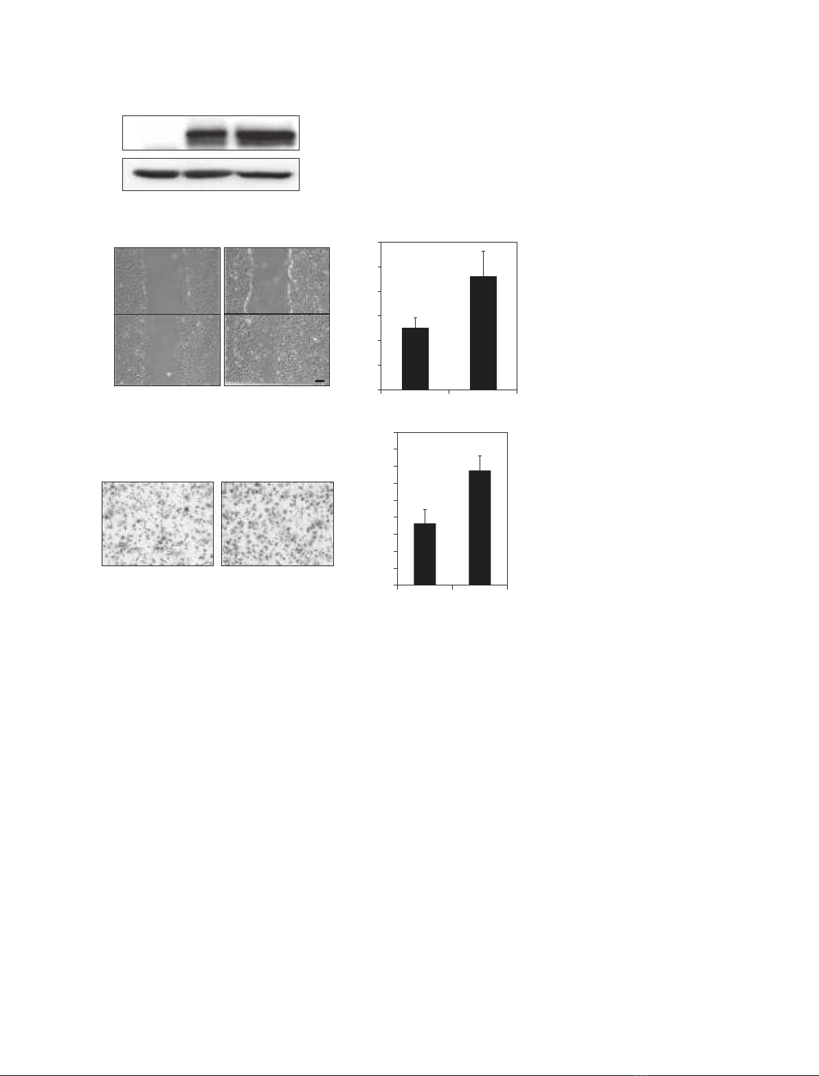
Integrin-mediated activation of Cdc42 is essential for cell polarization, whereas the integrin adaptor protein Cas is required for cell migration dur- ing wound healing. After phosphorylation on tyrosine residues, Cas recruits the adaptor proteins Crk and Nck to execute integrin-mediated signals. However, the mechanisms leading to Cdc42 activation and its relationship with Cas, Crk and Nck have not been elucidated clearly. In the present study, we demonstrate that Cas utilizes Nck2 to activate Cdc42 and induce cell polarization in response to wounding. By contrast, Cas recruits CrkII to activate Rac1 and promote the extension of cell protrusions needed for cell motility. These results indicate that Cas utilizes Nck2 and CrkII in a coordinated set of distinct pathways leading to cell migration.
(Received 14 April 2010, revised 1 June 2010, accepted 28 June 2010)
Structured digital abstract
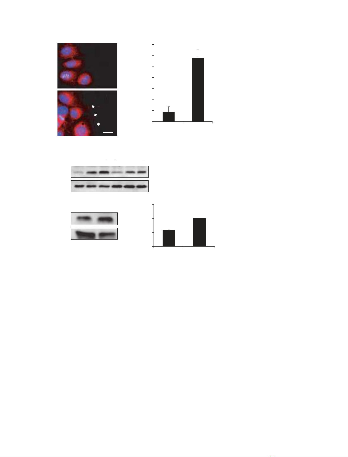
 l MINT-7909509: Cas (uniprotkb:Q61140) and Nck2 (uniprotkb:Q8BQ28) colocalize (MI:0403)
doi:10.1111/j.1742-4658.2010.07752.x
by fluorescence microscopy (MI:0416)
Introduction
polarity is determined by extracellular stimuli, such as chemoattractant gradients and cell–cell contact. Locali- zation and activation of Cdc42 in response to these environmental changes are key events leading to cell polarization [5,6].
The establishment of cell polarity is essential for a variety of cellular functions, such as cell division, dif- the molecular ferentiation and migration; however, mechanisms underlying cell polarization have not been elucidated thoroughly. Genetic and cell biological stud- ies have identified several molecules that are important for cell polarity. Among these proteins, Cdc42, a Rho family GTPase conserved in a wide range of organ- isms, has been found to play a pivotal role for the establishment of cell polarity [1–3]. In yeast, Cdc42 is required for polarized bud formation during cell divi- sion and morphological changes in response to phero- mone signaling [4]. In multicellular organisms, cell
Cas is a multiadaptor protein that regulates various signaling pathways in response to extracellular stimuli, including growth factors and integrin-mediated cell adhesion [7–9]. Cas was originally identified as a highly phosphorylated protein in cells transformed by v-Src and v-Crk [10,11]. Cas contains an N-terminal SH3 domain, proline-rich regions and a substrate domain with multiple tyrosine phosphorylation sites
Abbreviations CasKo, homozygous null Cas knockout; CasWt, CasKo transfected with wild-type Cas; DAPI, 4¢,6¢-diamino-2-phenylindole dihydrochloride; GST, glutathione S-transferase; PAK, p21-activated kinase; PBD, p21 binding domain; PIX, PAK-interacting guanine nucleotide exchange factor; PP2, 4-amino-5-(4-chlorophenyl)-7-(t-butyl)pyrazolo[3,4-D]pyramidine; siRNA, small interfering siRNA.
FEBS Journal 277 (2010) 3502–3513 ª 2010 The Authors Journal compilation ª 2010 FEBS
3502
K. Funasaka et al.
Cas ⁄ Nck2 regulates cell polarity
that associate with SH2 domains to direct protein interactions mediating signaling events leading to cell migration [12,13].
the wound healing assay, CasWt cells also migrated approximately 40% better than CasKo cells through a modified Boyden chamber (Fig. 1C).
Because cell polarization is an important prelude to migration [26], we examined the effects of Cas on cell polarization in response to wound healing. As shown in Fig. 2A, CasWt cells at the wound edge started to extend protrusions toward the free space within 4 h, and over 90% of the cells at the edge were polarized, with one side pointed toward the wound within 6 h. By contrast, < 10% of the CasKo cells at the wound edge displayed a polarized morphology 6 h after the wound was made.
Cas is ubiquitously expressed and its deletion in mice is embryonic lethal [14]. Fibroblasts derived from Cas-deficient mice showed cytoskeletal abnormalities and defects in cell migration and spreading, indicating an essential role of Cas for integrin-mediated signals [15]. Tyrosine phosphorylation of Cas is mostly medi- ated by the Src family kinases, and its phosphorylation is required for Cas-mediated cell migration and trans- formation [16–19]. Phosphorylated Cas recruits adap- tor proteins such as Crk and Nck [20–22]. Association of Crk with Cas enhances cell migration and spreading by activating Rac1 [23]. Nck is important for regulat- ing signals from cell surface receptors to the actin cystoskeleton, as well as for cell movement. A number of signaling molecules have been found to associate with Nck; however, the physiological importance of these interactions remains uncertain [24].
Measurement of protrusion length also indicated that Cas was required for the formation of cell protru- sions. As shown in Fig. 2B, CasWt cells exhibited cell protrusions with a length of 61 ± 23 lm (mean ± SD) by 3 h after wounding. This was almost twice the aver- age protrusion length exhibited by CasKo cells, which measured 35 ± 15 lm.
Microtubule elongation forms toward the leading edge of cells during wound healing [1]. Tubulin stain- ing indicates that Cas promoted this directional forma- tion of microtubules within 3 h after wounding. As shown in Fig. 2C, elongation of microtubules between the nucleus and wound was observed in over 80% of the CasWt cells on the wound edge. By contrast, < 10% of the CasKo cells displayed this directional organization of microtubules.
the
A wound-healing assay comprises a simple in vitro is experiment used to examine cell migration that enabled as a result of the release of physical con- straints. A scratch in the confluent monolayer initiates cell migration in the direction perpendicular to the scratch until the gap is filled with cells [3]. Several hours after the wound is made, cells on the edge of the wound develop a polarized morphology [1]. Polarized cells on the wound edge extend membrane protrusions and reorient the Golgi in the direction of migration [25]. Integrin-mediated activation of Cdc42 has been shown to be critical for this polarization during cell migration [1]; however, signaling molecules involved in the integrin-mediated activation of Cdc42 remain unknown. In the present study, we show that Cas utilizes Nck2 to regulate cell polarization and Cdc42 activity during cell migration in response to wound healing.
Results
Cas is required for the polarization of migrating cells
When cells are polarized for migration, the Golgi becomes oriented between the nucleus and the direc- tion of migration [3]. To examine the effects of Cas on Golgi orientation, the localization of the Golgi matrix protein, GM130 [27], was examined in CasKo and CasWt cells on the wound edge after wounding. As shown in Fig. 2D, polarized localization of the Golgi in CasKo cells was clearly delayed compared to that of CasWt cells. Approximately one-third of the Golgi was localized within a 120(cid:2) arc between the nucleus and the wound edge upon the wounding, which was the result of chance because cells were sectioned into three 120(cid:2) arcs. Three hours after wounding, approximately two-thirds of CasWt showed polarized localization of the Golgi, whereas < 40% of CasKo cells showed polarized localization of the Golgi.
Cas promotes Cdc42 activation and trafficking during wound healing
To examine the role of Cas in the establishment of cell polarity during cell migration, we performed a wound- healing assay using Cas deficient CasKo cells (homo- zygous null Cas knockout cells) and CasWt cells (generated by transfecting CasKo cells with wild-type Cas). Cas expression in CasWt cells was similar to that in Balb3T3 cells, and Cas was absent in CasKo cells (Fig. 1A). As shown in Fig. 1B, CasWt cells migrated faster than CasKo cells in this assay. In addition to
Cdc42 is a Rho GTPase that traffics to the leading edge of cell protrusions and regulates cell polarity dur- ing wound healing [1]. The effects of Cas on Cdc42 localization during wound healing were evaluated by
FEBS Journal 277 (2010) 3502–3513 ª 2010 The Authors Journal compilation ª 2010 FEBS
3503
K. Funasaka et al.
Cas ⁄ Nck2 regulates cell polarity
A
Balb3T3 CasKo CasWt
Cas
Actin
Wound healing assay (µm)
B
Distance of migration (24 h) CasKo CasWt 600
*
500 0 400
300
200 24 100 (h) 0 200 µm CasKo CasWt
l
450 400 Migration assay (3 h)
C
d e i f r e p s
l l
*
i
e c d e t a r g m
f o . o N
Fig. 1. Cas is essential for cell migration. (A) Western blot analysis of Cas in CasWt, CasKo and Balb3T3 cells. (B) Confluent monolayers of CasWt and CasKo cells were wounded with a pipette tip and incubated for 24 h. Data are the mean ± SD of the distance that leading edge of the monolayer traveled into the wound area in five randomly selected fields from three independent experiments (*P < 0.01); scale bar = 200 lm. (C) 5 · 104 CasKo and CasWt cells were loaded onto the upper surface of Boyden chambers, incubated for 3 h, fixed, and examined by microscopy. Cells that migrated to the lower surface of the chamber are shown as the mean ± SD from five randomly selected fields in three independent experiments (*P < 0.01).
in CasWt cells was almost twice that of CasKo cells (Fig. 3C).
Silencing of Cas in Balb3T3 cells inhibits cell polarization
immunofluorescence microscopy. As shown in Fig. 3A, whereas more than 50% of the CasWt cells at the wound edge contained Cdc42 localized on the leading edge, < 10% of the CasKo cells at the wound edge showed localization of Cdc42 on the leading edge. Thus, Cas is required for trafficking of Cdc42 to the leading edge of migrating cells.
To further evaluate the requirement of Cas for cell polarization, we used small interfering RNA (siRNA) to knockdown Cas expression in Balb3T3 cells. As shown in Fig. 4A, transfection with Cas siRNA effectively sup- pressed Cas expression. Three days after the transfection of either control or Cas siRNA, orientation of the Golgi during wound healing was examined by immunostain- ing. As shown in Fig. 4B, an average of 29 ± 3.8% of the cells transfected with Cas siRNA contained polar- ized Golgi by 3 h after wounding compared to an aver- age of 66 ± 3.2% seen in control transfectants.
In addition to intracellular location, the effects of Cas on Cdc42 activation were also examined. A previ- ous study demonstrated the activation of Cdc42 during wound healing [1]. Cdc42 activity was assessed by affinity precipitation of Cdc42-GTP with a glutathione S-transferase–p21-activated kinase–p21 binding domain (GST-PAK-PBD) fusion protein. As shown in Fig. 3B, wound-induced activation of Cdc42 was reduced in CasKo cells compared to CasWt cells. To further con- firm the reduced activation of Cdc42 in CasKo cells, we examined the activity of Cdc42 in both cell lines 3 h after wounding. Three independent experiments demonstrated that the Cdc42 activity 3 h after wounding
In addition to reducing cell polarization, Cas siRNA transfection also reduced Cdc42 activation and traf- ficking during wound healing. As shown in Fig. 4C,
FEBS Journal 277 (2010) 3502–3513 ª 2010 The Authors Journal compilation ª 2010 FEBS
3504
350 CasKo CasWt 300 250 200 150 100 50 0 CasKo CasWt
K. Funasaka et al.
Cas ⁄ Nck2 regulates cell polarity
6 (h) 0 2 4 A
CasKo
CasWt
100 µm
i
n o s u r t
)
o r p l l
*
m µ (
i
90 80 70 60 50 40 30 20 10 0
e c f o e c n a t s D
B C CasKo CasWt
20 µm CasKo CasWt
h t i
w s
l l
l
*
*
l
i g o g d e z i r a o p
e c f o e g a t n e c r e P
80 70 60 50 40 30 20 10 0
CasKo CasWt D CasKo CasWt
Fig. 2. Cas promotes wound-induced cell polarization. (A) Confluent monolayers of CasKo and CasWt cells were wounded and cells were incubated at 37 (cid:2)C with 5% CO2. Photographs were taken at the indicated time points (scale bar = 100 lm). (B) Three hours after wounding, the cells were fixed, immunostained with anti-a-tubulin serum and DAPI, and the length of the protrusions of wound edge cells was measured. Thirty cells in randomly selected fields were mea- sured in each of three independent experi- ments. Data are the distance (mean ± SD) between the leading edge and the nucleus (*P < 0.01). (C) Three hours after wounding, the cells were fixed and immunostained with anti-a-tubulin serum and DAPI (scale bar = 20 lm). (D) CasWt and CasKo cells were wounded, fixed and immunostained with anti-GM130 serum and DAPI at the indicated time points to evaluate the per- centage of cells with Golgi located in the 120(cid:2) arc facing the wound. One hundred cells were evaluated for Golgi localization in each of two independent experiments. Data are the mean ± SEM (*P < 0.01). Images on the right panel are representative images of immunostained cells 3 h after wounding. White lines indicate wound direction (green, GM130; blue, DAPI; scale bar = 20 lm).
cells transfected with Cas siRNA exhibited approxi- mately half of the Cdc42 activity found in control transfectants 3 h after wounding. Cdc42 was also evi- dent at the ends of cell protrusions on the wound edge in control transfectants, although it was not detected in cells transfected with Cas siRNA (Fig. 4D). Taken together with the results obtained from Cas knockout cells, these data indicate that Cas is an important com- ponent of the signaling cascade that directs cell polari- zation, Cdc42 activity and cell migration in response to wound healing.
phosphorylation was needed for the establishment of polarity during wound healing. As shown in Fig. 5A, tyrosine phosphorylation of Cas was induced by wounding, which was effectively suppressed by PP2 treatment. This inhibition of Cas phosphorylation by PP2 caused a decrease in cell elongation during wound healing. As shown in Fig. 5B, cells treated with PP2 did not extend protrusions into the wound area within 6 h after wounding. In addition, PP2 treatment reduced Golgi mobilization between the nucleus and wound edge to levels seen in CasKo cells (Fig. 5C). These data suggest that Src phosphorylates Cas to induce cell polarization and migration during wound healing.
Src kinase inhibition disrupts polarization of migrating cells
Nck2 is crucial for cell polarization and Cdc42 activation during wound healing
The Src tyrosine kinase phosphorylates Cas to pro- mote cell migration [18]. We employed a Src kinase [4-amino-5-(4-chlorophenyl)-7-(t-butyl)pyra- inhibitor zolo[3,4-d]pyramidine; PP2] to determine whether Cas
Crk and Nck are adaptor proteins that can associate with phosphorylated tyrosine residues of Cas [8]. Two
FEBS Journal 277 (2010) 3502–3513 ª 2010 The Authors Journal compilation ª 2010 FEBS
3505
20 µm 0 h 1 h 3 h 6 h
K. Funasaka et al.
Cas ⁄ Nck2 regulates cell polarity
70
A
h t i
i
60
w s
l l
CasKo 50
40
e g d e g n d a e l e h t n o d e z
i l
30
20
*
e c f o e g a t n e c r e P
a c o l 2 4 c d C
10 CasWt 0 CasKo CasWt
20 µm
CasKo
B
Time (h) 0 3 0 1 3 CasWt 1
Active Cdc42
Total Cdc42
1.5 CasWt CasKo
C
1 Active Cdc42
l
2 4 c d C e v i t c a
f o o i t a r e v i t a e R
0.5 Total Cdc42
Fig. 3. Cas promotes Cdc42 activation and intracellular trafficking during wound healing. (A) Three hours after wounding, cells were fixed and immunostained for Cdc42. DAPI was used to stain nuclei. Arrows indicate Cdc42 localized on the leading edge. Fifty cells on the wound edge in each of three independent experiments were evaluated for the localization of Cdc42. The percent- age of these cells with Cdc42 localized on the leading edge is presented as the mean ± SEM (*P < 0.01). (B) Forty scratches were made on the confluent monolayers of cells, and cells were lysed at the indicated time points to detect total Cdc42 and active, GTP bound, Cdc42. (C) Forty scratches were made and, 3 h later, cells were lysed to detect total and active Cdc42. Three independent experiments were performed and relative ratios of Cdc42 activity are shown as the mean ± SD. A representative result from the western blotting is shown.
Crk family members, CrkII and CrkL, can associate with phosphorylated Cas to regulate the actin cytoskel- eton, cell migration, invasion and survival [28,29]. The Nck family has two known members, Nck1 and Nck2, and both proteins can associate with phosphorylated Cas [20,22,24].
cells. As
Nck2 knockdown cells compared to either control or CrkII siRNA-transfected cells (Fig. 6D, E). Interest- ingly, the elongation of protrusions was reduced in CrkII knockdown cells but not in Nck2 knockdown cells (Fig. 6D, F). CrkII siRNA reduced CrkII expres- sion by approximately 50%, leading to a significant reduction in cell protrusion distance of approximately 30% compared to control cells (t-test: P < 0.01). These results indicate that Cas ⁄ CrkII association was required for the formation of protrusions, whereas Cas ⁄ Nck2 association was essential for the polariza- tion of cells.
As shown in Fig. 6A, Crk and Nck proteins were expressed to similar levels in CasKo and CasWt cells. We performed siRNA knockdown experiments to determine whether these proteins were involved in the Cas-mediated polarization of shown in Fig. 6B, transfection of specific siRNA to CasWt cells effectively suppressed the expression of target Crk or Nck proteins, but not other proteins. Cells transfected with Nck2 siRNA displayed significantly less polarized Golgi than other transfectants during wound healing, indicating that Nck2 played a critical role in the polar- ization of CasWt cells.
In addition to inhibiting orientation of the Golgi, cell protrusions were more randomly oriented in
To further confirm the role of Nck2 for cell polari- its localization and zation in cells expressing Cas, effects on Cdc42 activity during wound healing were examined. As shown in Fig. 7A, Nck2 co-localized with Cas on the leading edge of cells. By contrast, localization of Nck2 on the leading edge was not observed in CasKo cells (Fig. 7B), indicating that Cas was required for the polarized localization of Nck2.
FEBS Journal 277 (2010) 3502–3513 ª 2010 The Authors Journal compilation ª 2010 FEBS
3506
0 CasKo CasWt
K. Funasaka et al.
Cas ⁄ Nck2 regulates cell polarity
siRNA
A
Ctrl Cas
Cas
80
Actin
siRNA
B
70
h t i
60
w s
l l
l
50
40
*
l
30
i g o g d e z i r a o p
20
e c f o e g a t n e c r e P
10
0
Ctrl Cas
20 µm Ctrl Cas siRNA
siRNA
C
D
Ctrl Cas Active Cdc42
Total Cdc42 Ctrl siRNA
1.5
1
l
2 4 c d C e v i t c a
0.5
f o o i t a r e v i t a e R
Fig. 4. Silencing of Cas in Balb3T3 cells inhibits wound-induced cell polarization and activation of Cdc42. (A) Balb3T3 cells were transfected with either control or Cas siRNA and, 3 days later, cells were lysed and immunoblotted with anti-Cas serum. b-actin was used as a loading control. (B) Balb3T3 cells were transfected with either control or Cas siRNA and, 3 days later, cells were fixed 3 h after wounding and immuno- stained for GM130 to visualize the Golgi. One hundred cells were evaluated for the localization of the Golgi in each of three independent experiments. Data are the per- centage of cells (mean ± SEM, n = 300) dis- playing Golgi within the 120(cid:2) arc facing the wound (*P < 0.01). (C) Three days after siRNA transfection, Balb3T3 cells were scratched and then were examined 3 h later for Cdc42 activation. The relative activity of Cdc42 is indicated as a graph. (D) Three days after siRNA transfection, Balb3T3 cells were scratched and, 3 h later, cells were fixed and immunostained for Cdc42 expres- sion. Arrows indicate Cdc42 localized on the leading edge.
In addition to cell polarization, activation of Cdc42 during wound healing was dependent on Nck2. As shown in Fig. 7B, cells transfected with Nck2 siRNA displayed approximately 50% of the Cdc42 activity seen in control transfectants during wound healing, whereas depletion of CrkII did not affect Cdc42 acti- vation.
Discussion
which is an adaptor protein that mediates integrin sig- naling leading to cell migration, in cell polarization. In the present study, we found that Cas was essential for the polarization of migrating cells. Scratch-induced the elongation of protrusions and reorientation of Golgi were more prominent in cells that expressed Cas than in CasKo cells or cells treated with Cas siRNA. In addition, we found that activation and localization of Cdc42 on the leading edge of cells was disrupted in CasKo and Cas siRNA-transfected cells, indicating that Cas is crucial for the regulation of Cdc42 activity during cell polarization.
and wound healing
Polarization of cells in the direction of migration is required for the organized movement of cells during [5]. embryonic development Because integrin-mediated signaling pathways are cru- cial for cell polarization [1], we studied the role of Cas,
Multiple tyrosine residues in the substrate-binding domain of Cas are phosphorylated in response to vari- including integrin-mediated ous extracellular stimuli,
FEBS Journal 277 (2010) 3502–3513 ª 2010 The Authors Journal compilation ª 2010 FEBS
3507
Ctrl siRNA 0 Ctrl Cas siRNA 20 µm
K. Funasaka et al.
Cas ⁄ Nck2 regulates cell polarity
A
IP: Cas
Scratch (3 h) PP2 + – + + – –
pTyr
Cas
6 (h) 0 2 4
B
PP2 (10 µM)
Ctrl
100 µm
C
l
80
70
l
60
i g o g d e z i r a o p h t i
50
w s
l l
40
*
30
20
e c f o e g a t n e c r e P
10
Fig. 5. Src-mediated tyrosine phosphoryla- tion is required for wound-induced cell polar- ization. (A) CasWt cells were wounded and treated with 20 lM Src kinase inhibitor, PP2, for 3 h. Cell were lysed and immunoprecipi- tated with anti-Cas serum. Cells were immunoblotted with anti-phosphotyrosine and anti-Cas sera. (B) Confluent monolayers of CasWt cells were wounded and then incubated with dimethyl sulfoxide or PP2. Photographs were taken at the indicated time points (scale bar = 200 lm). (C) Wounded CasWt cells were incubated for 3 h with dimethyl sulfoxide or PP2 and immunostained with GM130 to visualize Golgi, and nuclei were stained with DAPI. Data are presented as the percentage of cells (mean ± SEM, n = 300) displaying Golgi that lied within the 120(cid:2) arc facing the wound. One hundred cells in each of three independent experiments were evaluated for Golgi localization (*P < 0.01).
adhesion. Among the tyrosine kinases required for integrin-mediated signal transduction, Src is critical for the phosphorylation of Cas [8]. PP2 treatment delayed the protrusion of cells toward the wound and dis- rupted reorientation of the Golgi in the direction of migration, which is consistent with the findings of pre- vious studies demonstrating that PP2 treatment dis- rupted polarization of astrocytes during migration [1]. Crk and Nck proteins are adaptor proteins that
associate with tyrosine-phosphorylated Cas through SH2 domains [20–22]. Interestingly, silencing of CrkII reduced the elongation of protrusions but did not disrupt the reorientation of the Golgi. Cas ⁄ CrkII asso- ciation regulates the activation of Rac via a functional cooperation with GTPase-activating protein DOCK180 [30–32]. Activation of Rac is essential for the for- mation of protrusions [1]; therefore, the Cas ⁄ CrkII pathway appears to regulate protrusion formation by
FEBS Journal 277 (2010) 3502–3513 ª 2010 The Authors Journal compilation ª 2010 FEBS
3508
0 Ctrl PP2
K. Funasaka et al.
Cas ⁄ Nck2 regulates cell polarity
siRNA
siRNA
A
B
Ctrl
Nck1
Ctrl
CrkII
CasKo CasWt
Nck1
CrkII
CrkII
Nck2
CrkL
CrkL
Actin
Actin
Nck1
siRNA
siRNA
Ctrl
Nck2
Ctrl
CrkL
Nck2
CrkL
Nck2
Actin
CrkII
Nck1
C
Actin
Actin
90
80
h t i
D
70
w s
l l
l
60
Ctrl siRNA
50
*
40
CrkII siRNA
Nck2 siRNA
l
30
i g o g d e z i r a o p
20
e c f o e g a t n e c r e P
10
0
Ctrl Nck1 Nck2 CrkII CrkL siRNA
20 µm
)
E
F
60
i
s
50
l l
i
40
*
30
m µ ( n o s u r t o r p l l
*
20
e c f o e g a t n e c r e P
10
s n o s u r t o r p l a n o i t c e r i d h t i
w
i
100 90 80 70 60 50 40 30 20 10 0
0
e c f o e c n a t s D
Ctrl CrkII Nck2 siRNA
Fig. 6. Nck2 is required for wound-induced cell polarization. (A) Expression of indicated proteins in CasKo and CasWt cells was examined by western blotting. (B) CasWt cells were transfected with the indicated siRNAs and, 3 days later, cells were lysed and expression of indicated proteins was evaluated by immunoblotting. (C) CasWt cells were transfected with the indicated siRNAs and, 3 days later, cells were fixed and immunostained with GM130 to visualize Golgi, and nuclei were stained with DAPI. The graph indicates the percentage of cells (mean ± SEM, n = 100) that have the Golgi in the 120(cid:2) arc facing the wound. (D) CasWt cells transfected with either Nck2 or CrkII siRNA were wounded and, 3 h later, cells were fixed and immunostained for a-tubulin and the nucleus. White lines indicate the wound direction (scale bar = 20 lm). (E) Data are presented as the percentage of cells (mean ± SEM, n = 150) displaying pro- trusions within the 60(cid:2) arc in the direction of migration. Fifty cells were counted in each of three independent experiments (*P < 0.01 compared to control and CrkII siRNA-transfected cells). (F) The length of the protrusions from cells on the wound edge was measured. Thirty cells were mea- sured in each of three independent experiments. Data are presented as distance (mean ± SD) between the leading edge and the nucleus (*P < 0.01 compared to control and Nck2 siRNA-transfected cells).
activating Rac during the wound-healing assay. In addition, knockdown of Nck2 expression resulted in randomly oriented protrusions and Golgi reorienta- tion, which indicates that the Cas ⁄ Nck2 pathway is essential for the establishment of cell polarity.
reported to specifically associate with Nck2. For exam- ple, Pinch1, which is an essential adaptor protein for integrin-mediated signaling, specifically interacts with the SH3 domain of Nck2 [33]. Signaling pathways spe- cifically regulated by Nck2 may mediate polarization; however, we cannot rule out the possibility that Nck2 is more abundantly expressed in CasWt cells and, thus, Nck2-knockdown resulted in a more significant disrup- tion of polarization than Nck1-knockdown did.
As shown in Fig. 8, these data suggest that Cas uti- lizes CrkII and Nck2 in parallel pathways to promote cell migration. Cas associates with Nck2 to activate Cdc42 and induce cell polarization. At the same time, Cas also associates with CrkII to induce Rac1 activa- tion, leading to cell protrusion and elongation.
Transfection of Nck1 siRNA into CasWt cells par- tially disrupted the reorientation of the Golgi. Nck1 the amino acid and Nck2 have 68% identity at sequence level and are considered to have redundant [24], although some proteins have been functions
We found that Nck2 was required for the activation of Cdc42 during wound healing. A previous study by Miyamoto et al. [34] reported that Nck1 was essential for the activation of Cdc42 by endothelin-1 stimula- tion. The same study also showed that the expression of a membrane-bound form of Nck1 was sufficient to activate Cdc42. These results suggest that there are
FEBS Journal 277 (2010) 3502–3513 ª 2010 The Authors Journal compilation ª 2010 FEBS
3509
K. Funasaka et al.
Cas ⁄ Nck2 regulates cell polarity
A
B
o K s a C
Nck2
t
W s a C
Cas Merge
20 µm 20 µm
1.5
C
l
2 4 c d C e v i t c a
f o o i t a r e v i t a e R
siRNA Ctrl Nck2 1 Active Cdc42 0.5 Total Cdc42 0 siRNA Ctrl Nck2 Ctrl CrkII siRNA Active Cdc42
Fig. 7. Nck2 is localized to the leading edge and is required for the activation of Cdc42. (A) Confluent monolayers of CasWt cells were wounded and, 3 h later, cells were fixed and immunostained for Cas and Nck2 (scale bars = 20 lm). (B) CasWt and CasKo cells were wounded and, 3 h later, cells were fixed and immunostained for Nck2. (C) CasWt cells were transfected with the indicated siRNAs and, 3 days later, cells were scratched and examined for Cdc42 activation. Three independent experiments were performed to measure Cdc42 activity in the absence of Nck2 and the graph indi- cates the relative activity of Cdc42 (mean ± SD).
Total Cdc42
Cas
Nck2
CrkII
Cdc42
Rac1
Polarization
Protrusion
Cell migration
Fig. 8. Schematic presentation of regulation of cell migration by Cas. Cas utilizes Nck2 to activate Cdc42 and induce cell polariza- tion. Cas also utilizes CrkII to augment Rac1 activity, leading to cell elongation. Acting together, these pathways result in cell migration.
biological activities, including actin cytoskeleton reor- ganization [35]. Recently, it was reported that PAK1 functions as a scaffold protein to regulate Cdc42 acti- vation [36]. In that case, PAK1 associates with both Gbc and PAK-interacting guanine nucleotide exchange to activate Cdc42 in response to factor a (aPIX) chemoattractants. bPIX, which has structural features similar to aPIX, has been reported to regulate Cdc42 activity during wound-healing assays [37,38]. The SH3 domain of bPIX associates with a nontypical proline- rich region of PAK1 [39], whereas the SH3 domain of Nck associates with the most N-terminal proline-rich region of PAK1 [40]. Because Cas associates with the SH2 domain of Nck, the protein complex of Cas– Nck2–PAK1–bPIX may play a role in Cdc42 activa- tion. Recent studies have also shown that Scrib, which is a multidomain scaffold protein, is localized to the leading edge of cells and regulates localization and activation of Cdc42 during cell polarization by inter- acting with bPIX [38,41].
important roles for Nck proteins in the regulation of Cdc42; however, the mechanism by which Nck pro- teins regulate Cdc42 activation has not been eluci- dated. PAK proteins are serine ⁄ threonine kinases that associate with Nck and are involved in a wide range of
In conclusion, in the present study, we have shown that Cas utilizes Nck2 to activate Cdc42 and induce cell polarization, whereas Cas also recruits CrkII to activate Rac1 to form cell protrusions and elongation for promotion of cell migration during wound healing.
FEBS Journal 277 (2010) 3502–3513 ª 2010 The Authors Journal compilation ª 2010 FEBS
3510
K. Funasaka et al.
Cas ⁄ Nck2 regulates cell polarity
Immunofluorescence analysis
Further studies will be required to elucidate more fully the roles of these focal adhesion proteins in cell polari- zation and migration.
Materials and methods
Cells, antibodies and reagents
Cells were cultured on glass coverslips coated with fibronec- tin. Confluent monolayers of cells were scratched with a pipette tip to achieve a wound of approximately 800 lm in width and incubated at 37 (cid:2)C with 5% CO2 for 3 h. Cells were fixed in 4% paraformaldehyde for 20 min, permeabi- lized with 0.5% Triton X-100 in NaCl ⁄ Pi for 5 min and incubated in 7% calf serum in NaCl ⁄ Pi for 30 min. Cells were incubated with primary antibody in NaCl ⁄ Pi for 1 h, washed with NaCl ⁄ Pi for 15 min, incubated with fluorescein isothiocyanate- or Alexa Fluor 594-labeled secondary anti- body in NaCl ⁄ Pi for 1 h, incubated with 4¢,6¢-diamino-2- phenylindole dihydrochloride (DAPI) for 5 min and then analyzed under a fluorescence microscope (BX60; Olympus, Tokyo, Japan).
Cdc42-activity assay
Cells from homozygous null Cas knockout mouse embryos were transfected with wild-type Cas (CasWt cells) or the parental transfection vector pBabeHygro (CasKo cells), selected for resistance to hygromycin, and maintained as described previously [14,18]. Clones were not taken for sub- sequent experiments to minimize potential effects of clonal variation. The antibodies used in the experiments were: anti-Cas, anti-GM130, anti-Nck1 and anti-Cdc42 sera (BD Transduction Laboratories, San Jose, CA, USA); anti-Crk serum (Cell Signaling, Danvers, MA, USA); anti-Nck2 serum (Millipore, Billerica, MA, USA); anti-CrkL serum (Santa Cruz Biotechnology, Santa Cruz, CA, USA); fluorescein isothiocyanate-conjugated anti-a-tubulin serum (Sigma, St Louis, MO, USA). PP2 was purchased from Funakoshi (Tokyo, Japan).
Cell migration assays
leading edge of
Forty scratches approximately 800 lm in width and the length of the dish were made on confluent monolayers of cells in 10 cm dishes. Cells were then incubated for 3 h, lysed with lysis buffer (Tris–HCl 25 mm, pH 7.4, NaCl 150 mm, MgCl2 10 mm, NP40 1%) with protease inhibitor cocktail (Roche Diagnostics, Basel, Switzerland) and centri- fuged at 21 880 g. for 20 min to remove cell debris. Cell incubated with GST-PAK-PBD (residues lysates were 67–150) fusion protein bound to glutathione-agarose beads for 1 h at 4 (cid:2)C. Beads were washed with lysis buffer four times and then subjected to western blotting with anti-Cdc42 serum to detect active Cdc42 protein bound to GST-PAK-PBD. Total Cdc42 protein was detected by immunoblotting total cell lysates.
Golgi reorientation measurements
surface of
Wound healing assays were performed by scratching con- fluent cell monolayers with a pipette tip and incubating at 37 (cid:2)C with 5% CO2. Twenty-four hours later, the distance that the monolayer traveled into the wound area was measured in five randomly selected fields from three independent experiments. To measure cell migration using Boyden chambers, 5 · 104 cells were seeded onto the upper surface of the chamber. The lower surface of the filter was coated with fibronectin. Three hours after seeding, cells were fixed with 70% metha- nol and stained with 0.5% of crystal violet. Cells that migrated to the lower the chambers were counted in five randomly selected fields from three inde- pendent experiments.
siRNA transfection
Measurement of Golgi reorientation was performed as described previously [3]. In brief, confluent cells that had been cultured on fibronectin-coated glass slides were scratched with a pipette tip and incubated for 3 h. Cells were fixed and stained for GM130 to visualize the Golgi. Cells on the wound edge were divided equally into three sectors, including the front sector between the nucleus and the leading edge. The Golgi in the front sector was deter- mined to be in the polarized position. One hundred cells in ten randomly selected fields were evaluated for Golgi locali- zation to determine the percentage of reoriented Golgi.
Measurement of protrusion orientation and length
Cells were transfected with each siRNA and, 3 days later when cells reached confluency, a scratch was made and the cells were fixed 3 h later. The cells were then stained with
siRNAs were designed and purchased from Sigma-Aldrich (St. Louis, MO, USA). The sequences of siRNAs were: Cas 5¢-UCAUUUGACUAAUAGUCUATT-3¢; Nck1 5¢-GGA UGAUUCCUGUCCCUUATT-3¢; Nck2 5¢-GGUCGCGA GGCUGUAUGUAGU-3¢; CrkL 5¢-CUUACUAGAUCCG UGAGUUAA-3¢; CrkII 5¢-GGAUCAACAGAAUCCCGA UTT-3¢; Control (designed to target luciferase) 5¢- CUUA CGCUGAGUACUUCGATT-3¢. Twenty nanomoles of siRNA was transfected into cells using Lipofectamine RNAiMAX (Invitrogen, Carlsbad, CA, USA) in accor- dance with the manufacturer’s instructions.
FEBS Journal 277 (2010) 3502–3513 ª 2010 The Authors Journal compilation ª 2010 FEBS
3511
K. Funasaka et al.
Cas ⁄ Nck2 regulates cell polarity
tyrosine-phosphorylated cellular proteins. Mol Cell Biol 9, 3951–3958.
12 Sakai R, Iwamatsu A, Hirano N, Ogawa S, Tanaka T, Mano H, Yazaki Y & Hirai H (1994) A novel signaling molecule, p130, forms stable complexes in vivo with v-Crk and v-Src in a tyrosine phosphorylation-dependent manner. EMBO J 13, 3748–3756.
13 Sakai R, Iwamatsu A, Hirano N, Ogawa S, Tanaka T,
anti-tubulin serum and DAPI nuclear stain. Protrusions of wound edge cells that had oriented within the 60(cid:2) arc in the direction of migration were regarded as directional protru- sions. Fifty cells in ten randomly selected fields were evalu- ated for directional protrusions in each of three independent experiments. To quantify the protrusions’ length, the dis- tance of the leading edge from the nuclei of wound edge cells was measured. In each of three independent experi- ments, 30 cells in randomly selected fields were evaluated to calculate the average length of these protrusions.
Nishida J, Yazaki Y & Hirai H (1994) Characterization, partial purification, and peptide sequencing of p130, the main phosphoprotein associated with v-Crk oncopro- tein. J Biol Chem 269, 32740–32746.
Acknowledgements
14 Honda H, Oda H, Nakamoto T, Honda Z, Sakai R,
Suzuki T, Saito T, Nakamura K, Nakao K, Ishikawa T et al. (1998) Cardiovascular anomaly, impaired actin bundling and resistance to Src-induced transformation in mice lacking p130Cas. Nat Genet 19, 361–365. 15 Honda H, Nakamoto T, Sakai R & Hirai H (1999)
We thank the members of the Division of Cancer Biol- ogy for helpful discussions and technical assistance. This research was funded in part by a grant from the Ministry of Education, Culture, Sports, Science and Technology of Japan.
References
p130(Cas), an assembling molecule of actin filaments, promotes cell movement, cell migration, and cell spreading in fibroblasts. Biochem Biophys Res Commun 262, 25–30.
16 Pellicena P & Miller WT (2001) Processive phosphoryla- tion of p130Cas by Src depends on SH3-polyproline interactions. J Biol Chem 276, 28190–28196.
1 Etienne-Manneville S & Hall A (2001) Integrin-medi- ated activation of Cdc42 controls cell polarity in migrating astrocytes through PKCzeta. Cell 106, 489–498.
2 Etienne-Manneville S & Hall A (2003) Rho GTPases in
cell biology. Nature 421, 753–756.
3 Nobes CD & Hall A (1999) Rho GTPases control
polarity, protrusion, and adhesion during cell move- ment. J Cell Biol 144, 1235–1244.
17 Huang J, Hamasaki H, Nakamoto T, Honda H, Hirai H, Saito M, Takato T & Sakai R (2002) Differential regulation of cell migration, actin stress fiber organiza- tion, and cell transformation by functional domains of Crk-associated substrate. J Biol Chem 277, 27265– 27272.
4 Chang F & Peter M (2003) Yeasts make their mark.
Nat Cell Biol 5, 294–299.
5 Etienne-Manneville S (2004) Cdc42 – the centre of
polarity. J Cell Sci 117, 1291–1300.
18 Goldberg GS, Alexander DB, Pellicena P, Zhang ZY, Tsuda H & Miller WT (2003) Src phosphorylates Cas on tyrosine 253 to promote migration of transformed cells. J Biol Chem 278, 46533–46540.
6 Fukata M, Nakagawa M & Kaibuchi K (2003)
19 Patwardhan P, Shen Y, Goldberg GS & Miller WT
Roles of Rho-family GTPases in cell polarisation and directional migration. Curr Opin Cell Biol 15, 590–597.
(2006) Individual Cas phosphorylation sites are dispens- able for processive phosphorylation by Src and anchor- age-independent cell growth. J Biol Chem 281, 20689– 20697.
7 Bouton AH, Riggins RB & Bruce-Staskal PJ (2001) Functions of the adapter protein Cas: signal conver- gence and the determination of cellular responses. Oncogene 20, 6448–6458.
20 Rivera GM, Antoku S, Gelkop S, Shin NY, Hanks SK, Pawson T & Mayer BJ (2006) Requirement of Nck adaptors for actin dynamics and cell migration stimu- lated by platelet-derived growth factor B. Proc Natl Acad Sci USA 103, 9536–9541.
8 Defilippi P, Di Stefano P & Cabodi S (2006) p130Cas: a versatile scaffold in signaling networks. Trends Cell Biol 16, 257–263.
9 O’Neill GM, Fashena SJ & Golemis EA (2000) Integrin signalling: a new Cas(t) of characters enters the stage. Trends Cell Biol 10, 111–119.
21 Burnham MR, Harte MT, Richardson A, Parsons JT & Bouton AH (1996) The identification of p130cas-bind- ing proteins and their role in cellular transformation. Oncogene 12, 2467–2472.
22 Schlaepfer DD, Broome MA & Hunter T (1997) Fibro-
10 Matsuda M, Mayer BJ, Fukui Y & Hanafusa H (1990) Binding of transforming protein, P47gag-crk, to a broad range of phosphotyrosine-containing proteins. Science 248, 1537–1539.
11 Reynolds AB, Kanner SB, Wang HC & Parsons JT
nectin-stimulated signaling from a focal adhesion kinase-c-Src complex: involvement of the Grb2, p130cas, and Nck adaptor proteins. Mol Cell Biol 17, 1702–1713.
(1989) Stable association of activated pp60src with two
FEBS Journal 277 (2010) 3502–3513 ª 2010 The Authors Journal compilation ª 2010 FEBS
3512
K. Funasaka et al.
Cas ⁄ Nck2 regulates cell polarity
23 Klemke RL, Leng J, Molander R, Brooks PC, Vuori K & Cheresh DA (1998) CAS ⁄ Crk coupling serves as a ‘‘molecular switch’’ for induction of cell migration. J Cell Biol 140, 961–972.
24 Buday L, Wunderlich L & Tamas P (2002) The Nck
33 Tu Y, Li F & Wu C (1998) Nck-2, a novel Src homol- ogy2 ⁄ 3-containing adaptor protein that interacts with the LIM-only protein PINCH and components of growth factor receptor kinase-signaling pathways. Mol Biol Cell 9, 3367–3382.
family of adapter proteins: regulators of actin cytoskele- ton. Cell Signal 14, 723–731.
34 Miyamoto Y, Yamauchi J, Mizuno N & Itoh H (2004) The adaptor protein Nck1 mediates endothelin A recep- tor-regulated cell migration through the Cdc42-depen- dent c-Jun N-terminal kinase pathway. J Biol Chem 279, 34336–34342.
25 Kupfer A, Louvard D & Singer SJ (1982) Polarization of the Golgi apparatus and the microtubule-organizing center in cultured fibroblasts at the edge of an experi- mental wound. Proc Natl Acad Sci USA 79, 2603–2607.
26 Hall A & Nobes CD (2000) Rho GTPases: molecular
35 Molli PR, Li DQ, Murray BW, Rayala SK & Kumar R (2009) PAK signaling in oncogenesis. Oncogene 28, 2545–2555.
switches that control the organization and dynamics of the actin cytoskeleton. Philos Trans R Soc Lond B Biol Sci 355, 965–970.
27 Nakamura N, Rabouille C, Watson R, Nilsson T, Hui
N, Slusarewicz P, Kreis TE & Warren G (1995) Charac- terization of a cis-Golgi matrix protein, GM130. J Cell Biol 131, 1715–1726.
28 Feller SM (2001) Crk family adaptors-signalling
36 Li Z, Hannigan M, Mo Z, Liu B, Lu W, Wu Y, Smrcka AV, Wu G, Li L, Liu M et al. (2003) Directional sensing requires G beta gamma-mediated PAK1 and PIX alpha- dependent activation of Cdc42. Cell 114, 215–227. 37 Cau J & Hall A (2005) Cdc42 controls the polarity of the actin and microtubule cytoskeletons through two distinct signal transduction pathways. J Cell Sci 118, 2579–2587.
complex formation and biological roles. Oncogene 20, 6348–6371.
29 Chodniewicz D & Klemke RL (2004) Regulation of
38 Osmani N, Vitale N, Borg JP & Etienne-Manneville S (2006) Scrib controls Cdc42 localization and activity to promote cell polarization during astrocyte migration. Curr Biol 16, 2395–2405.
integrin-mediated cellular responses through assembly of a CAS ⁄ Crk scaffold. Biochim Biophys Acta 1692, 63–76. 30 Kiyokawa E, Hashimoto Y, Kobayashi S, Sugimura H,
Kurata T & Matsuda M (1998) Activation of Rac1 by a Crk SH3-binding protein. DOCK180. Genes Dev 12, 3331–3336.
39 Mott HR, Nietlispach D, Evetts KA & Owen D (2005) Structural analysis of the SH3 domain of beta-PIX and its interaction with alpha-p21 activated kinase (PAK). Biochemistry 44, 10977–10983.
40 Bokoch GM, Wang Y, Bohl BP, Sells MA, Quilliam
LA & Knaus UG (1996) Interaction of the Nck adapter protein with p21-activated kinase (PAK1). J Biol Chem 271, 25746–25749.
31 Kiyokawa E, Hashimoto Y, Kurata T, Sugimura H & Matsuda M (1998) Evidence that DOCK180 up-regu- lates signals from the CrkII-p130(Cas) complex. J Biol Chem 273, 24479–24484.
32 Gu J, Sumida Y, Sanzen N & Sekiguchi K (2001) Lami- nin-10 ⁄ 11 and fibronectin differentially regulate inte- grin-dependent Rho and Rac activation via p130(Cas)- CrkII-DOCK180 pathway. J Biol Chem 276, 27090– 27097.
41 Dow LE, Elsum IA, King CL, Kinross KM, Richard- son HE & Humbert PO (2008) Loss of human Scribble cooperates with H-Ras to promote cell invasion through deregulation of MAPK signalling. Oncogene 27, 5988–6001.
FEBS Journal 277 (2010) 3502–3513 ª 2010 The Authors Journal compilation ª 2010 FEBS
3513